Hubbry Logo
Body fluidBody fluidMain
Open search
Body fluid
Community hub
Body fluid
logo
8 pages, 0 posts
0 subscribers
Be the first to start a discussion here.
Body fluid
Body fluid
from Wikipedia
Intracellular and extracellular fluid compartments. The extracellular fluid compartment is further subdivided into the interstitial fluid and the intravascular fluid compartments.

Body fluids, bodily fluids, or biofluids, sometimes body liquids, are liquids within the body of an organism.[1] In lean healthy adult men, the total body water is about 60% (60–67%) of the total body weight; it is usually slightly lower in women (52–55%).[2][3] The exact percentage of fluid relative to body weight is inversely proportional to the percentage of body fat. A lean 70 kg (150 lb) man, for example, has about 42 (42–47) liters of water in his body.

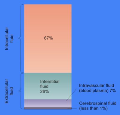
The total body of water is divided into fluid compartments,[1] between the intracellular fluid compartment (also called space, or volume) and the extracellular fluid (ECF) compartment (space, volume) in a two-to-one ratio: 28 (28–32) liters are inside cells and 14 (14–15) liters are outside cells.

The ECF compartment is divided into the interstitial fluid volume – the fluid outside both the cells and the blood vessels – and the intravascular volume (also called the vascular volume and blood plasma volume) – the fluid inside the blood vessels – in a three-to-one ratio: the interstitial fluid volume is about 12 liters; the vascular volume is about 4 liters.

The interstitial fluid compartment is divided into the lymphatic fluid compartment – about 2/3, or 8 (6–10) liters, and the transcellular fluid compartment (the remaining 1/3, or about 4 liters).[4]

The vascular volume is divided into the venous volume and the arterial volume; and the arterial volume has a conceptually useful but unmeasurable subcompartment called the effective arterial blood volume.[5]

Compartments by location

[edit]

Health

[edit]

Clinical samples

[edit]

Clinical samples are generally defined as non-infectious human or animal materials including blood, saliva, excreta, body tissue and tissue fluids, and also FDA-approved pharmaceuticals that are blood products.[7] In medical contexts, it is a specimen taken for diagnostic examination or evaluation, and for identification of disease or condition.[8]

See also

[edit]

References

[edit]

Further reading

[edit]
[edit]
Revisions and contributorsEdit on WikipediaRead on Wikipedia
from Grokipedia
Body fluids are the aqueous solutions present within the that serve as the primary medium for metabolic processes, transport, and waste elimination, comprising approximately 50-60% of an adult's total body weight and up to 75% in infants. These fluids are predominantly , with dissolved electrolytes, proteins, glucose, and other solutes, and are categorized into two main compartments: intracellular fluid (ICF), which occupies about 40% of body weight inside cells, and (ECF), accounting for roughly 20% outside cells. The ECF is further subdivided into intravascular fluid (plasma in blood vessels, ~5% of body weight), interstitial fluid (between cells, ~12-15%), and transcellular fluids (such as , , and ). The composition of body fluids varies by compartment to support specific functions; for instance, ICF is rich in , magnesium, and phosphates to facilitate cellular activities like reactions and , while ECF maintains higher sodium and levels to regulate osmotic balance and impulses. Intravascular fluids, including , transport oxygen, hormones, and nutrients throughout the body, while fluids enable the exchange of substances between blood and tissues. Transcellular fluids, though minor in volume, play critical roles in lubrication (e.g., in joints) and protection (e.g., cushioning the brain). Overall, these fluids maintain through mechanisms like and hydrostatic pressure, with daily fluid intake and output averaging 2,500 mL in adults to prevent imbalances that could lead to or . Body fluids are essential for physiological regulation, including pH balance, , and immune responses, and their osmolality is tightly controlled at around 286 mOsm/L to ensure proper cell function across compartments. Disruptions in fluid distribution, such as those caused by illness or injury, can result in conditions like or , underscoring the importance of in health. In clinical contexts, analysis of body fluids (e.g., , , or ) provides diagnostic insights into levels, infections, and organ function.

Definition and Overview

Definition

Body fluids are the liquids present within the bodies of living organisms, consisting primarily of water-based solutions that contain electrolytes, proteins, and other solutes essential for supporting vital life processes. These fluids, such as and , serve as the internal medium facilitating interactions between cells and their environment. Biologically, body fluids are crucial for enabling cellular functions, including the delivery of nutrients and oxygen to tissues, the removal of products, and the maintenance of a stable internal environment through . They regulate osmolality and hydrostatic pressure to prevent cellular swelling or shrinkage, ensuring optimal biochemical reactions and overall physiological balance. Without these fluids, processes like transport, signaling, and defense would be impossible, underscoring their indispensable role in sustaining life. In adults, body fluids typically constitute 50-60% of total body weight, though this varies by age, sex, and species; for instance, infants have a higher proportion around 75%, while it decreases to about 50% in older adults due to increased fat mass. Primarily composed of , these fluids reach 91-92% water content in plasma, the liquid component of , allowing for efficient dissolution and of solutes. From an evolutionary standpoint, body fluids originated in simple aquatic organisms where their composition closely mirrored surrounding to maintain osmotic balance, evolving over time in complex multicellular systems to adapt to diverse environments like freshwater and terrestrial habitats through mechanisms such as active ion transport and waste .

Historical Context

The understanding of body fluids has evolved significantly over millennia, beginning with theories that framed in terms of fluid balances. Around 400 BCE, and his followers proposed the theory of the four humors—blood, phlegm, yellow bile, and black bile—as the fundamental fluids constituting the body, with disease arising from their imbalance or . This humoral doctrine posited that these fluids, produced by specific organs and associated with the four elements (air, water, fire, earth), influenced and , forming the cornerstone of Western medicine for centuries. In the medieval and periods, Roman physician (c. 129–216 CE) refined Hippocratic ideas through animal dissections, linking the humors more explicitly to organ functions and emphasizing their role in nutrition and waste elimination. Galen's system described blood as formed in the liver from ingested food, then distributed via veins, while arterial blood arose from the heart's refinement process, integrating humoral balance with early anatomical observations. By the , human dissections by figures like in the 1540s challenged some Galenic assertions, revealing more accurate fluid pathways in organs such as the liver and kidneys, though humoral theory persisted. A pivotal advancement came in 1628 with William Harvey's demonstration of blood circulation, establishing the heart as a driving continuous fluid flow through closed vessels, which laid foundational principles for later fluid dynamics studies. The 19th century marked a shift toward experimental physiology, with Claude Bernard introducing the concept of the "milieu intérieur" in the 1850s, describing body fluids as a stable internal environment essential for cellular function amid external changes. Concurrently, Ivan Pavlov's late-19th-century investigations into digestive secretions, using fistulated dogs to measure gastric and pancreatic fluids, revealed neural and hormonal controls over fluid composition, earning him the 1904 Nobel Prize. Early 20th-century discoveries further illuminated fluid specificity and regulation. In 1901, identified the ABO blood groups through serological experiments on plasma and red cells, explaining transfusion incompatibilities and advancing blood as a distinct body fluid. Around the , Lawrence Henderson's physicochemical analyses of blood established key equilibria for acid-base and balance, quantifying how buffers like maintain fluid stability.

Classification and Types

Major Types

Body fluids are broadly categorized into major types based on their anatomical location and primary physiological roles, encompassing systemic circulating fluids and those that support specific protective or mechanical functions. These include , which serves as the central transport medium; , integral to immune surveillance and ; cerebrospinal fluid (CSF), essential for neural protection; , vital for joint mobility; and serous fluids, which lubricate visceral surfaces. Each type maintains distinct compositions and volumes tailored to its function, contributing to overall . Blood constitutes the primary circulating body fluid, comprising approximately 55% plasma—a watery matrix containing proteins, electrolytes, and nutrients—and 45% formed elements, including red blood cells, , and platelets. Its key role involves oxygen transport, with in red blood cells binding and delivering oxygen from the lungs to tissues throughout the body. In a typical , blood volume averages about 5 liters, varying slightly by body size and sex. Lymph is a translucent fluid originating from interstitial spaces, formed as excess drains into lymphatic capillaries. It transports immune cells, such as lymphocytes, back to the bloodstream and carries dietary fats absorbed in the intestines via specialized lacteals. As part of the , circulates through vessels and nodes, aiding in and preventing tissue . Cerebrospinal fluid (CSF) is a clear, colorless produced primarily in the of the brain's ventricles at a rate of about 500 ml per day, though total volume remains stable through continuous circulation and reabsorption. It cushions the brain and against mechanical shock, while also facilitating nutrient delivery and waste removal within the . In adults, CSF volume is approximately 150 ml, with about 125 ml in subarachnoid spaces and 25 ml in ventricles. Synovial fluid occupies the cavities of diarthrodial s, serving as a viscous to minimize friction between articular during movement. Its lubricating properties derive mainly from high-molecular-weight secreted by synovial cells, which also provides shock absorption. Normal volume per is small, typically ranging from 0.5 to 4 ml, sufficient for joint function without excess accumulation. Other major types include serous fluids, such as pleural fluid in the , pericardial fluid around the heart, and in the . These thin, watery secretions from mesothelial cells act as lubricants, enabling frictionless gliding of organs against surrounding structures during respiration, cardiac contraction, and visceral movement. Under normal conditions, volumes are minimal—often 15–50 ml for and similarly low for pleural and peritoneal—to maintain potential spaces without compression.
TypeApproximate Volume (Adult)Primary ComponentsPrimary Location
5 litersPlasma (55%), formed elements (45%)Vascular system (arteries, veins)
Varies (total ~2–4 L/day flow) fluid, immune cells, Lymphatic vessels and nodes
CSF150 mlWater, electrolytes, low proteinsVentricles and
Synovial0.5–4 ml per , synovial proteinsSynovial joint cavities
Serous (e.g., pleural, pericardial, peritoneal)5–50 ml per cavitySerous transudate, minimal cellsSerous membrane-lined cavities

Specialized Fluids

Specialized fluids in the body are secreted by specific glands or organs to support localized physiological processes, such as , , , and ocular maintenance. These fluids differ from systemic ones like or fluid by their targeted compositions and functions, often involving enzymes, acids, or protective elements tailored to particular environments. Digestive fluids facilitate the breakdown of nutrients in the . , produced by the salivary glands, contains the enzyme , which initiates by hydrolyzing complex carbohydrates into simpler sugars like and . Gastric juice, secreted by the stomach's parietal and chief cells, includes (HCl) for creating an acidic environment ( 1.5–3.5) that activates , a that begins protein by cleaving bonds. , synthesized by hepatocytes in the liver and stored in the , consists of bile salts, , and phospholipids that emulsify dietary fats, increasing their surface area for enzymatic action by pancreatic lipases. , released from acinar cells in the exocrine , is rich in proenzymes such as (which activates to for further protein hydrolysis), along with to neutralize gastric acidity in the . Urinary fluids arise from the kidneys' and modification processes, serving as a medium for waste elimination while reflecting renal . Urine formation begins with glomerular in the renal corpuscles, producing a filtrate similar to plasma (about 180 liters daily) that is cell- and protein-free, containing , electrolytes, glucose, and . This filtrate then undergoes tubular (e.g., reclaiming most and nutrients) and (e.g., adding ions or drugs) in the , , distal tubule, and collecting duct, resulting in concentrated final (typically 1–2 liters daily) whose composition—such as levels or pH—indicates function in maintaining . Reproductive fluids support viability and fetal development. Seminal fluid, contributed by the , , and bulbourethral glands, provides a nutrient-rich medium (including , prostaglandins, and enzymes) that nourishes and facilitates their transport through the female reproductive tract during . , surrounding the in the , offers mechanical protection against compression and , while allowing ; its volume peaks at approximately 800 milliliters at term (around 34–36 weeks gestation). Ocular fluids maintain the eye's structural integrity and optical clarity. Aqueous humor, a transparent fluid produced by the ciliary body's non-pigmented , circulates through the anterior and posterior chambers to nourish avascular tissues like the lens and , while its production and drainage via the regulate at about 15 mmHg. Vitreous humor, a gel-like substance filling the posterior chamber, comprises water, , and , providing structural support to the and lens while transmitting light without distortion.

Composition and Properties

Chemical Composition

Body fluids are predominantly composed of , which serves as the primary facilitating the dissolution and transport of solutes. Across various types, constitutes 92-99% of the total volume, enabling biochemical reactions and maintaining fluidity. In plasma, for instance, accounts for approximately 92% of the composition, while (CSF) and approach 99% under normal conditions. Electrolytes form a critical ionic component, contributing to osmotic balance and electrical neutrality. Common cations and anions include sodium (Na⁺), potassium (K⁺), chloride (Cl⁻), and bicarbonate (HCO₃⁻). In plasma, typical concentrations are 140 mM for Na⁺, 4 mM for K⁺, 103 mM for Cl⁻, and 24 mM for HCO₃⁻, with intracellular fluids showing inverted ratios such as higher K⁺ (around 140 mM). These ions maintain an osmolarity of about 280-300 mOsm/L across most fluids, preventing cellular swelling or shrinkage. Proteins and colloids add colloidal osmotic pressure and structural elements. In plasma, albumins (approximately 35-50 g/L) and globulins (20-35 g/L) predominate, regulating fluid distribution. Secretions like and contain mucins, which provide and . Organic molecules include carbohydrates such as glucose (4-6 mM in plasma), nitrogenous compounds like (2.5-7.5 mM) and (total ~2-3 mM), and dissolved gases including oxygen (O₂) and (CO₂). The of body fluids is tightly regulated, typically ranging from 7.35 to 7.45 in to support enzymatic function. The plays a central role, governed by the equilibrium: CO2+H2OH2CO3H++HCO3\text{CO}_2 + \text{H}_2\text{O} \rightleftharpoons \text{H}_2\text{CO}_3 \rightleftharpoons \text{H}^+ + \text{HCO}_3^- This system, catalyzed by , absorbs or releases H⁺ to stabilize . While commonalities exist, compositions vary slightly by fluid type; for example, CSF has higher protein levels (0.15-0.45 g/L) than (normally <0.15 g/L), reflecting barrier functions and filtration processes.

Physical Properties

Body fluids exhibit distinct physical properties that govern their movement, distribution, and interactions within physiological systems. Density varies among fluid types due to differences in cellular and solute content; for instance, has a density of approximately 1.06 g/mL, primarily attributable to the presence of erythrocytes and other cellular components. In contrast, (CSF) is less dense at about 1.007 g/mL, reflecting its acellular, aqueous composition similar to plasma ultrafiltrate. Viscosity, a measure of resistance to flow, also differs across body fluids. Human plasma viscosity at 37°C ranges from 1.2 to 1.3 cP, which is roughly 1.5 to 2 times that of (0.69 cP at the same temperature), influenced by plasma proteins such as fibrinogen and globulins. demonstrates non-Newtonian behavior, specifically shear-thinning, where its decreases under increasing shear rates to facilitate during movement. Osmolality, the concentration of solute particles per kilogram of solvent, is tightly maintained in body fluids at 280–300 mOsm/kg to avoid osmotic imbalances that could cause cellular swelling or shrinkage. This property contributes to Starling forces, where gradients driven by osmolality regulate fluid exchange across capillary walls. Surface tension plays a critical role in certain compartments, such as the alveoli, where reduces it from approximately 50 dynes/cm (without surfactant) to around 25 dynes/cm, preventing alveolar collapse during expiration. Blood flow dynamics in vessels are predominantly laminar rather than turbulent, enabling efficient circulation without excessive energy loss. This laminar flow is described by Poiseuille's law, which states that Q=πr4ΔP8ηLQ = \frac{\pi r^4 \Delta P}{8 \eta L}, where rr is the vessel , ΔP\Delta P is the difference, η\eta is fluid , and LL is vessel length; the equation highlights the profound influence of radius and viscosity on flow. Body fluids are maintained at a core of 37°C, which affects gas —most respiratory gases exhibit decreased at this compared to cooler conditions, impacting oxygen and in .

Physiological Functions

Transport and Exchange

Body fluids, particularly , serve as the primary medium for transporting essential nutrients and gases throughout the organism. Oxygen is carried by in red blood cells, where its binding follows a sigmoidal dissociation that plots hemoglobin saturation against partial of oxygen (pO₂). This enables efficient oxygen loading in the lungs at high pO₂ (around 100 mmHg, achieving ~97% saturation) and unloading in tissues at lower pO₂ (around 40 mmHg, dropping to ~75% saturation), optimizing delivery based on metabolic demand. Nutrients such as glucose and are dissolved in plasma or bound to carrier proteins, facilitating their distribution from sites of absorption, like the intestines, to peripheral tissues via convective flow in the bloodstream. Waste removal is another critical transport function, exemplified by the elimination of , a of . diffuses freely across glomerular capillaries in the kidneys, where it is filtered into Bowman's space at a rate determined by the (GFR), typically approximately 125 mL/min in healthy adults. This process clears about 50-60% of filtered , with the remainder reabsorbed in tubules, ensuring efficient nitrogenous excretion while maintaining plasma . Substances exchange between body fluid compartments and cells through passive and active mechanisms embedded in biological . , the primary passive process, follows Fick's , where the flux (J) of a is proportional to the concentration (ΔC) across the membrane and inversely proportional to the distance (Δx), expressed as: J=DΔCΔxJ = -D \frac{\Delta C}{\Delta x} Here, D is the diffusion coefficient, which varies by size, solubility, and membrane properties, allowing small, nonpolar gases like O₂ and CO₂ to cross rapidly. counters electrochemical gradients using energy, as seen in the Na⁺/K⁺-ATPase pump, which hydrolyzes one ATP to expel three Na⁺ ions from the cell and import two K⁺ ions, establishing a sodium essential for secondary of nutrients like glucose. At the capillary level, fluid and solute exchange between blood and interstitial spaces is governed by hydrostatic and oncotic pressures, quantified by the Starling equation for net filtration (J_v): Jv=Kf[(PcPi)σ(πcπi)]J_v = K_f \left[ (P_c - P_i) - \sigma (\pi_c - \pi_i) \right] where K_f is the filtration coefficient (reflecting capillary permeability and surface area), P_c and P_i are capillary and interstitial hydrostatic pressures, σ is the reflection coefficient (measuring protein permeability), and π_c and π_i are capillary and interstitial oncotic pressures. This balance typically results in net filtration at the arterial end (driven by higher P_c) and reabsorption at the venous end (favored by higher π_c from plasma proteins), regulating interstitial fluid volume. The complements exchange by excess —up to 3 L/day not reabsorbed by venules—and returning it to the bloodstream via ducts joining the subclavian veins, thereby preventing accumulation in tissues. capillaries, with their permeable endothelial flaps, facilitate uptake of proteins and cells alongside , maintaining overall .

Regulatory Roles

Body fluids play a crucial role in maintaining physiological balance through various regulatory mechanisms, including buffering, , immune defense, and , distribution, and osmotic stability. The in is the primary mechanism for pH regulation, where ions (HCO₃⁻) react with excess hydrogen ions (H⁺) to form (H₂CO₃), which dissociates into water and (CO₂) for rapid elimination via the lungs, thereby preventing or . This open system allows continuous adjustment, with the kidneys providing longer-term control by reabsorbing or excreting as needed. In regulation, sweat serves as an evaporative coolant, where secretion from eccrine glands absorbs during from the skin surface, accounting for up to 22% of total loss under normal conditions and becoming the dominant mechanism during stress or exercise. Blood flow redistribution further aids by altering cutaneous circulation: increases skin blood flow to promote dissipation, while conserves core by reducing peripheral flow, ensuring internal stability. Body fluids contribute to immune function primarily through plasma proteins, which include antibodies (immunoglobulins) that neutralize pathogens by binding to antigens and the —a cascade of over 30 proteins that enhances , lyses microbes, and amplifies to mount an effective innate and adaptive defense. These soluble components in enable rapid systemic responses to infections without relying solely on cellular elements. Lubrication and protection are provided by specialized fluids that minimize friction and cushion vital structures. in joint cavities, rich in and lubricin, acts as a viscous to reduce shear forces on articular during movement, while also delivering nutrients via . Serous fluids, secreted by mesothelial cells lining pleural, pericardial, and peritoneal cavities, form thin lubricating layers that prevent and abrasion between organs and surrounding tissues during respiration and . (CSF) in the provides shock absorption by suspending the and in a buoyant medium, reducing mechanical stress from head movements or impacts and protecting neural tissue from compressive forces. Body fluids facilitate distribution as the primary medium for endocrine signaling, with hormones secreted by glands diffusing into interstitial fluid and then entering the bloodstream for transport to distant target cells, enabling coordinated regulation of , growth, and . Osmotic and volume stability in body fluids prevent or overhydration by maintaining concentrations and fluid distribution across compartments. Extracellular fluids, particularly plasma, regulate osmolarity through sodium and , where shifts in osmotic gradients drive water movement to equalize pressures and sustain cell volume, averting cellular shrinkage in or swelling in overhydration. This balance ensures overall fluid , with total comprising about 60% in adults, dynamically adjusted to support circulatory and tissue integrity.

Compartments and Distribution

Intracellular vs. Extracellular

Body fluids in humans are primarily divided into two major compartments: the intracellular fluid (ICF) and the (ECF). The ICF constitutes approximately two-thirds of total , amounting to about 28 liters in a 70 kg adult male, while the ECF makes up the remaining one-third, or roughly 14 liters. These proportions reflect the fluid's distribution within and outside cells, with the ICF filling the of all cells and serving as the medium for intracellular processes. The chemical compositions of the ICF and ECF differ markedly, particularly in major cations, to support distinct physiological roles. The ICF has a high concentration of about 140 mM and a low sodium concentration of around 10 mM, whereas the ECF exhibits the opposite: high sodium at 140 mM and low at 4 mM. These ionic gradients are maintained by the selective permeability of cell membranes, which incorporate channels and pumps to regulate the passage of solutes, and by the Donnan equilibrium, arising from the presence of non-diffusible charged proteins inside cells that influence distribution across the membrane. Total , the sum of ICF and ECF volumes, is typically measured using dilution techniques such as administration of deuterium oxide, a stable tracer that equilibrates throughout all before sampling and analysis. This method provides an accurate estimate, revealing variations by age and sex; for instance, total body water comprises about 60% of body weight in adult males but only 50% in adult females due to differences in body fat and muscle mass. In newborns, the percentage is higher, around 75-80%, decreasing progressively with age. Functionally, the ICF supports cellular , housing enzymes and organelles essential for energy production and biochemical reactions within cells. In contrast, the ECF facilitates systemic exchange, transporting nutrients, oxygen, and waste between cells and organs; it includes subcompartments like plasma and fluid, though these are not detailed here. This compartmentalization ensures osmotic balance and prevents unchecked mixing, with water freely diffusing across membranes to equalize osmotic pressures while ions remain segregated.

Subcompartments by Location

The (ECF) is spatially organized into distinct subcompartments that facilitate targeted physiological interactions: the intravascular plasma, the surrounding tissue cells, and the transcellular fluids within epithelial-lined cavities. These subcompartments collectively account for approximately one-third of total in a typical 70-kg adult , totaling about 14 L. Plasma, confined to the vascular system, comprises roughly 3 L and represents the fluid matrix for blood cells and solutes, enabling systemic transport. This volume equates to about 5% of body weight and is dynamically maintained through interactions with other compartments. fluid, the largest ECF subcompartment at approximately 11 L, bathes individual cells and tissues, providing a medium for local delivery, waste removal, and signaling; it constitutes the bulk of the non-vascular ECF and serves as an extension of the broader ECF milieu, differing from intracellular fluid in its extracellular positioning. Transcellular fluids form a minor portion of the ECF, totaling around 1 L (about 2.5% of total body water), and are sequestered in specialized cavities such as the (CSF) in the (~150 mL), lubricating major joints (e.g., 2–4 mL in the ), and other serous secretions. The subcompartments are interconnected via the vascular and lymphatic systems, distinguishing vascular (plasma) from non-vascular ( and transcellular) regions. Fluid exchange between plasma and spaces occurs across endothelium through small pores and endothelial clefts, driven by hydrostatic and oncotic pressures as described by the Starling equation; this results in a daily flux of approximately 20 L of fluid filtering from capillaries into the , with direct of most and lymphatic vessels draining the excess (~2–4 L/day) back to the circulation. Transcellular fluids, while more isolated, communicate indirectly with fluid through epithelial barriers, maintaining their specialized compositions. Renal dynamics further link these compartments, as the kidneys filter about 180 L of plasma-derived fluid daily through glomerular , with nearly all to preserve volume balance across the ECF. Organ-specific transcellular fluids occupy defined anatomical niches, supporting localized functions like cushioning and lubrication. Peritoneal fluid fills the abdominal cavity (~20–50 mL normally), facilitating organ mobility; pleural fluid lines the thoracic cavities (~5–15 mL per hemithorax), reducing friction during respiration; and pericardial fluid envelops the heart (~15–50 mL), minimizing motion-related wear. These volumes are tightly regulated to prevent interference with organ mechanics, with any shifts occurring via subtle exchanges with adjacent interstitial spaces.

Regulation and Homeostasis

Mechanisms of Balance

The maintains through a dynamic equilibrium between intake and output, ensuring stable volume and osmolarity across compartments. This is achieved via integrated physiological processes that adjust movement in response to internal and external cues, preventing or overhydration. Key mechanisms include renal processing, gastrointestinal absorption, insensible losses, neural feedback via , and capillary exchange governed by Starling forces. Renal regulation plays a central role in by modulating water excretion through glomerular filtration, tubular , and . In the kidneys, approximately 180 liters of plasma are filtered daily at the , with over 99% of the water reabsorbed in the , , distal tubule, and collecting duct to conserve volume. is fine-tuned by antidiuretic hormone (ADH), which increases permeability in the collecting duct by inserting water channels into the apical of principal cells, facilitating of water back into the bloodstream. of solutes like ions and further influences water retention, allowing the kidneys to produce volumes ranging from 0.5 to 20 liters per day depending on hydration status. Gastrointestinal absorption contributes to fluid intake, primarily through the ingestion of water and aqueous foods, with an average daily input of about 2 liters from beverages and diet, supplemented by metabolic production. This absorbed enters the bloodstream via the intestinal mucosa, where it is osmotically drawn into enterocytes alongside nutrients, balancing systemic needs. Output is matched accordingly, with fecal water loss typically minimal at 100-200 mL per day under normal conditions. Insensible losses represent passive evaporation from the skin and lungs, accounting for 0.5-1 liter per day in adults and contributing to ongoing turnover without conscious regulation. Through the skin, of occurs at a rate of 300-400 mL daily, while respiratory evaporation in the lungs adds a similar amount as humidified air is exhaled. These losses increase with environmental factors like heat or low but are essential for and . Feedback loops, particularly the thirst mechanism, ensure proactive adjustment of intake by detecting changes in plasma osmolarity. Osmoreceptors in the sense elevations in blood solute concentration, triggering sensations that prompt consumption to restore balance; this response activates within minutes of a 1-2% increase in osmolarity. The integrates these signals to maintain euvolemia, coordinating with renal mechanisms for efficient correction. At the capillary level, forces govern the distribution of fluid between intravascular and interstitial spaces, balancing and to prevent . Net pressure is determined by the interplay of hydrostatic pressures (pushing fluid out) and oncotic pressures (pulling fluid in), with arterial-end hydrostatic pressure favoring outflow (~35 mmHg) and venous-end oncotic pressure (~25 mmHg) promoting inflow. This equilibrium results in minimal net fluid gain or loss across most capillary beds, with lymphatics reclaiming any excess interstitial fluid. Overall daily fluid balance is expressed as total intake equaling total output, typically around 2.5-3 liters in adults: intake from beverages and food (~2 L), metabolic oxidation (~0.3 L); output via (~1.5 L), insensible losses (~0.9 L), and (~0.1 L). This equation underscores the body's capacity to adapt, with renal output adjusting most flexibly to maintain .

Hormonal and Neural Control

Body fluid dynamics are tightly regulated by hormonal signals from the endocrine system and neural inputs from the , which coordinate adjustments in renal , vascular tone, and fluid retention to maintain . Antidiuretic hormone (ADH), also known as , is synthesized in the and released from the gland in response to increased or decreased . ADH acts on V2 receptors in the renal collecting ducts to insert water channels, thereby enhancing water reabsorption and concentrating urine to conserve body water. Deficiency of ADH leads to , characterized by excessive dilute urine output and potential . Aldosterone, a secreted by the of the , promotes sodium retention and potassium excretion in the distal renal tubules and collecting ducts. This action indirectly facilitates water retention by , helping to expand volume. Aldosterone release is primarily stimulated through the renin-angiotensin-aldosterone system (RAAS), where low renal triggers juxtaglomerular cells in the to secrete renin, which cleaves circulating angiotensinogen into I. I is then converted to II by (ACE), predominantly in the lungs; II stimulates aldosterone secretion while also inducing to raise . In opposition, (ANP), secreted by atrial myocytes in response to atrial stretch from elevated , inhibits sodium reabsorption in the renal collecting ducts and suppresses aldosterone and renin release, thereby promoting and to reduce fluid overload. Neural control integrates with these hormonal pathways via located in the carotid sinuses and , which detect stretch from arterial changes and relay signals through the glossopharyngeal and vagus nerves to the nucleus tractus solitarius in the brainstem. Decreased activates sympathetic outflow from the rostral ventrolateral medulla, enhancing of arterioles and venules to redistribute fluid toward the central circulation. Sympathetic nerves also innervate the kidneys and , stimulating renin release and catecholamine secretion to amplify RAAS activation and vascular tone. Disruptions in these controls can impair ; for instance, from excessive aldosterone production causes sodium retention, volume expansion, and , often resistant to standard treatments.

Clinical and Health Aspects

Sampling and Analysis

Body fluid sampling in clinical settings involves standardized procedures to obtain specimens for diagnostic analysis while minimizing risks to patients. Blood sampling is one of the most common methods, typically performed via , where a needle is inserted into a , often in the , to collect for routine tests such as complete counts or chemistry panels. For assessing gases and acid-base balance, arterial blood sampling, or arterial sticks, is used, usually from the , employing a heparinized to prevent clotting without interfering with measurements. Anticoagulants like EDTA () are commonly added to tubes for hematological analyses, as it chelates calcium to inhibit clotting while preserving cell morphology. Cerebrospinal fluid (CSF) collection requires a , a procedure where a needle is inserted into the subarachnoid space between the L3-L4 or L4-L5 vertebrae to access CSF for evaluating infections or bleeding. The patient is positioned laterally or seated with the spine flexed to widen intervertebral spaces, and up to 10-20 mL of CSF is typically withdrawn into sterile tubes for sequential analysis. Contraindications include local or systemic at the puncture site, such as skin abscesses or bacteremia, to avoid introducing pathogens into the or causing . Urine sampling employs non-invasive techniques for assessing renal function and metabolic disorders. The clean-catch midstream method involves cleansing the genital area and collecting the middle portion of the stream to reduce contamination from , ideal for routine . For quantitative evaluations of analytes like protein or clearance, a 24-hour collection is performed, where all voided over 24 hours is gathered in a refrigerated container, starting after discarding the first morning void and ending with the next day's first void. Initial screening often uses tests, which detect parameters such as (normal 4.5-8.0) and glucose (negative in healthy individuals) through colorimetric reactions on reagent strips. Other body fluids are sampled via targeted aspirations. is obtained through , a sterile needle insertion into the space (e.g., ), yielding 1-5 mL for diagnosing or by analyzing , cell count, and crystals. Pleural fluid collection via involves ultrasound-guided needle insertion into the pleural space, typically removing 5-10 mL for diagnostic purposes to evaluate effusions for or , though larger volumes up to 1 L may be therapeutic. These procedures generally yield small volumes of 1-10 mL to suffice for needs while limiting patient discomfort. Laboratory analysis of body fluids employs precise techniques to quantify components. measurement, such as sodium, , and , relies on ion-selective electrodes (ISEs), which generate a potential difference proportional to activity in the sample via a selective , enabling rapid, automated assessment in clinical analyzers. For cellular and protein evaluation, is standard: wet mounts or stained smears under light identify cells (e.g., leukocytes indicating ) and crystals, while protein quantification uses methods like or to detect abnormalities such as elevated globulins in inflammatory fluids. Safety and ethical considerations are paramount in body fluid sampling. Informed consent must be obtained prior to procedures, explaining risks like bleeding or infection, benefits, and alternatives, ensuring patient autonomy in line with institutional review board guidelines. All collections adhere to sterile techniques, including skin antisepsis with chlorhexidine, use of sterile needles and containers, and gloving to prevent contamination or nosocomial infections. Reference ranges guide interpretation; for instance, normal plasma sodium concentration is 135-145 mmol/L, deviations from which may signal fluid imbalances requiring further clinical correlation.

Associated Disorders

Disorders of body fluids encompass a range of pathological conditions arising from imbalances in fluid , composition, or distribution, often leading to significant clinical consequences. , also known as volume depletion, occurs when there is a loss of (ECF), commonly caused by conditions such as , , excessive sweating, fever, or inadequate oral intake. Symptoms include , dry , decreased output, , , , and in severe cases, with and impaired organ . can be classified as isotonic, involving proportional loss of and solutes leading to ECF volume reduction without major osmotic shifts, or hypertonic, characterized by greater water loss relative to sodium, resulting in and cellular shrinkage. Edema represents an excess of ECF in the spaces, manifesting as due to increased interstitial fluid volume. Common causes include , which impairs venous return and elevates hydrostatic , and from conditions like or , reducing plasma and promoting fluid leakage into tissues. is categorized as pitting, where leaves a persistent indentation in the skin, often seen in cardiac or venous causes, versus non-pitting, which lacks indentation and is typical of lymphatic obstruction or . Electrolyte disorders frequently disrupt body fluid , with defined as serum sodium concentration below 135 mEq/L, potentially causing neurological symptoms such as , , seizures, and due to . , characterized by serum exceeding 5 mM, arises from renal failure, , or medications, leading to cardiac arrhythmias, , and potentially fatal conduction abnormalities like peaked T waves or on ECG. Acid-base imbalances in body fluids can profoundly affect regulation. Metabolic acidosis features low (HCO3-) levels, often below 22 mEq/L, as seen in where ketone production overwhelms buffering capacity, resulting in symptoms like , fatigue, and confusion. Respiratory alkalosis, conversely, involves reduced partial pressure of carbon dioxide (PaCO2) due to , leading to elevated and symptoms including , paresthesias, and . Specific abnormalities in distinct body fluids highlight localized pathologies. In (CSF), bacterial is associated with elevated (WBC) counts, typically exceeding 1,000 cells/mm³, predominantly neutrophils, indicating acute inflammation and aiding diagnosis via . in crystal-induced arthritis, such as or pseudogout, reveals diagnostic crystals: needle-shaped monosodium urate in or rhomboid in pseudogout, often accompanied by high WBC counts and inflammatory changes. Treatment of these disorders focuses on restoring fluid and balance. For and , intravenous (IV) fluids such as 0.9% normal saline are administered to replenish volume and correct sodium deficits, with monitoring to avoid overcorrection. In cases of from , diuretics promote fluid excretion, while for imbalances like , stabilizers like and insulin-glucose infusions shift intracellularly. Severe renal failure causing persistent fluid and derangements necessitates dialysis to remove excess solutes and fluid, preventing complications like arrhythmias or .

References

Add your contribution
Related Hubs
User Avatar
No comments yet.