Hubbry Logo
search
logo
2248241

Spirochaete

logo
Community Hub0 Subscribers
Read side by side
from Wikipedia

Spirochaetes
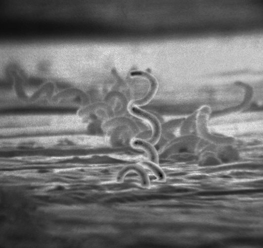
Treponema pallidum, a spirochaete which causes syphilis
Scientific classification Edit this classification
Domain: Bacteria
Kingdom: Pseudomonadati
Phylum: Spirochaetota
Garrity and Holt 2021[3]
Class: Spirochaetia
Paster 2020[1]: 471–563 [2]
Orders
Synonyms

Spirochaetota:

  • "Protozoobacteriales" Prévot 1958
  • "Spirochaetae" Cavalier-Smith 2002
  • "Spirochaetaeota" Oren et al. 2015
  • "Spirochaetes" Garrity and Holt 2001
  • "Spirochaetota" Whitman et al. 2018

Spirochaetia:

  • Spirochaetes Cavalier-Smith 2002
  • "Spirochaetia" Cavalier-Smith 2020
  • "Leptospiria" Cavalier-Smith 2020
Cross section of a spirochaete cell
Endoflagella Components. Legend: Fig. 1: A cross-section of a typical spirochete cell showing endoflagella located in the periplasm between the inner cytoplasmic membrane and the outer membrane. Periplasm, consisting of a gel-like matrix, provides a semi-stable medium to secure endoflagella during rotation. The axial filament, indicated in red, is composed of bundles of endoflagella.
Fig. 2: A side-view of a spirochete cell which shows two axial filaments in opposing motion. One axial filament rotates in a clockwise orientation; an adjacent axial filament rotates in a counter-clockwise orientation. Rotation of the endoflagella creates torsion and drives the corkscrew rotation of the cell.
Fig. 3: An expanded view of the cellular membranes that surround endoflagellum. Both the inner and outer membrane contain a phospholipid bi-layer, with non-polar fatty acid chains in-ward of polar phosphorus heads. Peptidoglycan, the cell wall, provides structure in bacterial microorganisms. Axial filaments are superior to the peptidoglycan.

A spirochaete (/ˈsprˌkt/)[4] or spirochete is a member of the phylum Spirochaetota (also called Spirochaetes[5] /ˌsprˈktz/), which contains distinctive diderm (double-membrane) Gram-negative bacteria, most of which have long, helically coiled (corkscrew-shaped or spiraled, hence the name) cells.[6] Spirochaetes are chemoheterotrophic in nature, with lengths between 3 and 500 μm and diameters around 0.09 to at least 3 μm.[7]

Spirochaetes are distinguished from other bacterial phyla by the location of their flagella, called endoflagella, or periplasmic flagella, which are sometimes called axial filaments.[8][9] Endoflagella are anchored at each end (pole) of the bacterium within the periplasmic space (between the inner and outer membranes) where they project backwards to extend the length of the cell.[10] These cause a twisting motion which allows the spirochaete to move. When reproducing, a spirochaete will undergo asexual transverse binary fission. Most spirochaetes are free-living and anaerobic, but there are numerous exceptions. Spirochaete bacteria are diverse in their pathogenic capacity and the ecological niches that they inhabit, as well as molecular characteristics including guanine-cytosine content and genome size.[11][12]

Pathogenicity

[edit]

Many organisms within the Spirochaetota phylum cause prevalent diseases. Pathogenic members of this phylum include the following:

Salvarsan, the first partially organic synthetic antimicrobial drug in medical history, was effective against spirochaetes and primarily used to cure syphilis. Additionally, oral spirochaetes are known to play a significant role in the pathogenesis of human periodontal disease.[17]

Taxonomy and molecular signatures

[edit]

The class currently consists of 14 validly named genera across four orders and five families.[18][19][20] The orders Brachyspirales, Brevinematales and Leptospirales each contain a single family, Brachyspiraceae, Brevinemataceae and Leptospiraceae, respectively. The Spirochaetales order harbours two families, Spirochaetaceae and Borreliaceae. Molecular markers in the form of conserved signature indels (CSIs) and CSPs have been found specific for each of the orders, with the exception of Brevinimetales, that provide a reliable means to demarcate these clades from one another within the diverse phylum.[19] Additional CSIs have been found exclusively shared by each family within the Spirochaetales. These molecular markers are in agreement with the observed phylogenetic tree branching of two monophyletic clades within the Spirochaetales order.[19] CSIs have also been found that further differentiate taxonomic groups within the Borreliaceae family that further delineate evolutionary relationships that are in accordance with physical characteristics such as pathogenicity (viz. Borrelia emend. Borreliella gen. nov.).[21] However, this study has been criticized, and other studies using different approaches do not support the proposed split.[22] The new naming system for the Lyme and relapsing fever Borrelia has not been adopted by the scientific literature.[22]

A CSI has also been found exclusively shared by all Spirochaetota species.[19] This CSI is a three-amino-acid insert in the flagellar basal body rod protein FlgC which is an important part of the unique endoflagellar structure shared by Spirochaetota species.[23] Given that the CSI is exclusively shared by members within this phylum, it has been postulated that it may be related to the characteristic flagellar properties observed among Spirochaetota species.[19][23]

Historically, all families belonging to the Spirochaetota phylum were assigned to a single order, the Spirochaetales.[11][12] However, the current taxonomic view is more connotative of accurate evolutionary relationships. The distribution of a CSI is indicative of shared ancestry within the clade for which it is specific. It thus functions as a synapomorphic characteristic, so that the distributions of different CSIs provide the means to identify different orders and families within the phylum and so justify the phylogenetic divisions.[19]

Phylogeny

[edit]
16S rRNA based LTP_10_2024[24][25][26] 120 marker proteins based GTDB 09-RS220[27][28][29]
Brevinematia
Spirochaetia
"Exilispirales"
"Exilispiraceae"

Exilispira

Brachyspirales
Spirochaetales
Marispirochaetaceae

Marispirochaeta

Sediminispirochaetaceae

Sediminispirochaeta

Spirochaeta thermophila

Salinispiraceae

Spirochaeta lutea

Salinispira

Spirochaetaceae

Spirochaeta

Alkalispirochaetaceae

Spirochaeta halophila

Alkalispirochaeta

Spirochaeta aurantia

Spirochaetaceae 2

Spirochaeta cellobiosiphila

"Leptospirae"
Leptospiria
Turneriellales
Turneriellaceae

Turneriella

Leptospirales
"Euspirochaetae"
"Exilispiria"
"Exilispirales"
"Exilispiraceae"

Exilispira

Spirochaetia
"Entomospirales"
"Entomospiraceae"

Entomospira

DSM‑17781
DSM‑17781

Spirochaeta cellobiosiphila

Spirochaetales_E
"Thiospirochaetaceae"

Thiospirochaeta

DSM‑2461

Spirochaeta species-group 2

Spirochaetaceae_B

Oceanispirochaeta

Spirochaetales_A
Spirochaetaceae_A

Spirochaeta thermophila

"Salinispirales"
"Salinispiraceae"

Spirochaeta lutea

Salinispira

DSM‑8902

Spirochaeta africana

"Alkalispirochaetaceae"

Alkalispirochaeta

"Marispirochaetales"
Marispirochaetaceae

Marispirochaeta

"Sediminispirochaetales"
Sediminispirochaetaceae

Sediminispirochaeta

Sphaerochaetales

Taxonomy

[edit]

The currently accepted taxonomy is based on the List of Prokaryotic names with Standing in Nomenclature (LPSN)[30] and National Center for Biotechnology Information (NCBI).[31]

  • Phylum Spirochaetota Garrity and Holt 2021
    • Genus ?"Spirosymplokos" Guerrero et al. 1993
    • Class Leptospiria Chuvochina et al. 2024
      • Order Turneriellales Chuvochina et al. 2024
        • Family Turneriellaceae Chuvochina et al. 2024
      • Order Leptospirales Gupta et al. 2014
    • Class Brachyspiria Chuvochina et al. 2024
    • Class Brevinematia Chuvochina et al. 2024
      • Order Brevinematales Gupta et al. 2014
        • Family "Longinemataceae" Karnachuk et al. 2021
          • Genus ?"Longinema" corrig. Karnachuk et al. 2021
        • Family Brevinemataceae Paster 2012
        • Family Thermospiraceae Ben Ali Gam et al. 2023
    • Class Spirochaetia Paster 2020
      • Order Spirochaetales Buchanan 1917
      • Order "Exilispirales" Pallen, Rodriguez-R & Alikhan 2022
        • Family "Exilispiraceae" Pallen, Rodriguez-R & Alikhan 2022
      • Order Borreliales Chuvochina et al. 2024
        • Family Borreliaceae Gupta et al. 2014
          • Genus Borrelia Swellengrebel 1907 (relapsing fever Borrelia, reptile-associated Borrelia and Echidna-associated Borrelia)
          • Genus Borreliella Adeolu & Gupta 2015 (Lyme disease Borrelia)
          • Genus ?Cristispira Gross 1910
      • Order "Entomospirales" Pallen, Rodriguez-R & Alikhan 2022
        • Family "Entomospiraceae" Pallen, Rodriguez-R & Alikhan 2022
          • Genus Entomospira Grana-Miraglia et al. 2024 non Enderlein 1917
      • Order "Marispirochaetales" Pallen, Rodriguez-R & Alikhan 2022
        • Family Marispirochaetaceae Chuvochina et al. 2024
      • Order "Sediminispirochaetales" Pallen, Rodriguez-R & Alikhan 2022
        • Family Sediminispirochaetaceae Chuvochina et al. 2024
      • Order Sphaerochaetales Chuvochina et al. 2024
      • Order Treponematales Chuvochina et al. 2024

See also

[edit]

References

[edit]
[edit]
Revisions and contributorsEdit on WikipediaRead on Wikipedia
from Grokipedia
Spirochaetes constitute a distinct phylum of Gram-negative bacteria, known as Spirochaetota (previously Spirochaetes), renowned for their unique helical or spiral-shaped morphology and motility propelled by endoflagella, which are internal flagella housed within the periplasmic space between the inner cytoplasmic and outer membranes.[1][2] These diderm prokaryotes, typically slender and flexible with cell dimensions ranging from 0.1 to 3 μm in width and 3.5 to 500 μm in length, exhibit either tightly coiled corkscrew forms or flat-wave serpentine shapes, enabling them to navigate viscous environments effectively.[3][1] The phylum encompasses diverse genera, including Treponema, Borrelia, Leptospira, and Spirochaeta, unified by molecular signatures such as conserved indels in proteins like FlgC, though they vary in lifestyle from free-living saprophytes to obligate parasites.[1] Motility arises from the rotation of these periplasmic flagella (also called axial filaments), which overlap in the cell's central region and generate torque up to 4000 pN nm—significantly higher than in externally flagellated bacteria like Escherichia coli—allowing translational, rotational, and undulating movements at speeds of 7–15 μm/s.[2] Unlike typical flagella in other bacteria, spirochete endoflagella are sequestered internally, providing a structural advantage for propulsion through dense media such as mucosal surfaces or biofilms.[2][3] Spirochaetes inhabit a broad range of ecological niches, from anaerobic marine sediments and freshwater soils to the gastrointestinal tracts of animals and the bloodstreams of vertebrates, with many species demonstrating aerobic, microaerophilic, or anaerobic metabolisms.[1] Taxonomically, the phylum Spirochaetota comprises the class Spirochaetia, which includes several orders such as Spirochaetales, Leptospirales, Brachyspirales, Brevinematales, and Winmispirales (as of 2025), and families including Spirochaetaceae and Leptospiraceae, reflecting phylogenomic revisions to better capture genetic diversity.[1][4] Notably pathogenic members include Treponema pallidum, the causative agent of syphilis; Borrelia burgdorferi, responsible for Lyme disease; and Leptospira interrogans, which causes leptospirosis, highlighting the phylum's medical significance despite the majority being non-pathogenic.[1][3]

Characteristics

Morphology

Spirochaetes are characterized by their distinctive helical or spiral-shaped cells, which exhibit irregular coiling and flexibility. These bacteria typically measure 5–250 μm in length and 0.1–3 μm in diameter, though some species can reach up to 500 μm long. The helical morphology arises from the rigid spiral of the protoplasmic cylinder, the core cellular component enclosed by the cell envelope, allowing the cells to maintain a corkscrew-like form under electron microscopy.[5] The cell wall of spirochaetes is flexible and diderm-like, consisting of an outer sheath (outer membrane), a thin peptidoglycan layer in the periplasmic space, and an inner cytoplasmic membrane. This structure lacks the thickness of peptidoglycan found in typical Gram-positive bacteria, resulting in poor reactivity to Gram staining and classification as Gram-indeterminate or weakly Gram-negative.[5] Electron microscopy reveals the peptidoglycan as a loosely cross-linked mesh, often containing unique amino acids like L-ornithine in free-living species such as Spirochaeta stenostrepta.[5] The outer sheath is lipoprotein-rich and protects the underlying protoplasmic cylinder, while the absence of surface structures like pili or fimbriae is a consistent ultrastructural feature across the phylum.[5] Endoflagella, also known as axial filaments or periplasmic flagella, are flagellar structures inserted subterminally at the poles of the cell and wrapped helically around the protoplasmic cylinder within the periplasmic space. These filaments, typically 15–20 nm in diameter, overlap in the cell center in many species and consist of a hook, filament, and basal body similar to external bacterial flagella.[5] High-resolution electron micrographs demonstrate their enclosure by the outer sheath, distinguishing them from external flagella in other bacteria.[2] Morphological variations occur among genera, reflecting adaptations to different environments. For instance, Borrelia species, such as B. burgdorferi, display loose, irregular coils or flat-wave shapes with wavelengths of approximately 2.8 μm and cell widths of 0.3 μm.[2] In contrast, Treponema species exhibit tighter, more regular helices with fine spirals, as seen in T. pallidum, which measures 6–15 μm in length and 0.1–0.2 μm in diameter.[5] These differences in coiling amplitude and pitch are evident in ultrastructural studies and correlate with the number and arrangement of endoflagella, such as 14–22 per cell in Borrelia.[2]

Motility

Spirochaetes exhibit a distinctive form of motility powered by the rotation of their endoflagella, which are located within the periplasmic space and drive the cell body to twist and translate in a propagating helical wave pattern.[6] This rotation generates propulsion without external flagella, allowing the bacteria to achieve swimming speeds of up to 15–20 μm/s, particularly in viscous environments where such motion is advantageous for navigating through mucus or biofilms.[7] The endoflagella function as internal rotary motors, anchored at the cell poles and overlapping in the periplasm, which enables the flexible, corkscrew-shaped cell body to deform and propel forward efficiently.[6] The motility of spirochaetes encompasses several distinct modes, including translational movement, spinning, and flexing, all orchestrated by the coordinated rotation of the endoflagella. In translational mode, the cell body advances linearly through undulation; spinning involves rotation around the long axis for reorientation; and flexing allows bending to navigate obstacles. These modes rely on the endoflagella acting as enclosed motors that transmit torque to the protoplasmic cylinder without direct environmental contact, distinguishing spirochaete locomotion from that of other flagellated bacteria.[6] Certain free-living spirochaetes, such as species of Leptospira and Spirosymplokos, demonstrate chemotaxis and phototaxis, enabling directed movement toward favorable chemical gradients or light sources, respectively. These behaviors are mediated by flagellar motor proteins, including components like CheY for signal transduction and the stator-rotor complex (MotA/MotB and FliG) for modulating rotation direction and speed in response to environmental stimuli. For instance, Spirosymplokos from microbial mats exhibits positive phototaxis, altering direction toward illuminated areas, which may aid in positioning within light-variable habitats.[8] Spirochaete motility adapts to different environments, with enhanced creeping observed on solid surfaces compared to free swimming in aqueous fluids.[9] On surfaces, the endoflagella facilitate adhesion and lateral crawling at reduced speeds, while in fluids, the helical wave propagation supports rapid, three-dimensional navigation; this versatility is evident in species like Leptospira, where surface motility enables host tissue penetration.[9] Such environmental responsiveness underscores the endoflagella's role in both propulsion and adaptation.[6] Experimental studies have visualized these dynamics using dark-field microscopy, which reveals the characteristic undulating and corkscrew motions of live spirochaetes, such as Treponema and Leptospira species, without staining interference.[10] These observations confirm the endoflagella-driven deformations, showing how rotation induces planar or helical waves that propagate from one cell end to the other, achieving net displacement.[10]

Physiology

Spirochaetes are predominantly chemoorganotrophic bacteria, relying on organic compounds for carbon and energy sources. Their metabolism is primarily fermentative or involves anaerobic respiration, with many species exhibiting obligate or facultative anaerobiosis; however, limited aerobic capabilities are observed in certain taxa, such as some members of the Spirochaetales, which can tolerate oxygen or function as microaerophiles.[11] For instance, fermentative pathways in genera like Treponema and Borrelia produce end products such as acetate, ethanol, and hydrogen gas from carbohydrates or amino acids, enabling survival in low-oxygen environments. These bacteria often depend on host-derived or environmental nutrients for growth, including amino acids and simple sugars, due to incomplete biosynthetic pathways. Many spirochaetes, particularly host-associated species like Treponema pallidum, lack genes for de novo synthesis of essential amino acids and certain vitamins, such as B vitamins (e.g., thiamine and biotin), necessitating exogenous supply for proliferation. This auxotrophy underscores their adaptation to nutrient-rich niches, where they scavenge pre-formed compounds rather than synthesizing them independently.[12] Cell division in spirochaetes occurs via binary fission, a process conserved across the phylum, resulting in transverse division that maintains their helical morphology. Generation times vary widely by species and environmental conditions, typically ranging from 4 to 33 hours; for example, Borrelia burgdorferi exhibits a doubling time of 12–24 hours in culture, while Treponema pallidum requires 30–33 hours. These rates reflect their adaptation to specific growth constraints, such as nutrient availability and temperature. Spirochaetes employ various stress responses to maintain viability under adverse conditions, particularly those affecting the cell envelope. Osmotic regulation involves mechanisms like the production of compatible solutes or adjustments in membrane composition to counter hypo- or hyperosmotic stress, as seen in Borrelia species where envelope integrity is crucial for survival. Antibiotic resistance is frequently linked to the unique cell envelope structure, including the absence of lipopolysaccharide in some genera (e.g., Borrelia), which confers intrinsic resistance to polymyxins by limiting drug penetration. Regulatory proteins like DksA further modulate transcriptional responses to stressors, including starvation and antimicrobial exposure, enhancing envelope stability. Genomic features significantly influence spirochaete physiology, with genomes typically ranging from 0.8 to ~5 Mb in size, reflecting streamlined metabolic capabilities and reliance on external nutrients in many species. For example, the genome of Treponema pallidum is approximately 1.1 Mb, encoding limited biosynthetic genes, while free-living species like Spirochaeta may approach 3–4 Mb with broader metabolic versatility. GC content varies considerably (24–66 mol%), correlating with ecological niches; low GC in Borrelia (around 28%) associates with AT-biased codon usage suited to host environments, whereas higher values in aquatic spirochaetes support diverse enzymatic functions.[11] These genomic traits underpin their physiological constraints and adaptations.

Ecology

Habitats

Spirochaetes are prevalent in various aquatic environments, including freshwater sediments, marine waters, and sulfidic muds, where free-living species such as those in the genus Spirochaeta often inhabit biofilms and microbial mats.[13][14] These bacteria thrive in nutrient-rich, moist settings like ponds, rivers, marshes, and coastal sediments, contributing to organic matter decomposition in these ecosystems.[15] Many spirochaetes exhibit adaptations to extreme conditions, particularly in anaerobic or low-oxygen sediments where obligately or facultatively anaerobic species predominate.[15] They are also found in high-salinity habitats, such as hypersaline ponds and soda lakes, with species like Spirochaeta halophila and Spirochaeta americana demonstrating tolerance to elevated salt concentrations through osmotic adaptations.[16][17] Their helical morphology and motility enable colonization of these challenging niches, facilitating movement through viscous sediments.[13] In microbial communities, spirochaetes play key roles in syntrophic associations, particularly in anaerobic digesters where certain cluster II spirochaetes participate in acetate oxidation coupled with hydrogenotrophic methanogenesis to enhance methane production.[18] Similarly, in termite hindguts, non-pathogenic symbiotic spirochaetes form dense populations that aid in lignocellulose degradation and nitrogen fixation, supporting host nutrition without causing disease.[19][20] Spirochaetes exhibit a global distribution, with higher diversity observed in tropical and temperate zones due to favorable warm, moist conditions that support their proliferation in sediments and waters.[21] They are ubiquitous across continents, from marine coastal areas to inland freshwater systems, reflecting their broad ecological adaptability.[22]

Interactions with Hosts

Spirochaetes form symbiotic and commensal associations with diverse hosts, contributing to microbial community stability and host physiology without inducing pathology. In mutualistic interactions, Treponema species in the hindguts of lower termites play a key role in lignocellulose degradation, converting complex carbohydrates into acetate via acetogenesis, which serves as a primary energy source for the host's nutrition on nitrogen-poor wood diets.[20] These spirochaetes also facilitate nitrogen fixation and lignin phenolic degradation, enhancing overall digestive efficiency through synergistic enzyme activity with host and other symbionts.[20] Commensal spirochaetes are integral to animal microbiomes, particularly in oral and gastrointestinal habitats. In humans, Treponema phylotypes constitute part of the normal oral flora, comprising over 49 taxa that maintain low-abundance presence across healthy sites like the gingiva and saliva, supporting community balance without disease association.[23] Similarly, in livestock such as cattle, novel Treponema isolates from the gastrointestinal tract, including the rumen and colon, cluster as distinct phylotypes that coexist as non-pathogenic symbionts, diversifying the microbial ecosystem.[24] Evolutionary adaptations enable spirochaetes to colonize host niches effectively, including biofilm formation on mucosal surfaces for persistence and protection. Proteins like CheWS, a SAM-binding sensor with a CheR-like domain, integrate chemotaxis signals to regulate pleomorphic states and biofilm development, allowing spirochaetes such as Treponema denticola to form mature biofilms in response to host molecules and symbionts, thereby facilitating stable host associations.[25] In non-human hosts, Cristispira species exemplify commensal symbiosis, residing abundantly in the crystalline styles of marine and freshwater mollusks' digestive tracts, where their motility and helical morphology support harmless integration into the host's microflora.[26] Rumen spirochaetes, including Treponema-like genera, similarly inhabit ruminant foreguts, contributing to fermentation without detriment. Recent findings from 2023–2025 indicate that habitat loss and urbanization disrupt these balances, reducing spirochaete abundance in wildlife guts—such as Treponema in rural versus urban-adapted populations—leading to dysbiosis and diminished microbial diversity tied to altered environmental pressures.[27]

Classification

Phylogeny

The phylum Spirochaetes represents a deep-branching, monophyletic lineage within the domain Bacteria, as established by phylogenetic analyses of 16S rRNA gene sequences that consistently place it as a distinct phylum separate from other major bacterial groups.[28] These analyses highlight its ancient origin, with the phylum's radiation predating many other bacterial lineages and showing no close affiliation to superphyla like PVC or Terrabacteria in standard 16S rRNA trees, though some phylogenomic studies suggest loose associations with Bacteroidetes in broader groupings.[29] The monophyly of Spirochaetes is further corroborated by the presence of conserved signature indels—unique insertions or deletions in proteins such as HSP60 and RNA polymerase subunits—that are exclusive to all members of the phylum and absent in other bacteria.[1] Phylogenetic reconstructions using multi-locus sequence analysis of concatenated proteins, including up to 75 conserved genes from genome-sequenced representatives, resolve the internal structure of Spirochaetes into major clades corresponding to the orders Spirochaetales, Leptospirales, Brachyspirales, and Brevinematales.[29] These clades exhibit deep branching patterns, with robust support for the monophyletic nature of the phylum, with bootstrap values exceeding 90% across key nodes, and reveal a basal position for Spirochaetales relative to the other orders.[30] Horizontal gene transfer (HGT) has significantly influenced the evolutionary trajectory of Spirochaetes, with genomic evidence indicating multiple acquisitions of genes from distantly related bacteria, particularly those involved in motility structures like the endoflagellar apparatus.[31] For instance, comparative genomics of genera like Sphaerochaeta shows extensive HGT from Clostridia-like donors, contributing to variations in flagellar and chemotaxis systems that underpin the phylum's characteristic helical swimming.[32] These events likely occurred throughout the phylum's history, enhancing adaptability without disrupting core phylogenetic signals.[33] Recent metagenomic studies from 2022 to 2025 have expanded the known phylogenetic diversity of Spirochaetes by identifying new uncultured lineages within existing orders, such as novel taxa in Spirochaetales from marine environments.[34] These discoveries, derived from high-throughput sequencing of environmental samples, also reveal spirochaete presence in hypersaline microbial mats.[35] Such findings underscore the phylum's ecological breadth and suggest that uncultured groups may represent basal branches predating the divergence of known orders. As of November 2025, the taxonomy reflects ongoing refinements based on these genomic insights, with no major new orders proposed since 2024.[36]

Taxonomy

The phylum Spirochaetota Garrity and Holt 2021 encompasses a diverse group of helical bacteria within the domain Bacteria and kingdom Pseudomonadati, with the class Spirochaetia Paster and Dewhirst 2000 as its sole class.[36][37] This class is subdivided into five orders: Spirochaetales Buchana 1917 (Approved Lists 1980), which includes predominantly free-living species; Leptospirales Garrity and Holt 2021, containing pathogenic forms; Borreliales Chuvochina et al. 2024, also featuring pathogenic taxa; Brachyspirales Garrity et al. 2021; and Brevinematales Garrity et al. 2021. The order Spirochaetales is the largest, housing the family Spirochaetaceae Swellengrebel 1907 (Approved Lists 1980), while Leptospirales includes the family Leptospiraceae Hovind-Hougen 1979 (Approved Lists 1980), and Borreliales comprises the family Borreliaceae Gupta et al. 2013.[38][39] Key genera illustrate the phylum's diversity: Spirochaeta Ehrenberg 1835 (etymology: Greek spira, coil, and chaite, hair; type genus of the phylum, with type species S. plicatilis Ehrenberg 1835 and type strain DSM 5503), encompassing free-living aquatic species; Treponema Schaudinn 1905 (etymology: Greek trepo, to turn, and nema, thread; type species T. pallidum (Schaudinn 1905) Schaudinn 1905, with type strain not applicable due to unculturability, but reference strain Nichols used); Leptospira Noguchi 1917 (etymology: Greek leptos, fine, and spira, coil; type species L. interrogans (Stimson 1907) Noguchi 1917, type strain RGA); and Borrelia Swellengrebel 1907 (etymology: named after Amédée Borrel; type species B. anserina (Sakharoff 1891) Swellengrebel 1907, type strain not specified, but reference strains like B31 for B. burgdorferi).[40][41] The taxonomy of Spirochaetota originated in the 19th century with Christian Gottfried Ehrenberg's 1835 description of Spirochaeta plicatilis from freshwater environments, establishing the foundational genus. A pivotal advancement occurred in 1905 when Fritz Schaudinn and Erich Hoffmann identified Treponema pallidum as the syphilis agent, distinguishing it from other spirochaetes via microscopy and linking it to human disease.[42] Early classifications grouped spirochaetes under a single order Spirochaetales, but 20th-century studies using serological and morphological criteria refined families like Spirochaetaceae and Leptospiraceae. Contemporary taxonomy has been reshaped by phylogenomic analyses and whole-genome sequencing, enabling precise delineation of monophyletic clades and resolving ambiguities in earlier schemes.[1] A 2013 proposal restructured the phylum into major lineages based on conserved protein sequences from 15 genomes, supporting the separation of pathogenic and free-living groups. As of 2025, over 100 validly named species are recognized across the phylum, each defined by type strains deposited in culture collections like DSMZ or ATCC to ensure nomenclatural stability and reproducibility.[36] Recent changes include the 2024 establishment of the order Borreliales, driven by genomic data from over 50 Borrelia strains that highlighted distinct evolutionary divergence from other spirochaetes, reclassifying the genus Borrelia and its relatives accordingly.[39]

Molecular Signatures

Spirochaetes are characterized by several molecular signatures that serve as reliable markers for their identification and phylogenetic classification. Conserved signature indels (CSIs) in widely distributed proteins provide synapomorphic traits unique to the phylum and its major clades. For instance, a 3-amino-acid insertion in the FlgC flagellar basal-body rod protein is exclusively shared by all sequenced spirochaetes, acting as a molecular marker for the entire phylum.[29] Additional CSIs delineate families and genera; the family Spirochaetaceae possesses seven CSIs, including a 15-amino-acid insertion in phosphoribosylpyrophosphate synthetase, while the family Leptospiraceae features five CSIs, such as an 8-amino-acid insertion in the 50S ribosomal protein L14.[29] These indels, identified through comparative genomic analyses, offer high specificity for taxonomic delineation independent of 16S rRNA-based methods.[29] The 16S rRNA gene sequences of spirochaetes exhibit signature motifs within their nine hypervariable regions (V1-V9) that distinguish major orders, enabling precise phylogenetic placement. These motifs, combined with conserved domains, facilitate the differentiation of orders like Spirochaetales, Brachyspirales, and Leptospirales through sequence comparisons.[29] For example, alignments of nearly complete 16S rRNA sequences reveal clade-specific nucleotide patterns that correlate with branching orders in phylogenetic trees constructed from multiple spirochaete representatives.[29] Genome architecture in spirochaetes includes distinctive features, particularly in pathogenic genera. Borrelia species possess a linear chromosome, a rare trait among bacteria that typically have circular chromosomes, alongside numerous linear and circular plasmids that encode essential virulence factors such as those involved in host adaptation and immune evasion.[43] These plasmids are critical for infectivity, as demonstrated by studies showing that loss of specific plasmids correlates with reduced virulence in Borrelia burgdorferi.[44] Proteomic signatures, notably the flaB gene encoding the flagellar filament core protein FlaB, are pivotal for diagnostics. The flaB gene's sequence variability allows for species-specific PCR amplification, offering higher sensitivity than 16S rRNA targets for detecting low-abundance spirochaetes in clinical samples.[45] Multiple flaB paralogs in spirochaetes contribute to flagellar structure and motility, with their protein products serving as serological markers in assays for pathogens like Borrelia.[46] Recent advances in CRISPR-based methods have enhanced rapid detection of spirochaetes in complex samples, including metagenomes. A CRISPR/Cas12a system targeting Borrelia sensu lato DNA achieves high sensitivity in human samples, detecting as few as 10 copies of pathogen DNA even in the presence of host genomic material, outperforming traditional qPCR in speed and specificity.[47] This approach, developed in 2025, facilitates point-of-care identification and metagenomic screening for spirochaete pathogens in environmental or clinical metagenomes.[47]

Pathogenicity

Human Pathogens

Spirochaetes encompass several genera that are significant human pathogens, primarily through direct transmission via sexual contact, arthropod vectors, or environmental exposure to contaminated water or soil. The most prominent include Treponema pallidum subsp. pallidum, the causative agent of syphilis; Borrelia burgdorferi sensu stricto, responsible for Lyme disease; and Leptospira interrogans, which causes leptospirosis. These bacteria exploit their unique helical morphology and motility to invade tissues and disseminate systemically, often evading early immune detection.[48][49][50][51] Treponema pallidum subsp. pallidum causes syphilis, a chronic multisystem infection transmitted primarily through sexual contact with infectious lesions during the primary or secondary stages, or congenitally from mother to fetus via the placenta. The disease progresses through distinct stages: the primary stage features a painless chancre at the site of inoculation, typically appearing 10–90 days post-exposure and healing without treatment; the secondary stage, occurring weeks to months later, involves a disseminated rash, mucous membrane lesions, and systemic symptoms like fever and lymphadenopathy; a latent phase follows, which may last years; and the tertiary stage can manifest as gummatous lesions, cardiovascular complications, or neurosyphilis affecting the central nervous system. Congenital syphilis arises from untreated maternal infection, leading to fetal risks including stillbirth, neonatal death, or developmental abnormalities.[48][49][52][53] Lyme disease, caused by Borrelia burgdorferi sensu stricto in North America, is transmitted through the bite of infected blacklegged ticks (Ixodes scapularis), with transmission requiring 36–48 hours of attachment. Early localized symptoms include erythema migrans, a characteristic expanding rash at the bite site in 70–80% of cases, accompanied by flu-like symptoms such as fever and fatigue; dissemination can lead to early neurologic manifestations like facial palsy or meningitis (neuroborreliosis), cardiac involvement, or arthritis. If untreated, late-stage disease may cause chronic arthritis, peripheral neuropathy, or encephalopathy.[50][54][55] Leptospirosis, primarily due to Leptospira interrogans serovars, is a zoonotic infection acquired through contact with urine from infected animals (e.g., rodents, livestock) contaminating water or soil, often during flooding or recreational activities in endemic areas. The illness presents in two phases: an acute septicemic phase with fever, headache, and myalgia, followed by an immune phase with potential organ involvement; severe cases progress to Weil's disease, characterized by jaundice, renal failure, hemorrhagic vasculitis, and high mortality (5–15%). Transmission is waterborne, with bacteria entering via cuts or mucous membranes, and outbreaks are common in tropical regions post-rainfall.[51][56][57] Key virulence factors among pathogenic spirochaetes include adhesins that facilitate host cell attachment and tissue invasion, such as the Erp and Rev proteins in Borrelia species, which bind extracellular matrix components. Borrelia burgdorferi employs antigenic variation through the vlsE locus, generating diverse surface lipoproteins to evade adaptive immunity, allowing persistent infection and relapse. These mechanisms, combined with motility enabling penetration of endothelial barriers, contribute to dissemination and chronicity.[58][59][60] Diagnosis of spirochaete infections relies on serological tests for antibody detection (e.g., treponemal and nontreponemal assays for syphilis, two-tier EIA followed by Western blot for Lyme disease) and molecular methods like PCR for direct pathogen identification, particularly in early or severe cases. Treatment involves antibiotics: penicillin G is the first-line for all stages of syphilis, administered parenterally; doxycycline or amoxicillin for early Lyme disease, with intravenous ceftriaxone for neuroborreliosis; and doxycycline or penicillin for leptospirosis, with supportive care for severe manifestations. Early intervention prevents progression, though post-treatment syndromes may occur in Lyme disease.[52][61][56] Epidemiologically, syphilis affects an estimated 8 million adults aged 15–49 globally in 2022, with projections reaching 9 million cases by 2025, driven by increases in the Americas and among men who have sex with men. Lyme disease sees approximately 476,000 diagnosed cases annually in the United States alone, with over 89,000 reported in 2023, primarily in the Northeast and Midwest; global estimates exceed 500,000 cases when including Europe. Leptospirosis causes over 1 million cases worldwide each year, resulting in nearly 60,000 deaths, with highest burdens in tropical Asia and the Americas.[49][62][63][51]

Non-Human Pathogens

Spirochaetes of the genus Borrelia cause significant diseases in various non-human animals, including canine Lyme disease and avian borreliosis. In dogs, Borrelia burgdorferi sensu lato, transmitted primarily by Ixodes ticks, leads to Lyme borreliosis characterized by fever, lethargy, lameness, and polyarthritis, with potential progression to renal involvement in severe cases.[64] This infection is prevalent in endemic areas, affecting companion animals and contributing to veterinary care costs through diagnosis and treatment with antibiotics like doxycycline.[65] In birds, particularly poultry such as chickens, Borrelia anserina causes avian borreliosis or fowl spirochaetosis, an acute septicemic disease transmitted by argasid ticks like Argas persicus, resulting in high mortality rates due to anemia, weight loss, and sudden death.[66] Experimental studies have demonstrated reduced egg production and growth in infected broiler breeders, underscoring its economic impact on avian industries.[67] Leptospira species pose a major threat to livestock, especially through bovine leptospirosis, where serovars like Leptospira interrogans serovar Hardjo induce reproductive failures including abortions, stillbirths, and weak calves.[68] Infected cattle often show subclinical infections, but outbreaks can lead to herd-level losses in milk production and fertility, with abortions typically occurring in the third trimester as a manifestation of chronic disease.[69] Rodents, particularly rats, serve as primary reservoirs, shedding leptospires in urine and contaminating water sources that facilitate transmission to cattle via mucosal exposure.[70] A systematic review of European cases reported abortion as the most common sign in 58.6% of affected herds, highlighting the disease's role in economic burdens for dairy and beef operations.[70] Brachyspira species are key pathogens in enteric diseases of production animals, notably swine dysentery caused by Brachyspira hyodysenteriae in pigs. This anaerobic spirochaete colonizes the large intestine, producing mucohemorrhagic diarrhea, weight loss, and dehydration, with lesions confined to the cecum and spiral colon, leading to growth retardation and mortality in grower-finisher pigs.[71] Formerly classified under Serpulina, Brachyspira pilosicoli and related species cause intestinal spirochaetosis in birds, including laying hens and broilers, resulting in wet litter, reduced feed efficiency, and diarrhea without gross lesions but with histopathological evidence of epithelial attachment.[72] In poultry, infection prevalence can reach high levels in intensive systems, contributing to decreased egg production and increased condemnations at processing.[73] Many spirochaete pathogens maintain zoonotic reservoirs in wildlife, amplifying transmission risks to domestic animals and influencing population dynamics. For instance, small mammals like white-footed mice serve as competent reservoirs for Borrelia burgdorferi, sustaining tick populations and potentially altering rodent behavior and survival rates in infected ecosystems.[74] Control measures in veterinary medicine emphasize vaccination, such as polyvalent inactivated vaccines for leptospirosis in cattle that provide serovar-specific immunity and reduce abortion incidence by up to 80% in herds.[75] Experimental oral vaccines targeting reservoir hosts, like bait-delivered formulations for mice against Lyme borreliosis, have shown promise in interrupting transmission cycles and mitigating ecological impacts on wildlife biodiversity.[76] In swine, antimicrobial prophylaxis and biosecurity limit Brachyspira spread, while in poultry, improved hygiene and selective breeding enhance resistance to intestinal spirochaetosis.[77] These interventions not only protect animal health but also curb spillover effects on broader ecosystems.

References

User Avatar
No comments yet.