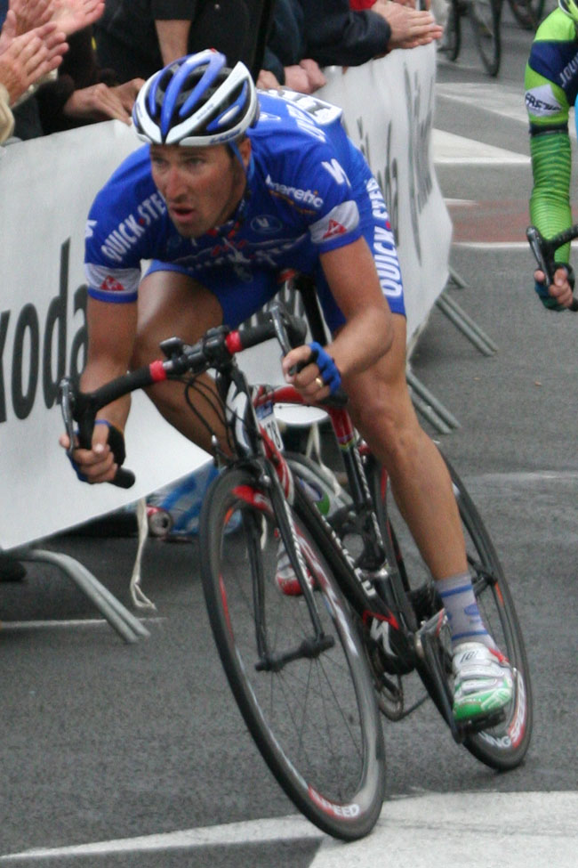

Exercise physiology
View on Wikipedia

Exercise physiology is the physiology of physical exercise. It is one of the allied health professions, and involves the study of the acute responses and chronic adaptations to exercise. Exercise physiologists are the highest qualified exercise professionals and utilise education, lifestyle intervention and specific forms of exercise to rehabilitate and manage acute and chronic injuries and conditions.
Understanding the effect of exercise involves studying specific changes in muscular, cardiovascular, and neurohormonal systems that lead to changes in functional capacity and strength due to endurance training or strength training.[2] The effect of training on the body has been defined as the reaction to the adaptive responses of the body arising from exercise[3] or as "an elevation of metabolism produced by exercise".[4]
Exercise physiologists study the effect of exercise on pathology, and the mechanisms by which exercise can reduce or reverse disease progression.
History
[edit]British physiologist Archibald Hill introduced the concepts of maximal oxygen uptake and oxygen debt in 1922.[5][6] Hill and German physician Otto Meyerhof shared the 1922 Nobel Prize in Physiology or Medicine for their independent work related to muscle energy metabolism.[7] Building on this work, scientists began measuring oxygen consumption during exercise. Notable contributions were made by Henry Taylor at the University of Minnesota, Scandinavian scientists Per-Olof Åstrand and Bengt Saltin in the 1950s and 60s, the Harvard Fatigue Laboratory, German universities, and the Copenhagen Muscle Research Centre among others.[8][9]
In some countries it is a Primary Health Care Provider. Accredited Exercise Physiologists (AEP's) are university-trained professionals who prescribe exercise-based interventions to treat various conditions using dose response prescriptions specific to each individual.[citation needed]
Energy expenditure
[edit]Humans have a high capacity to expend energy for many hours during sustained exertion. For example, one individual cycling at a speed of 26.4 km/h (16.4 mph) through 8,204 km (5,098 mi) over 50 consecutive days expended a total of 1,145 MJ (273,850 kcal; 273,850 dieter calories) with an average power output of 173.8 W.[10]
Skeletal muscle burns 90 mg (0.5 mmol) of glucose each minute during continuous activity (such as when repetitively extending the human knee),[11] generating ≈24 W of mechanical energy, and since muscle energy conversion is only 22–26% efficient,[12] ≈76 W of heat energy. Resting skeletal muscle has a basal metabolic rate (resting energy consumption) of 0.63 W/kg[13] making a 160 fold difference between the energy consumption of inactive and active muscles. For short duration muscular exertion, energy expenditure can be far greater: an adult human male when jumping up from a squat can mechanically generate 314 W/kg. Such rapid movement can generate twice this amount in nonhuman animals such as bonobos,[14] and in some small lizards.[15]
This energy expenditure is very large compared to the basal resting metabolic rate of the adult human body. This rate varies somewhat with size, gender and age but is typically between 45 W and 85 W.[16] [17] Total energy expenditure (TEE) due to muscular expended energy is much higher and depends upon the average level of physical work and exercise done during the day.[18] Thus exercise, particularly if sustained for very long periods, dominates the energy metabolism of the body. Physical activity energy expenditure correlates strongly with the gender, age, weight, heart rate, and VO2 max of an individual, during physical activity.[19]
Metabolic changes
[edit]
Rapid energy sources
[edit]Energy needed to perform short lasting, high intensity bursts of activity is derived from anaerobic metabolism within the cytosol of muscle cells, as opposed to aerobic respiration which utilizes oxygen, is sustainable, and occurs in the mitochondria. The quick energy sources consist of the phosphocreatine (PCr) system, fast glycolysis, and adenylate kinase. All of these systems re-synthesize adenosine triphosphate (ATP), which is the universal energy source in all cells. The most rapid source, but the most readily depleted of the above sources is the PCr system which utilizes the enzyme creatine kinase. This enzyme catalyzes a reaction that combines phosphocreatine and adenosine diphosphate (ADP) into ATP and creatine. This resource is short lasting because oxygen is required for the resynthesis of phosphocreatine via mitochondrial creatine kinase. Therefore, under anaerobic conditions, this substrate is finite and only lasts between approximately 10 to 30 seconds of high intensity work. Fast glycolysis, however, can function for approximately 2 minutes prior to fatigue, and predominantly uses intracellular glycogen as a substrate. Glycogen is broken down rapidly via glycogen phosphorylase into individual glucose units during intense exercise. Glucose is then oxidized to pyruvate and under anaerobic conditions is reduced to lactic acid. This reaction oxidizes NADH to NAD, thereby releasing a hydrogen ion, promoting acidosis. For this reason, fast glycolysis can not be sustained for long periods of time.[citation needed]
Plasma glucose
[edit]Plasma glucose is said to be maintained when there is an equal rate of glucose appearance (entry into the blood) and glucose disposal (removal from the blood). In the healthy individual, the rates of appearance and disposal are essentially equal during exercise of moderate intensity and duration; however, prolonged exercise or sufficiently intense exercise can result in an imbalance leaning towards a higher rate of disposal than appearance, at which point glucose levels fall producing the onset of fatigue. Rate of glucose appearance is dictated by the amount of glucose being absorbed at the gut as well as liver (hepatic) glucose output. Although glucose absorption from the gut is not typically a source of glucose appearance during exercise, the liver is capable of catabolizing stored glycogen (glycogenolysis) as well as synthesizing new glucose from specific reduced carbon molecules (glycerol, pyruvate, and lactate) in a process called gluconeogenesis. The ability of the liver to release glucose into the blood from glycogenolysis is unique, since skeletal muscle, the other major glycogen reservoir, is incapable of doing so. Unlike skeletal muscle, liver cells contain the enzyme glycogen phosphatase, which removes a phosphate group from glucose-6-P to release free glucose. In order for glucose to exit a cell membrane, the removal of this phosphate group is essential. Although gluconeogenesis is an important component of hepatic glucose output, it alone cannot sustain exercise. For this reason, when glycogen stores are depleted during exercise, glucose levels fall and fatigue sets in. Glucose disposal, the other side of the equation, is controlled by the uptake of glucose by the working skeletal muscles. During exercise, despite decreased insulin concentrations, muscle increases GLUT4 translocation and glucose uptake. The mechanism for increased GLUT4 translocation is an area of ongoing research.[citation needed]
glucose control: As mentioned above, insulin secretion is reduced during exercise, and does not play a major role in maintaining normal blood glucose concentration during exercise, but its counter-regulatory hormones appear in increasing concentrations. Principle among these are glucagon, epinephrine, and growth hormone. All of these hormones stimulate liver (hepatic) glucose output, among other functions. For instance, both epinephrine and growth hormone also stimulate adipocyte lipase, which increases non-esterified fatty acid (NEFA) release. By oxidizing fatty acids, this spares glucose utilization and helps to maintain blood sugar level during exercise.[citation needed]
Exercise for diabetes: Exercise is a particularly potent tool for glucose control in those who have diabetes mellitus. In a situation of elevated blood glucose (hyperglycemia), moderate exercise can induce greater glucose disposal than appearance, thereby decreasing total plasma glucose concentrations. As stated above, the mechanism for this glucose disposal is independent of insulin, which makes it particularly well-suited for people with diabetes. In addition, there appears to be an increase in sensitivity to insulin for approximately 12–24 hours post-exercise. This is particularly useful for those who have type II diabetes and are producing sufficient insulin but demonstrate peripheral resistance to insulin signaling. However, during extreme hyperglycemic episodes, people with diabetes should avoid exercise due to potential complications associated with ketoacidosis. Exercise could exacerbate ketoacidosis by increasing ketone synthesis in response to increased circulating NEFA's.[citation needed]
Type II diabetes is also intricately linked to obesity, and there may be a connection between type II diabetes and how fat is stored within pancreatic, muscle, and liver cells. Likely due to this connection, weight loss from both exercise and diet tends to increase insulin sensitivity in the majority of people.[20] In some people, this effect can be particularly potent and can result in normal glucose control. Although nobody is technically cured of diabetes, individuals can live normal lives without the fear of diabetic complications; however, regain of weight would assuredly result in diabetes signs and symptoms.[citation needed]
Oxygen
[edit]Vigorous physical activity (such as exercise or hard labor) increases the body's demand for oxygen. The first-line physiologic response to this demand is an increase in heart rate, breathing rate, and depth of breathing.[citation needed]
Oxygen consumption (VO2) during exercise is best described by the Fick Equation: VO2=Q x (a-vO2diff), which states that the amount of oxygen consumed is equal to cardiac output (Q) multiplied by the difference between arterial and venous oxygen concentrations. More simply put, oxygen consumption is dictated by the quantity of blood distributed by the heart as well as the working muscle's ability to take up the oxygen within that blood; however, this is a bit of an oversimplification. Although cardiac output is thought to be the limiting factor of this relationship in healthy individuals, it is not the only determinant of VO2 max. That is, factors such as the ability of the lung to oxygenate the blood must also be considered. Various pathologies and anomalies cause conditions such as diffusion limitation, ventilation/perfusion mismatch, and pulmonary shunts that can limit oxygenation of the blood and therefore oxygen distribution. In addition, the oxygen carrying capacity of the blood is also an important determinant of the equation. Oxygen carrying capacity is often the target of exercise (ergogenic aids) aids used in endurance sports to increase the volume percentage of red blood cells (hematocrit), such as through blood doping or the use of erythropoietin (EPO). Furthermore, peripheral oxygen uptake is reliant on a rerouting of blood flow from relatively inactive viscera to the working skeletal muscles, and within the skeletal muscle, capillary to muscle fiber ratio influences oxygen extraction.[citation needed]
Dehydration
[edit]Dehydration refers both to hypohydration (dehydration induced prior to exercise) and to exercise-induced dehydration (dehydration that develops during exercise). The latter reduces aerobic endurance performance and results in increased body temperature, heart rate, perceived exertion, and possibly increased reliance on carbohydrate as a fuel source. Although the negative effects of exercise-induced dehydration on exercise performance were clearly demonstrated in the 1940s, athletes continued to believe for years thereafter that fluid intake was not beneficial. More recently, negative effects on performance have been demonstrated with modest (<2%) dehydration, and these effects are exacerbated when the exercise is performed in a hot environment. The effects of hypohydration may vary, depending on whether it is induced through diuretics or sauna exposure, which substantially reduce plasma volume, or prior exercise, which has much less impact on plasma volume. Hypohydration reduces aerobic endurance, but its effects on muscle strength and endurance are not consistent and require further study.[21] Intense prolonged exercise produces metabolic waste heat, and this is removed by sweat-based thermoregulation. A male marathon runner loses each hour around 0.83 L in cool weather and 1.2 L in warm (losses in females are about 68 to 73% lower).[22] People doing heavy exercise may lose two and half times as much fluid in sweat as urine.[23] This can have profound physiological effects. Cycling for 2 hours in the heat (35 °C) with minimal fluid intake causes body mass decline by 3 to 5%, blood volume likewise by 3 to 6%, body temperature to rise constantly, and in comparison with proper fluid intake, higher heart rates, lower stroke volumes and cardiac outputs, reduced skin blood flow, and higher systemic vascular resistance. These effects are largely eliminated by replacing 50 to 80% of the fluid lost in sweat.[22][24]
Other
[edit]- Plasma catecholamine concentrations increase 10-fold in whole body exercise.[25]
- Ammonia is produced by exercised skeletal muscles from ADP (the precursor of ATP) by purine nucleotide deamination and amino acid catabolism of myofibrils.[26]
- interleukin-6 (IL-6) increases in blood circulation due to its release from working skeletal muscles.[27] This release is reduced if glucose is taken, suggesting it is related to energy depletion stresses.[28]
- Sodium absorption is affected by the release of interleukin-6 as this can cause the secretion of arginine vasopressin which, in turn, can lead to exercise-associated dangerously low sodium levels (hyponatremia). This loss of sodium in blood plasma can result in swelling of the brain. This can be prevented by awareness of the risk of drinking excessive amounts of fluids during prolonged exercise.[29][30]
Brain
[edit]At rest, the human brain receives 15% of total cardiac output, and uses 20% of the body's energy consumption.[31] The brain is normally dependent for its high energy expenditure upon aerobic metabolism. The brain as a result is highly sensitive to failure of its oxygen supply with loss of consciousness occurring within six to seven seconds,[32] with its EEG going flat in 23 seconds.[33] Therefore, the brain's function would be disrupted if exercise affected its supply of oxygen and glucose.
Protecting the brain from even minor disruption is important since exercise depends upon motor control. Because humans are bipeds, motor control is needed for keeping balance. For this reason, brain energy consumption is increased during intense physical exercise due to the demands in the motor cognition needed to control the body.[34]
Exercise Physiologists treat a range of neurological conditions including (but not limited to): Parkinson's, Alzheimer's, Traumatic Brain Injury, Spinal Cord Injury, Cerebral Palsy and mental health conditions.[citation needed]
Cerebral oxygen
[edit]Cerebral autoregulation usually ensures the brain has priority to cardiac output, though this is impaired slightly by exhaustive exercise.[35] During submaximal exercise, cardiac output increases and cerebral blood flow increases beyond the brain's oxygen needs.[36] However, this is not the case for continuous maximal exertion: "Maximal exercise is, despite the increase in capillary oxygenation [in the brain], associated with a reduced mitochondrial O2 content during whole body exercise"[37] The autoregulation of the brain's blood supply is impaired particularly in warm environments[38]
Glucose
[edit]In adults, exercise depletes the plasma glucose available to the brain: short intense exercise (35 min ergometer cycling) can reduce brain glucose uptake by 32%.[39]
At rest, energy for the adult brain is normally provided by glucose but the brain has a compensatory capacity to replace some of this with lactate. Research suggests that this can be raised, when a person rests in a brain scanner, to about 17%,[40] with a higher percentage of 25% occurring during hypoglycemia.[41] During intense exercise, lactate has been estimated to provide a third of the brain's energy needs.[39][42] There is evidence that the brain might, however, in spite of these alternative sources of energy, still suffer an energy crisis since IL-6 (a sign of metabolic stress) is released during exercise from the brain.[26][34]
Hyperthermia
[edit]Humans use sweat thermoregulation for body heat removal, particularly to remove the heat produced during exercise. Moderate dehydration as a consequence of exercise and heat is reported to impair cognition.[43][44] These impairments can start after body mass lost that is greater than 1%.[45] Cognitive impairment, particularly due to heat and exercise is likely to be due to loss of integrity to the blood brain barrier.[46] Hyperthermia can also lower cerebral blood flow,[47][48] and raise brain temperature.[34]
Fatigue
[edit]Intense activity
[edit]Researchers once attributed fatigue to a build-up of lactic acid in muscles.[49] However, this is no longer believed.[50][51] Rather, lactate may stop muscle fatigue by keeping muscles fully responding to nerve signals.[52] The available oxygen and energy supply, and disturbances of muscle ion homeostasis are the main factors determining exercise performance, at least during brief very intense exercise.[citation needed]
Each muscle contraction involves an action potential that activates voltage sensors, and so releases Ca2+ ions from the muscle fibre's sarcoplasmic reticulum. The action potentials that cause this also require ion changes: Na influxes during the depolarization phase and K effluxes for the repolarization phase. Cl− ions also diffuse into the sarcoplasm to aid the repolarization phase. During intense muscle contraction, the ion pumps that maintain homeostasis of these ions are inactivated and this (with other ion related disruption) causes ionic disturbances. This causes cellular membrane depolarization, inexcitability, and so muscle weakness.[53] Ca2+ leakage from type 1 ryanodine receptor) channels has also been identified with fatigue.[54]

Endurance failure
[edit]After intense prolonged exercise, there can be a collapse in body homeostasis. Some famous examples include:
- Dorando Pietri in the 1908 Summer Olympic men's marathon ran the wrong way and collapsed several times.[citation needed]
- Jim Peters in the marathon of the 1954 Commonwealth Games staggered and collapsed several times, and though he had a five-kilometre (three-mile) lead, failed to finish. Though it was formerly believed that this was due to severe dehydration, more recent research suggests it was the combined effects upon the brain of hyperthermia, hypertonic hypernatraemia associated with dehydration, and possibly hypoglycaemia.[55]
- Gabriela Andersen-Schiess in the woman's marathon at the Los Angeles 1984 Summer Olympics in the race's final 400 meters, stopping occasionally and shown signs of heat exhaustion. Though she fell across the finish line, she was released from medical care only two hours later.[citation needed]
Central governor
[edit]Tim Noakes, based on an earlier idea by the 1922 Nobel Prize in Physiology or Medicine winner Archibald Hill[56] has proposed the existence of a central governor. In this, the brain continuously adjusts the power output by muscles during exercise in regard to a safe level of exertion. These neural calculations factor in prior length of strenuous exercise, the planned duration of further exertion, and the present metabolic state of the body. This adjusts the number of activated skeletal muscle motor units, and is subjectively experienced as fatigue and exhaustion. The idea of a central governor rejects the earlier idea that fatigue is only caused by mechanical failure of the exercising muscles ("peripheral fatigue"). Instead, the brain models[57] the metabolic limits of the body to ensure that whole body homeostasis is protected, in particular that the heart is guarded from hypoxia, and an emergency reserve is always maintained.[58][59][60][61] The idea of the central governor has been questioned since 'physiological catastrophes' can and do occur suggesting that if it did exist, athletes (such as Dorando Pietri, Jim Peters and Gabriela Andersen-Schiess) can override it.[62]
Other factors
[edit]Exercise fatigue has also been suggested to be affected by:
- brain hyperthermia[63]
- glycogen depletion in brain cells[42][64]
- depletion of muscle and liver glycogen (see "hitting the wall")[65]
- reactive oxygen species impairing skeletal muscle function[66]
- reduced level of glutamate secondary to uptake of ammonia in the brain[26]
- Fatigue in diaphragm and abdominal respiratory muscles limiting breathing[67]
- Impaired oxygen supply to muscles[68]
- Ammonia effects upon the brain[26]
- Serotonin pathways in the brain[69]
Cardiac biomarkers
[edit]Prolonged exercise such as marathons can increase cardiac biomarkers such as troponin, B-type natriuretic peptide (BNP), and ischemia-modified (aka MI) albumin. This can be misinterpreted by medical personnel as signs of myocardial infarction, or cardiac dysfunction. In these clinical conditions, such cardiac biomarkers are produced by irreversible injury of muscles. In contrast, the processes that create them after strenuous exertion in endurance sports are reversible, with their levels returning to normal within 24-hours (further research, however, is still needed).[70][71][72]
Human adaptations
[edit]Humans are specifically adapted to engage in prolonged strenuous muscular activity (such as efficient long distance bipedal running).[73] This capacity for endurance running may have evolved to allow the running down of game animals by persistent slow but constant chase over many hours.[74]
Central to the success of this is the ability of the human body to effectively remove muscle heat waste. In most animals, this is stored by allowing a temporary increase in body temperature. This allows them to escape from animals that quickly speed after them for a short duration (the way nearly all predators catch their prey). Humans, unlike other animals that catch prey, remove heat with a specialized thermoregulation based on sweat evaporation. One gram of sweat can remove 2,598 J of heat energy.[75] Another mechanism is increased skin blood flow during exercise that allows for greater convective heat loss that is aided by our upright posture. This skin based cooling has resulted in humans acquiring an increased number of sweat glands, combined with a lack of body fur that would otherwise stop air circulation and efficient evaporation.[76] Because humans can remove exercise heat, they can avoid the fatigue from heat exhaustion that affects animals chased in a persistent manner, and so eventually catch them.[77]
Selective breeding experiments with rodents
[edit]Rodents have been specifically bred for exercise behavior or performance in several different studies.[78] For example, laboratory rats have been bred for high or low performance on a motorized treadmill with electrical stimulation as motivation.[79] The high-performance line of rats also exhibits increased voluntary wheel-running behavior as compared with the low-capacity line.[80] In an experimental evolution approach, four replicate lines of laboratory mice have been bred for high levels of voluntary exercise on wheels, while four additional control lines are maintained by breeding without regard to the amount of wheel running.[81] These selected lines of mice also show increased endurance capacity in tests of forced endurance capacity on a motorized treadmill.[82] However, in neither selection experiment have the precise causes of fatigue during either forced or voluntary exercise been determined.[citation needed]
Exercise-induced muscle pain
[edit]Physical exercise may cause pain both as an immediate effect that may result from stimulation of free nerve endings by low pH, as well as a delayed onset muscle soreness. The delayed soreness is fundamentally the result of ruptures within the muscle, although apparently not involving the rupture of whole muscle fibers.[83]
Muscle pain can range from a mild soreness to a debilitating injury depending on intensity of exercise, level of training, and other factors.[84]
There is some preliminary evidence to suggest that moderate intensity continuous training has the ability to increase someone's pain threshold.[85]
Education in exercise physiology
[edit]Accreditation programs exist with professional bodies in most developed countries, ensuring the quality and consistency of education. In Canada, one may obtain the professional certification title – Certified Exercise Physiologist for those working with clients (both clinical and non clinical) in the health and fitness industry. In Australia, one may obtain the professional certification title - Accredited Exercise Physiologist (AEP) through the professional body Exercise and Sports Science Australia (ESSA). In Australia, it is common for an AEP to also have the qualification of an Accredited Exercise Scientist (AES). The premiere governing body is the American College of Sports Medicine.[citation needed]
An exercise physiologist's area of study may include but is not limited to biochemistry, bioenergetics, cardiopulmonary function, hematology, biomechanics, skeletal muscle physiology, neuroendocrine function, and central and peripheral nervous system function. Furthermore, exercise physiologists range from basic scientists, to clinical researchers, to clinicians, to sports trainers.[citation needed]
Colleges and universities offer exercise physiology as a program of study on various different levels, including undergraduate, graduate degrees and certificates, and doctoral programs. The basis of Exercise Physiology as a major is to prepare students for a career in the field of health sciences. A program that focuses on the scientific study of the physiological processes involved in physical or motor activity, including sensorimotor interactions, response mechanisms, and the effects of injury, disease, and disability. Includes instruction in muscular and skeletal anatomy; molecular and cellular basis of muscle contraction; fuel utilization; neurophysiology of motor mechanics; systemic physiological responses (respiration, blood flow, endocrine secretions, and others); fatigue and exhaustion; muscle and body training; physiology of specific exercises and activities; physiology of injury; and the effects of disabilities and disease. Careers available with a degree in Exercise Physiology can include: non-clinical, client-based work; strength and conditioning specialists; cardiopulmonary treatment; and clinical-based research.[86]
In order to gauge the multiple areas of study, students are taught processes in which to follow on a client-based level. Practical and lecture teachings are instructed in the classroom and in a laboratory setting. These include:
- Health and risk assessment: In order to safely work with a client on the job, you must first be able to know the benefits and risks associated with physical activity. Examples of this include knowing specific injuries the body can experience during exercise, how to properly screen a client before their training begins, and what factors to look for that may inhibit their performance.[citation needed]
- Exercise testing: Coordinating exercise tests in order to measure body compositions, cardiorespiratory fitness, muscular strength/endurance, and flexibility. Functional tests are also used in order to gain understanding of a more specific part of the body. Once the information is gathered about a client, exercise physiologists must also be able to interpret the test data and decide what health-related outcomes have been discovered.[citation needed]
- Exercise prescription: Forming training programs that best meet an individual's health and fitness goals. Must be able to take into account different types of exercises, the reasons/goal for a client's workout, and pre-screened assessments. Knowing how to prescribe exercises for special considerations and populations is also required. These may include age differences, pregnancy, joint diseases, obesity, pulmonary disease, etc.[87]
Curriculum
[edit]The curriculum for exercise physiology includes biology, chemistry, and applied sciences. The purpose of the classes selected for this major is to have a proficient understanding of human anatomy, human physiology, and exercise physiology. Includes instruction in muscular and skeletal anatomy; molecular and cellular basis of muscle contraction; fuel utilization; neurophysiology of motor mechanics; systemic physiological responses (respiration, blood flow, endocrine secretions, and others); fatigue and exhaustion; muscle and body training; physiology of specific exercises and activities; physiology of injury; and the effects of disabilities and disease. Not only is a full class schedule needed to complete a degree in Exercise Physiology, but a minimum amount of practicum experience is required and internships are recommended.[88]
See also
[edit]References
[edit]- ^ Capostagno, B; Lambert, M. I; Lamberts, R. P (2016). "A Systematic Review of Submaximal Cycle Tests to Predict, Monitor, and Optimize Cycling Performance". International Journal of Sports Physiology and Performance. 11 (6): 707–714. doi:10.1123/ijspp.2016-0174. PMID 27701968.
- ^ Awtry, Eric H.; Balady, Gary J. (2007). "Exercise and Physical Activity". In Topol, Eric J. (ed.). Textbook of Cardiovascular Medicine (3rd ed.). Lippincott Williams & Wilkins. p. 83. ISBN 978-0-7817-7012-5.
- ^ Bompa, Tudor O.; Haff, G. Gregory (2009) [1983]. "Basis for Training". Periodization: Theory and Methodology of Training (5th ed.). Champaign, Illinois: Human Kinetics. pp. 12–13. ISBN 978-0-7360-8547-2.[permanent dead link]
- ^ Lee, Buddy (2010). Jump Rope Training (2nd ed.). Human Kinetics. p. 207. ISBN 978-0-7360-8978-4.
- ^ Hale, Tudor (2008-02-15). "History of developments in sport and exercise physiology: A. V. Hill, maximal oxygen uptake, and oxygen debt". Journal of Sports Sciences. 26 (4): 365–400. doi:10.1080/02640410701701016. ISSN 0264-0414. PMID 18228167. S2CID 33768722.
- ^ Bassett, D. R.; Howley, E. T. (1997). "Maximal oxygen uptake: "classical" versus "contemporary" viewpoints". Medicine & Science in Sports & Exercise. 29 (5): 591–603. doi:10.1097/00005768-199705000-00002. ISSN 0195-9131. PMID 9140894.
- ^ "The Nobel Prize in Physiology or Medicine 1922". NobelPrize.org. Retrieved 2018-10-11.
- ^ Seiler, Stephen (2011). "A Brief History of Endurance Testing in Athletes" (PDF). Sportscience. 15 (5).
- ^ "History of Exercise Physiology". Human Kinetics Europe. Retrieved 2018-10-11.
- ^ Gianetti, G; Burton, L; Donovan, R; Allen, G; Pescatello, LS (2008). "Physiologic and psychological responses of an athlete cycling 100+ miles daily for 50 consecutive days". Current Sports Medicine Reports. 7 (6): 343–7. doi:10.1249/JSR.0b013e31818f0670. PMID 19005357.. This individual while exceptional was not physiologically extraordinary since he was described as "subelite" due to his not being "able to adjust power output to regulate energy expenditure as occurs with elite athletes during ultra-cycling events" page 347.
- ^ Richter, EA; Kiens, B; Saltin, B; Christensen, NJ; Savard, G (1988). "Skeletal muscle glucose uptake during dynamic exercise in humans: Role of muscle mass". The American Journal of Physiology. 254 (5 Pt 1): E555–61. doi:10.1152/ajpendo.1988.254.5.E555. PMID 3284382.
- ^ Bangsbo, J (1996). "Physiological factors associated with efficiency in high intensity exercise". Sports Medicine. 22 (5): 299–305. doi:10.2165/00007256-199622050-00003. PMID 8923647. S2CID 23080799.
- ^ Elia, M. (1992) "Energy expenditure in the whole body". Energy metabolism. Tissue determinants and cellular corollaries. 61–79 Raven Press New York. ISBN 978-0-88167-871-0
- ^ Scholz, MN; d'Août, K; Bobbert, MF; Aerts, P (2006). "Vertical jumping performance of bonobo (Pan paniscus) suggests superior muscle properties". Proceedings: Biological Sciences. 273 (1598): 2177–84. doi:10.1098/rspb.2006.3568. PMC 1635523. PMID 16901837.
- ^ Curtin NA, Woledge RC, Aerts P (2005). "Muscle directly meets the vast power demands in agile lizards". Proceedings: Biological Sciences. 272 (1563): 581–4. doi:10.1098/rspb.2004.2982. PMC 1564073. PMID 15817432.
{{cite journal}}: CS1 maint: multiple names: authors list (link) - ^ Henry, CJ (2005). "Basal metabolic rate studies in humans: Measurement and development of new equations". Public Health Nutrition. 8 (7A): 1133–52. doi:10.1079/phn2005801. PMID 16277825.
- ^ Henry 2005 provides BMR formula various ages given body weight: those for BMR aged 18–30 in MJ/day (where mass is body weight in kg) are: male BMR = 0.0669 mass + 2.28; females BMR = 0.0546 mass + 2.33; 1 MJ per day = 11.6 W. The data providing these formula hide a high variance: for men weighing 70 kg, measured BMR is between 50 and 110 W, and women weighing 60 kg, between 40 W and 90 W.
- ^ Torun, B (2005). "Energy requirements of children and adolescents". Public Health Nutrition. 8 (7A): 968–93. doi:10.1079/phn2005791. PMID 16277815.
- ^ Keytel, L.R. (March 2005). "Prediction of energy expenditure from heart rate monitoring during submaximal exercise" (PDF). Journal of Sports Sciences. 23 (3): 289–97. doi:10.1080/02640410470001730089. PMID 15966347. S2CID 14267971. Archived from the original (PDF) on 16 April 2015. Retrieved 16 April 2015.
- ^ Boutcher, Stephen H. (2011). "High-Intensity Intermittent Exercise and Fat Loss". Journal of Obesity. 2011 868305. doi:10.1155/2011/868305. PMC 2991639. PMID 21113312.
- ^ Barr, SI (1999). "Effects of dehydration on exercise performance". Canadian Journal of Applied Physiology. 24 (2): 164–72. doi:10.1139/h99-014. PMID 10198142.
- ^ a b Cheuvront SN, Haymes EM (2001). "Thermoregulation and marathon running: biological and environmental influences". Sports Med. 31 (10): 743–62. doi:10.2165/00007256-200131100-00004. PMID 11547895. S2CID 45969661.
- ^ Porter, AM (2001). "Why do we have apocrine and sebaceous glands?". Journal of the Royal Society of Medicine. 94 (5): 236–7. doi:10.1177/014107680109400509. PMC 1281456. PMID 11385091.
- ^ González-Alonso, J; Mora-Rodríguez, R; Below, PR; Coyle, EF (1995). "Dehydration reduces cardiac output and increases systemic and cutaneous vascular resistance during exercise". Journal of Applied Physiology. 79 (5): 1487–96. doi:10.1152/jappl.1995.79.5.1487. PMID 8594004.
- ^ Holmqvist, N; Secher, NH; Sander-Jensen, K; Knigge, U; Warberg, J; Schwartz, TW (1986). "Sympathoadrenal and parasympathetic responses to exercise". Journal of Sports Sciences. 4 (2): 123–8. doi:10.1080/02640418608732108. PMID 3586105.
- ^ a b c d Nybo, L; Dalsgaard, MK; Steensberg, A; Møller, K; Secher, NH (2005). "Cerebral ammonia uptake and accumulation during prolonged exercise in humans". The Journal of Physiology. 563 (Pt 1): 285–90. doi:10.1113/jphysiol.2004.075838. PMC 1665558. PMID 15611036.
- ^ Febbraio, MA; Pedersen, BK (2002). "Muscle-derived interleukin-6: Mechanisms for activation and possible biological roles". FASEB Journal. 16 (11): 1335–47. doi:10.1096/fj.01-0876rev. PMID 12205025. S2CID 14024672.
- ^ Febbraio, MA; Steensberg, A; Keller, C; Starkie, RL; Nielsen, HB; Krustrup, P; Ott, P; Secher, NH; Pedersen, BK (2003). "Glucose ingestion attenuates interleukin-6 release from contracting skeletal muscle in humans". The Journal of Physiology. 549 (Pt 2): 607–12. doi:10.1113/jphysiol.2003.042374. PMC 2342952. PMID 12702735.
- ^ Siegel, AJ; Verbalis, JG; Clement, S; Mendelson, JH; Mello, NK; Adner, M; Shirey, T; Glowacki, J; et al. (2007). "Hyponatremia in marathon runners due to inappropriate arginine vasopressin secretion". The American Journal of Medicine. 120 (5): 461.e11–7. doi:10.1016/j.amjmed.2006.10.027. PMID 17466660.
- ^ Siegel, AJ (2006). "Exercise-associated hyponatremia: Role of cytokines". The American Journal of Medicine. 119 (7 Suppl 1): S74–8. doi:10.1016/j.amjmed.2006.05.012. PMID 16843089.
- ^ Lassen, NA (1959). "Cerebral blood flow and oxygen consumption in man". Physiological Reviews. 39 (2): 183–238. doi:10.1152/physrev.1959.39.2.183. PMID 13645234. S2CID 29275804.
- ^ Rossen R, Kabat H, Anderson JP (1943). "Acute arrest of cerebral circulation in man". Archives of Neurology & Psychiatry. 50 (5): 510–28. doi:10.1001/archneurpsyc.1943.02290230022002.
- ^ Todd, MM; Dunlop, BJ; Shapiro, HM; Chadwick, HC; Powell, HC (1981). "Ventricular fibrillation in the cat: A model for global cerebral ischemia". Stroke: A Journal of Cerebral Circulation. 12 (6): 808–15. doi:10.1161/01.STR.12.6.808. PMID 7303071.
- ^ a b c Secher, NH; Seifert, T; Van Lieshout, JJ (2008). "Cerebral blood flow and metabolism during exercise: Implications for fatigue". Journal of Applied Physiology. 104 (1): 306–14. doi:10.1152/japplphysiol.00853.2007. PMID 17962575.
- ^ Ogoh, S; Dalsgaard, MK; Yoshiga, CC; Dawson, EA; Keller, DM; Raven, PB; Secher, NH (2005). "Dynamic cerebral autoregulation during exhaustive exercise in humans". American Journal of Physiology. Heart and Circulatory Physiology. 288 (3): H1461–7. doi:10.1152/ajpheart.00948.2004. PMID 15498819.
- ^ Ide, K; Horn, A; Secher, NH (1999). "Cerebral metabolic response to submaximal exercise". Journal of Applied Physiology. 87 (5): 1604–8. CiteSeerX 10.1.1.327.7515. doi:10.1152/jappl.1999.87.5.1604. PMID 10562597.
- ^ Secher, NH; Seifert, T; Van Lieshout, JJ (2008). "Cerebral blood flow and metabolism during exercise: Implications for fatigue". Journal of Applied Physiology. 104 (1): 306–14. doi:10.1152/japplphysiol.00853.2007. PMID 17962575. page 309
- ^ Watson, P; Shirreffs, SM; Maughan, RJ (2005). "Blood-brain barrier integrity may be threatened by exercise in a warm environment". American Journal of Physiology. Regulatory, Integrative and Comparative Physiology. 288 (6): R1689–94. doi:10.1152/ajpregu.00676.2004. PMID 15650123.
- ^ a b Kemppainen, J; Aalto, S; Fujimoto, T; Kalliokoski, KK; Långsjö, J; Oikonen, V; Rinne, J; Nuutila, P; Knuuti, J (2005). "High intensity exercise decreases global brain glucose uptake in humans". The Journal of Physiology. 568 (Pt 1): 323–32. doi:10.1113/jphysiol.2005.091355. PMC 1474763. PMID 16037089.
- ^ Smith, D; Pernet, A; Hallett, WA; Bingham, E; Marsden, PK; Amiel, SA (2003). "Lactate: A preferred fuel for human brain metabolism in vivo". Journal of Cerebral Blood Flow and Metabolism. 23 (6): 658–64. doi:10.1097/01.WCB.0000063991.19746.11. PMID 12796713.
- ^ Lubow, JM; Piñón, IG; Avogaro, A; Cobelli, C; Treeson, DM; Mandeville, KA; Toffolo, G; Boyle, PJ (2006). "Brain oxygen utilization is unchanged by hypoglycemia in normal humans: Lactate, alanine, and leucine uptake are not sufficient to offset energy deficit". American Journal of Physiology. Endocrinology and Metabolism. 290 (1): E149 – E153. doi:10.1152/ajpendo.00049.2005. PMID 16144821. S2CID 8297686.
- ^ a b Dalsgaard, MK (2006). "Fuelling cerebral activity in exercising man". Journal of Cerebral Blood Flow and Metabolism. 26 (6): 731–50. doi:10.1038/sj.jcbfm.9600256. PMID 16395281. S2CID 24976326.
- ^ Baker, LB; Conroy, DE; Kenney, WL (2007). "Dehydration impairs vigilance-related attention in male basketball players". Medicine & Science in Sports & Exercise. 39 (6): 976–83. doi:10.1097/mss.0b013e3180471ff2. PMID 17545888. S2CID 25267863.
- ^ Cian, C; Barraud, PA; Melin, B; Raphel, C (2001). "Effects of fluid ingestion on cognitive function after heat stress or exercise-induced dehydration". International Journal of Psychophysiology. 42 (3): 243–51. doi:10.1016/S0167-8760(01)00142-8. PMID 11812391.
- ^ Sharma, VM; Sridharan, K; Pichan, G; Panwar, MR (1986). "Influence of heat-stress induced dehydration on mental functions". Ergonomics. 29 (6): 791–9. doi:10.1080/00140138608968315. PMID 3743537.
- ^ Maughan, RJ; Shirreffs, SM; Watson, P (2007). "Exercise, heat, hydration and the brain". Journal of the American College of Nutrition. 26 (5 Suppl): 604S – 612S. doi:10.1080/07315724.2007.10719666. PMID 17921473. S2CID 27256788.
- ^ Nybo, L; Møller, K; Volianitis, S; Nielsen, B; Secher, NH (2002). "Effects of hyperthermia on cerebral blood flow and metabolism during prolonged exercise in humans". Journal of Applied Physiology. 93 (1): 58–64. doi:10.1152/japplphysiol.00049.2002. PMID 12070186.
- ^ Nybo, L; Nielsen, B (2001). "Middle cerebral artery blood velocity is reduced with hyperthermia during prolonged exercise in humans". The Journal of Physiology. 534 (Pt 1): 279–86. doi:10.1111/j.1469-7793.2001.t01-1-00279.x. PMC 2278686. PMID 11433008.
- ^ Hermansen, L (1981). "Effect of Metabolic Changes on Force Generation in Skeletal Muscle During Maximal Exercise". Ciba Foundation Symposium 82 - Human Muscle Fatigue: Physiological Mechanisms. Novartis Foundation Symposia. Vol. 82. pp. 75–88. doi:10.1002/9780470715420.ch5. ISBN 978-0-470-71542-0. PMID 6913479.
{{cite book}}:|work=ignored (help) - ^ Brooks, GA (2001). "Lactate doesn't necessarily cause fatigue: Why are we surprised?". The Journal of Physiology. 536 (Pt 1): 1. doi:10.1111/j.1469-7793.2001.t01-1-00001.x. PMC 2278833. PMID 11579151.
- ^ Gladden, LB (2004). "Lactate metabolism: A new paradigm for the third millennium". The Journal of Physiology. 558 (Pt 1): 5–30. doi:10.1113/jphysiol.2003.058701. PMC 1664920. PMID 15131240.
- ^ Pedersen TH, Nielsen OB, Lamb GD, Stephenson DG (2004). "Intracellular acidosis enhances the excitability of working muscle". Science. 305 (5687): 1144–7. Bibcode:2004Sci...305.1144P. doi:10.1126/science.1101141. PMID 15326352. S2CID 24228666.
- ^ McKenna, MJ; Bangsbo, J; Renaud, JM (2008). "Muscle K+, Na+, and Cl disturbances and Na+-K+ pump inactivation: Implications for fatigue". Journal of Applied Physiology. 104 (1): 288–95. doi:10.1152/japplphysiol.01037.2007. PMID 17962569. S2CID 25190764.
- ^ Bellinger, AM; Reiken, S; Dura, M; Murphy, PW; Deng, SX; Landry, DW; Nieman, D; Lehnart, SE; et al. (2008). "Remodeling of ryanodine receptor complex causes "leaky" channels: A molecular mechanism for decreased exercise capacity". Proceedings of the National Academy of Sciences of the United States of America. 105 (6): 2198–202. Bibcode:2008PNAS..105.2198B. doi:10.1073/pnas.0711074105. PMC 2538898. PMID 18268335.
- ^ Noakes, T; Mekler, J; Pedoe, DT (2008). "Jim Peters' collapse in the 1954 Vancouver Empire Games marathon". South African Medical Journal. 98 (8): 596–600. PMID 18928034.
- ^ Hill A. V.; Long C. N. H.; Lupton H. (1924). "Muscular exercise, lactic acid and the supply and utilisation of oxygen. Parts I–III". Proc. R. Soc. Lond. 97 (679): 438–475. doi:10.1098/rspb.1924.0037.
- ^ St Clair Gibson, A; Baden, DA; Lambert, MI; Lambert, EV; Harley, YX; Hampson, D; Russell, VA; Noakes, TD (2003). "The conscious perception of the sensation of fatigue". Sports Medicine. 33 (3): 167–76. doi:10.2165/00007256-200333030-00001. PMID 12656638. S2CID 34014572.
- ^ Noakes, TD; St Clair Gibson, A; Lambert, EV (2005). "From catastrophe to complexity: A novel model of integrative central neural regulation of effort and fatigue during exercise in humans: Summary and conclusions". British Journal of Sports Medicine. 39 (2): 120–4. doi:10.1136/bjsm.2003.010330. PMC 1725112. PMID 15665213.
- ^ Noakes, TD; Peltonen, JE; Rusko, HK (2001). "Evidence that a central governor regulates exercise performance during acute hypoxia and hyperoxia". The Journal of Experimental Biology. 204 (Pt 18): 3225–34. doi:10.1242/jeb.204.18.3225. PMID 11581338.
- ^ Noakes, TD (2000). "Physiological models to understand exercise fatigue and the adaptations that predict or enhance athletic performance". Scandinavian Journal of Medicine & Science in Sports. 10 (3): 123–45. doi:10.1034/j.1600-0838.2000.010003123.x. PMID 10843507. S2CID 23103331.
- ^ St Clair Gibson, A; Lambert, ML; Noakes, TD (2001). "Neural control of force output during maximal and submaximal exercise". Sports Medicine. 31 (9): 637–50. doi:10.2165/00007256-200131090-00001. PMID 11508520. S2CID 1111940.
- ^ Esteve-Lanao, J; Lucia, A; Dekoning, JJ; Foster, C (2008). Earnest, Conrad P. (ed.). "How do humans control physiological strain during strenuous endurance exercise?". PLOS ONE. 3 (8) e2943. Bibcode:2008PLoSO...3.2943E. doi:10.1371/journal.pone.0002943. PMC 2491903. PMID 18698405.
- ^ Nybo, L (2008). "Hyperthermia and fatigue". Journal of Applied Physiology. 104 (3): 871–8. doi:10.1152/japplphysiol.00910.2007. PMID 17962572.
- ^ Dalsgaard, MK; Secher, NH (2007). "The brain at work: A cerebral metabolic manifestation of central fatigue?". Journal of Neuroscience Research. 85 (15): 3334–9. doi:10.1002/jnr.21274. PMID 17394258. S2CID 23623274.
- ^ Smyth, Barry (2021-05-19). "How recreational marathon runners hit the wall: A large-scale data analysis of late-race pacing collapse in the marathon". PLOS ONE. 16 (5) e0251513. Bibcode:2021PLoSO..1651513S. doi:10.1371/journal.pone.0251513. ISSN 1932-6203. PMC 8133477. PMID 34010308.
- ^ Ferreira, LF; Reid, MB (2008). "Muscle-derived ROS and thiol regulation in muscle fatigue". Journal of Applied Physiology. 104 (3): 853–60. doi:10.1152/japplphysiol.00953.2007. PMID 18006866.
- ^ Romer, LM; Polkey, MI (2008). "Exercise-induced respiratory muscle fatigue: Implications for performance". Journal of Applied Physiology. 104 (3): 879–88. doi:10.1152/japplphysiol.01157.2007. PMID 18096752.
- ^ Amann, M; Calbet, JA (2008). "Convective oxygen transport and fatigue" (PDF). Journal of Applied Physiology. 104 (3): 861–70. doi:10.1152/japplphysiol.01008.2007. hdl:10553/6567. PMID 17962570. S2CID 22648694.
- ^ Newsholme, EA; Blomstrand, E (1995). "Tryptophan, 5-Hydroxytryptamine and a Possible Explanation for Central Fatigue". Fatigue. Advances in Experimental Medicine and Biology. Vol. 384. pp. 315–20. doi:10.1007/978-1-4899-1016-5_25. ISBN 978-1-4899-1018-9. PMID 8585461.
- ^ Scharhag, J; George, K; Shave, R; Urhausen, A; Kindermann, W (2008). "Exercise-associated increases in cardiac biomarkers". Medicine & Science in Sports & Exercise. 40 (8): 1408–15. doi:10.1249/MSS.0b013e318172cf22. PMID 18614952.
- ^ Lippi, G; Schena, F; Salvagno, GL; Montagnana, M; Gelati, M; Tarperi, C; Banfi, G; Guidi, GC (2008). "Influence of a half-marathon run on NT-proBNP and troponin T". Clinical Laboratory. 54 (7–8): 251–4. PMID 18942493.
- ^ Kolata, Gina (2008-11-27). "The Lab Says Heart Attack, but the Patient Is Fine". The New York Times. ISSN 0362-4331. Retrieved 2023-02-08.
- ^ Bramble, DM; Lieberman, DE (2004). "Endurance running and the evolution of Homo" (PDF). Nature. 432 (7015): 345–52. Bibcode:2004Natur.432..345B. doi:10.1038/nature03052. PMID 15549097. S2CID 2470602.
- ^ Carrier, David R. (1984). "The Energetic Paradox of Human Running and Hominid Evolution". Current Anthropology. 25 (4): 483–495. doi:10.1086/203165. S2CID 15432016.
- ^ Snellen, JW; Mitchell, D; Wyndham, CH (1970). "Heat of evaporation of sweat". Journal of Applied Physiology. 29 (1): 40–4. doi:10.1152/jappl.1970.29.1.40. PMID 5425034.
- ^ Lupi, O (2008). "Ancient adaptations of human skin: Why do we retain sebaceous and apocrine glands?". International Journal of Dermatology. 47 (7): 651–4. doi:10.1111/j.1365-4632.2008.03765.x. PMID 18613867. S2CID 32885875.
- ^ Liebenberg, Louis (2006). "Persistence Hunting by Modern Hunter-Gatherers". Current Anthropology. 47 (6): 1017–1026. doi:10.1086/508695. S2CID 224793846.
- ^ Feder, ME; Garland Jr, T; Marden, JH; Zera, AJ (2010). "Locomotion in response to shifting climate zones: Not so fast" (PDF). Annual Review of Physiology. 72: 167–90. doi:10.1146/annurev-physiol-021909-135804. PMID 20148672. S2CID 36520695. Archived from the original (PDF) on 2023-10-12. Retrieved 2011-10-31.
- ^ Koch, L. G.; Britton, S. L. (2001). "Artificial selection for intrinsic aerobic endurance running capacity in rats". Physiological Genomics. 5 (1): 45–52. CiteSeerX 10.1.1.325.7411. doi:10.1152/physiolgenomics.2001.5.1.45. PMID 11161005. S2CID 2340159.
- ^ Waters, RP; Renner, KJ; Pringle, RB; Summers, CH; Britton, SL; Koch, LG; Swallow, JG (2008). "Selection for aerobic capacity affects corticosterone, monoamines and wheel-running activity". Physiology & Behavior. 93 (4–5): 1044–54. doi:10.1016/j.physbeh.2008.01.013. PMC 2435267. PMID 18304593.
- ^ Swallow, JG; Carter, PA; Garland Jr, T (1998). "Artificial selection for increased wheel-running behavior in house mice". Behavior Genetics. 28 (3): 227–37. doi:10.1023/A:1021479331779. PMID 9670598. S2CID 18336243.
- ^ Meek, TH; Lonquich, BP; Hannon, RM; Garland Jr, T (2009). "Endurance capacity of mice selectively bred for high voluntary wheel running". The Journal of Experimental Biology. 212 (18): 2908–17. doi:10.1242/jeb.028886. PMID 19717672.
- ^ Nosaka, Ken (2008). "Muscle Soreness and Damage and the Repeated-Bout Effect". In Tiidus, Peter M (ed.). Skeletal muscle damage and repair. Human Kinetics. pp. 59–76. ISBN 978-0-7360-5867-4.
- ^ Cheung, Karoline; Hume, Patria A.; Maxwell, Linda (2012-10-23). "Delayed Onset Muscle Soreness". Sports Medicine. 33 (2): 145–164. doi:10.2165/00007256-200333020-00005. ISSN 0112-1642. PMID 12617692. S2CID 26525519.
- ^ Hakansson, S.; Jones, M. D.; Ristov, M.; Marcos, L.; Clark, T.; Ram, A.; Morey, R.; Franklin, A.; McCarthy, C. (2018). "Intensity-dependent effects of aerobic training on pressure pain threshold in overweight men: A randomized trial". European Journal of Pain. 22 (10): 1813–1823. doi:10.1002/ejp.1277. hdl:1959.4/unsworks_83572. ISSN 1532-2149. PMID 29956398. S2CID 49602409.
- ^ Davis, Paul. "Careers in Exercise Physiology". Archived from the original on 2018-01-03. Retrieved 2012-04-18.
- ^ American College of Sports Medicine (2010). ACSM's guidelines for exercise testing and prescription (8th ed.). Philadelphia: Lippincott Williams & Wilkins. ISBN 978-0-7817-6903-7.
- ^ University, Ohio. "Class Requirements".
External links
[edit] Media related to Exercise physiology at Wikimedia Commons

| Types | |||
|---|---|---|---|
| Science | |||
| Organizations | |||
| Business | |||
| Communication | |||
| Culture | |||
| Equipment | |||
| Politics | |||
| Sport by region |
| ||


Physiology types | ||
|---|---|---|
| Animals |  | |
| Plants | ||
| Cells | ||
| Related topics | ||