Hubbry Logo
Sterol regulatory element-binding proteinSterol regulatory element-binding proteinMain
Open search
Sterol regulatory element-binding protein
Community hub
Sterol regulatory element-binding protein
logo
8 pages, 0 posts
0 subscribers
Be the first to start a discussion here.
Sterol regulatory element-binding protein
from Wikipedia
Sterol regulatory element-binding transcription factor 1
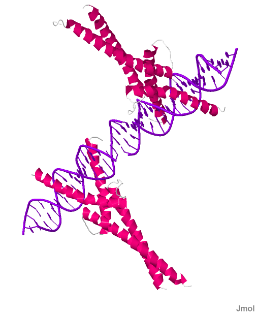
X-ray crystallography of Sterol Regulatory Element Binding Protein 1A with polydeoxyribonucleotide.[1]
Identifiers
SymbolSREBF1
NCBI gene6720
HGNC11289
OMIM184756
PDB1am9
RefSeqNM_004176
UniProtP36956
Other data
LocusChr. 17 p11.2
Search for
StructuresSwiss-model
DomainsInterPro
sterol regulatory element-binding transcription factor 2
Identifiers
SymbolSREBF2
NCBI gene6721
HGNC11290
OMIM600481
RefSeqNM_004599
UniProtQ12772
Other data
LocusChr. 22 q13
Search for
StructuresSwiss-model
DomainsInterPro

Sterol regulatory element-binding proteins (SREBPs) are transcription factors that bind to the sterol regulatory element DNA sequence TCACNCCAC.[2] Mammalian SREBPs are encoded by the genes SREBF1 and SREBF2. SREBPs belong to the basic-helix-loop-helix leucine zipper class of transcription factors.[3] Unactivated SREBPs are attached to the nuclear envelope and endoplasmic reticulum membranes. In cells with low levels of sterols, SREBPs are cleaved to a water-soluble N-terminal domain that is translocated to the nucleus. These activated SREBPs then bind to specific sterol regulatory element DNA sequences, thus upregulating the synthesis of enzymes involved in sterol biosynthesis.[4][5] Sterols in turn inhibit the cleavage of SREBPs and therefore synthesis of additional sterols is reduced through a negative feed back loop.

Isoforms

[edit]

Mammalian genomes have two separate SREBP genes (SREBF1 and SREBF2):

  • SREBP-1 expression produces two different isoforms, SREBP-1a and -1c. These isoforms differ in their first exons owing to the use of different transcriptional start sites for the SREBP-1 gene. SREBP-1c was also identified in rats as ADD-1. SREBP-1c is responsible for regulating the genes required for de novo lipogenesis.[6]
  • SREBP-2 regulates the genes of cholesterol metabolism.[6]

Function

[edit]

SREB proteins are indirectly required for cholesterol biosynthesis and for uptake and fatty acid biosynthesis. These proteins work with asymmetric sterol regulatory element (StRE). SREBPs have a structure similar to E-box-binding helix-loop-helix (HLH) proteins. However, in contrast to E-box-binding HLH proteins, an arginine residue is replaced with tyrosine making them capable of recognizing StREs and thereby regulating membrane biosynthesis.[7]

Mechanism of action

[edit]
caption
SREBP activation by proteolytic cleavage. SREBP precursors are retained in the ERTooltip endoplasmic reticulum membranes through a tight association with SCAPTooltip SREBP cleavage-activating protein and a protein of the INSIGTooltip insulin-induced gene protein family. Under the appropriate conditions, SCAP dissociates from INSIG and escorts the SREBP precursors from the ER to the Golgi apparatus. Once there, two proteases, S1PTooltip site-1 protease and S2PTooltip site-2 protease, sequentially cleave the precursor protein, releasing the mature form of SREBPs into the cytoplasm. The mature form then migrates to the nucleus, where it activates the promoter of genes involved in cholesterol uptake or in cholesterol synthesis. SREBP processing can be controlled by the cellular sterol content.

Animal cells maintain proper levels of intracellular lipids (fats and oils) under widely varying circumstances (lipid homeostasis).[8][9][10] For example, when cellular cholesterol levels fall below the level needed, the cell makes more of the enzymes necessary to make cholesterol. A principal step in this response is to make more of the mRNA transcripts that direct the synthesis of these enzymes. Conversely, when there is enough cholesterol around, the cell stops making those mRNAs and the level of the enzymes falls. As a result, the cell quits making cholesterol once it has enough.

A notable feature of this regulatory feedback machinery was first observed for the SREBP pathway - regulated intramembrane proteolysis (RIP). Subsequently, RIP was found to be used in almost all organisms from bacteria to human beings and regulates a wide range of processes ranging from development to neurodegeneration.

A feature of the SREBP pathway is the proteolytic release of a membrane-bound transcription factor, SREBP. Proteolytic cleavage frees it to move through the cytoplasm to the nucleus. Once in the nucleus, SREBP can bind to specific DNA sequences (the sterol regulatory elements or SREs) that are found in the control regions of the genes that encode enzymes needed to make lipids. This binding to DNA leads to the increased transcription of the target genes.

The ~120 kDa SREBP precursor protein is anchored in the membranes of the endoplasmic reticulum (ER) and nuclear envelope by virtue of two membrane-spanning helices in the middle of the protein. The precursor has a hairpin orientation in the membrane, so that both the amino-terminal transcription factor domain and the COOH-terminal regulatory domain face the cytoplasm. The two membrane-spanning helices are separated by a loop of about 30 amino acids that lies in the lumen of the ER. Two separate, site-specific proteolytic cleavages are necessary for release of the transcriptionally active amino-terminal domain. These cleavages are carried out by two distinct proteases, called site-1 protease (S1P) and site-2 protease (S2P).

In addition to S1P and S2P, the regulated release of transcriptionally active SREBP requires the cholesterol-sensing protein SREBP cleavage-activating protein (SCAP), which forms a complex with SREBP owing to interaction between their respective carboxy-terminal domains. SCAP, in turn, can bind reversibly with another ER-resident membrane protein, INSIG. In the presence of sterols, which bind to INSIG and SCAP, INSIG and SCAP also bind one another. INSIG always stays in the ER membrane and thus the SREBP-SCAP complex remains in the ER when SCAP is bound to INSIG. When sterol levels are low, INSIG and SCAP no longer bind. Then, SCAP undergoes a conformational change that exposes a portion of the protein ('MELADL') that signals it to be included as cargo in the COPII vesicles that move from the ER to the Golgi apparatus. In these vesicles, SCAP, dragging SREBP along with it, is transported to the Golgi. The regulation of SREBP cleavage employs a notable feature of eukaryotic cells, subcellular compartmentalization defined by intracellular membranes, to ensure that cleavage occurs only when needed.

Once in the Golgi apparatus, the SREBP-SCAP complex encounters active S1P. S1P cleaves SREBP at site-1, cutting it into two halves. Because each half still has a membrane-spanning helix, each remains bound in the membrane. The newly generated amino-terminal half of SREBP (which is the ‘business end' of the molecule) then goes on to be cleaved at site-2 that lies within its membrane-spanning helix. This is the work of S2P, an unusual metalloprotease. This releases the cytoplasmic portion of SREBP, which then travels to the nucleus where it activates transcription of target genes (e.g. LDL receptor gene)

Regulation

[edit]

Absence of sterols activates SREBP, thereby increasing cholesterol synthesis.[11]

Insulin, cholesterol derivatives, T3 and other endogenous molecules have been demonstrated to regulate the SREBP1c expression, particularly in rodents. Serial deletion and mutation assays reveal that both SREBP (SRE) and LXR (LXRE) response elements are involved in SREBP-1c transcription regulation mediated by insulin and cholesterol derivatives. Peroxisome proliferation-activated receptor alpha (PPARα) agonists enhance the activity of the SREBP-1c promoter via a DR1 element at -453 in the human promoter. PPARα agonists act in cooperation with LXR or insulin to induce lipogenesis.[12]

A medium rich in branched-chain amino acids stimulates expression of the SREBP-1c gene via the mTORC1/S6K1 pathway. The phosphorylation of S6K1 was increased in the liver of obese db/db mice. Furthermore, depletion of hepatic S6K1 in db/db mice with the use of an adenovirus vector encoding S6K1 shRNA resulted in down-regulation of SREBP-1c gene expression in the liver as well as a reduced hepatic triglyceride content and serum triglyceride concentration.[13]

mTORC1 activation is not sufficient to stimulate hepatic SREBP-1c in the absence of Akt signaling, revealing the existence of an additional downstream pathway also required for this induction which is proposed to involve mTORC1-independent Akt-mediated suppression of INSIG-2a, a liver-specific transcript encoding the SREBP-1c inhibitor INSIG2.[14]

FGF21 has been shown to repress the transcription of sterol regulatory element binding protein 1c (SREBP-1c). Overexpression of FGF21 ameliorated the up-regulation of SREBP-1c and fatty acid synthase (FAS) in HepG2 cells elicited by FFAs treatment. Moreover, FGF21 could inhibit the transcriptional levels of the key genes involved in processing and nuclear translocation of SREBP-1c, and decrease the protein amount of mature SREBP-1c. Unexpectedly, overexpression of SREBP-1c in HepG2 cells could also inhibit the endogenous FGF21 transcription by reducing FGF21 promoter activity.[15]

SREBP-1c has also been shown to upregulate in a tissue specific manner the expression of PGC1alpha expression in brown adipose tissue.[16]

Nur77 is suggested to inhibit LXR and downstream SREBP-1c expression modulating hepatic lipid metabolism.[17]

History

[edit]

The SREBPs were elucidated in the laboratory of Nobel laureates Michael Brown and Joseph Goldstein at the University of Texas Southwestern Medical Center in Dallas. Their first publication on this subject appeared in October 1993.[3][18]

References

[edit]
[edit]
Revisions and contributorsEdit on WikipediaRead on Wikipedia
from Grokipedia
Sterol regulatory element-binding proteins (SREBPs) are a family of membrane-bound transcription factors that regulate lipid homeostasis in vertebrate cells by directly activating the expression of more than 30 genes dedicated to the synthesis and uptake of cholesterol, fatty acids, triglycerides, and phospholipids, as well as the production of NADPH required for these biosynthetic pathways. These proteins function as precursors embedded in the endoplasmic reticulum (ER) and nuclear envelope, each consisting of approximately 1,150 amino acids organized into three domains: an N-terminal basic helix-loop-helix leucine zipper (bHLH-Zip) domain for DNA binding and dimerization (~480 amino acids), two transmembrane segments connected by a short luminal loop (~30 amino acids), and a C-terminal regulatory domain (~590 amino acids) that interacts with escort and retention proteins. The SREBP family includes three isoforms encoded by two genes: SREBP-1a and SREBP-1c (produced from the SREBF1 gene on chromosome 17p11.2 via alternative promoters and splicing, with SREBP-1a being a more potent transcriptional activator and SREBP-1c preferentially promoting ) and SREBP-2 (encoded by the SREBF2 gene on chromosome 22q13, which primarily drives biosynthesis). Activation occurs through a -dependent proteolytic mechanism: when levels are low, the SREBP cleavage-activating protein (SCAP) escorts the SREBP precursor from the ER to the Golgi apparatus, where sequential cleavages by site-1 protease (S1P) and site-2 protease (S2P) release the mature N-terminal domain, which then translocates to the nucleus to bind regulatory elements (SREs) in target gene promoters. Key target genes include HMGCR and HMGCS1 for synthesis (preferentially by SREBP-2), FASN, ACLY, and SCD1 for and production (preferentially by SREBP-1c), and genes like GPAT for synthesis. Regulation of SREBPs extends beyond sensing to include transcriptional feed-forward loops (via SREs in their own promoters), insulin-mediated enhancement of SREBP-1c expression and processing through and LXR pathways, and modulation by polyunsaturated fatty acids (PUFAs) that stabilize Insig proteins to inhibit cleavage. In addition to their core role in hepatic —where they support the production of lipoproteins and acids for systemic distribution—SREBPs influence insulin signaling by linking nutrient availability to , contribute to innate immunity by suppressing excessive (e.g., via SREBP-2 regulation of IL-1β), and are dysregulated in diseases such as nonalcoholic , , and various cancers, where they promote tumor growth through lipid accumulation and oncogenic signaling.

Molecular Structure and Isoforms

Overall Structure

Sterol regulatory element-binding proteins (SREBPs) exist primarily as inactive precursor forms that are embedded in the membranes of the (ER) and . These precursors are approximately 120 kDa in size and adopt a hairpin-like , with both the N-terminal and C-terminal domains projecting into the and a short loop of about 31 facing the ER lumen. This membrane-bound configuration maintains the precursor in an inactive state until levels trigger its processing. The overall architecture of SREBP precursors is organized into three distinct domains. The N-terminal domain, comprising roughly 480 , functions in DNA binding and transcriptional activation. This is followed by a central hydrophobic region of about 80 that includes two transmembrane segments, each approximately 22 long, which anchor the protein to the ER . The C-terminal domain, spanning around 590 , serves as a regulatory region that interacts with other proteins to control precursor stability and processing. All SREBP isoforms share this tripartite organization, though they differ in alternative usage that affects the length of the N-terminal activation domain. The N-terminal domain contains a basic helix-loop-helix (bHLH-LZ) motif responsible for dimerization and sequence-specific DNA binding. The basic region within this motif, spanning residues 320–333 in SREBP-1a, makes direct contacts with the sterol regulatory element (SRE) DNA sequence (5'-ATCACCCCAC-3') via key such as His328, Glu332, and Arg336, which form hydrogen bonds with specific bases in the major groove. This binding interface enables the dimeric SREBP to recognize and interact with target gene promoters. The bHLH-LZ motif exhibits strong evolutionary conservation across the SREBP family and in homologs from diverse species, including and fungi, underscoring its fundamental role in lipid homeostasis regulation. This conservation extends to the basic region's DNA-contacting residues, which are preserved to maintain specificity for SRE-like sequences.

Isoforms and Expression

Sterol regulatory element-binding proteins (SREBPs) exist in three principal isoforms: SREBP-1a, SREBP-1c, and SREBP-2. The SREBP-1a and SREBP-1c isoforms are transcribed from the single SREBF1 gene, located on human chromosome 17p11.2, through the use of alternative promoters and alternative splicing of the first exon. In contrast, SREBP-2 is encoded by the separate SREBF2 gene on human chromosome 22q13.2. These isoforms share a common modular architecture, including an N-terminal domain for transcriptional activation, a central basic helix-loop-helix leucine zipper (bHLH-LZ) DNA-binding domain, and a C-terminal regulatory domain, but they differ significantly in their transactivation potential and functional tuning. A key structural distinction between SREBP-1a and SREBP-1c lies in their domains. SREBP-1a features a longer acidic encoded by 1a, which confers potent transcriptional capability, whereas SREBP-1c utilizes 1c, resulting in a shorter domain with four unique at the and reduced strength—approximately 10-fold weaker than that of SREBP-1a. SREBP-2, like SREBP-1a, possesses a prolonged but exhibits sequence variations that preferentially direct it toward activating genes involved in biosynthesis rather than . These differences arise primarily from the alternative first s and contribute to isoform-specific regulatory roles without altering the shared bHLH-LZ or membrane-spanning regions. Expression patterns of SREBP isoforms are tissue-specific and reflect their specialized contributions to . SREBP-1a is ubiquitously expressed at low basal levels across most tissues, including the intestine and , but is notably scarce in the liver. In contrast, SREBP-1c predominates in lipogenic tissues such as the liver, , and , where it drives under nutrient-replete conditions. SREBP-2, meanwhile, maintains high expression levels in nearly all tissues to sustain , often co-occurring with SREBP-1c in the liver and other tissues but serving as the primary isoform for cholesterogenic processes. The expression of SREBP-1c is particularly responsive to hormonal signals, with its promoter containing insulin-responsive elements that mediate transcriptional induction in insulin-sensitive tissues like liver and adipose. This insulin-dependent regulation enhances SREBP-1c mRNA levels, linking nutrient availability to lipogenic , whereas SREBP-1a and SREBP-2 promoters show less direct sensitivity to insulin.

Biological Functions

Role in Lipid Homeostasis

Sterol regulatory element-binding proteins (SREBPs) serve as master transcription factors that orchestrate the and cellular uptake of essential , including , fatty acids, and triglycerides, thereby maintaining to avert both from excess accumulation and deficiency that impairs integrity. These proteins respond to intracellular levels, activating genes that balance lipid production with demand, ensuring adequate supplies for biogenesis, , and signaling while preventing pathological imbalances. Among the isoforms, SREBP-1 predominantly regulates and , particularly in nutrient-replete conditions such as the fed state, where it promotes to store excess energy. In contrast, SREBP-2 primarily governs synthesis and the uptake of () , fine-tuning levels to support cellular proliferation and . This isoform-specific division allows precise control over distinct lipid pathways, with both contributing to overall lipid equilibrium. SREBPs integrate regulation with broader metabolic cues, notably through insulin-mediated activation that links glucose availability to lipogenic , ensuring production aligns with surplus. At the cellular level, they prevent (ER) stress arising from dysregulation by sustaining balanced composition, a function critical for and cellular resilience. Evolutionarily conserved from orthologs regulating to mammalian SREBPs adapting to complex environments, these factors underscore an ancient mechanism for adaptation across eukaryotes.00085-1)

Key Target Genes

Sterol regulatory element-binding proteins (SREBPs) activate the transcription of more than 30 genes involved in biosynthesis and uptake, as identified through analyses and later confirmed by ChIP-seq studies that revealed broader genomic occupancy, including hundreds of potential targets in hepatic . In the biosynthetic pathway, SREBPs upregulate key enzymes and receptors, such as HMGCR (3-hydroxy-3-methylglutaryl-CoA reductase), the rate-limiting enzyme in synthesis; SQLE ( epoxidase), which converts to ; FDPS (farnesyl diphosphate synthase), involved in isoprenoid production; and LDLR ( receptor), which facilitates uptake from extracellular sources.80213-5) These targets collectively ensure cellular by promoting both and import. For fatty acid and triglyceride synthesis, SREBPs target genes encoding lipogenic enzymes, including FASN (), which assembles long-chain fatty acids; ACACA ( alpha), the rate-limiting enzyme providing ; SCD (stearoyl-CoA desaturase), which introduces double bonds into fatty acids; and GPAT (glycerol-3-phosphate acyltransferase), an initial step in triglyceride assembly.80213-5) Additionally, INSIG1 (insulin-induced gene 1) serves as a feedback regulator, inhibiting SREBP activation when levels are sufficient. Isoform specificity modulates this gene regulation: SREBP-2 preferentially activates cholesterol-related genes like HMGCR and LDLR, while SREBP-1c favors lipogenic targets such as FASN and ACACA, ensuring coordinated responses to cellular demands.80213-5) SREBPs bind regulatory elements (SREs) in the promoters of these targets to drive their expression.

Activation and Mechanism of Action

Proteolytic Processing

The maturation of SREBP from its inactive membrane-bound precursor to its active form occurs through a tightly regulated two-step intramembrane in the Golgi apparatus. The precursor SREBP, embedded in the (ER) membrane as a complex with SREBP cleavage-activating protein (SCAP), is first escorted by SCAP to the Golgi under low conditions via COPII-coated vesicles. In the Golgi, site-1 protease (S1P), a membrane-bound of the family, performs the initial cleavage at the luminal loop of the SREBP precursor, generating an intermediate fragment of approximately 70 kDa that remains anchored in the membrane. This step exposes a cleavage site within the for the subsequent action of site-2 protease (S2P). The second cleavage is carried out by S2P, a metalloprotease embedded in the Golgi membrane, which cleaves within the transmembrane of the intermediate, releasing the mature N-terminal domain (NTD) of approximately 68 kDa into the . Both S1P and S2P reside in the Golgi membranes; S1P's is oriented toward the lumen, while S2P's catalytic site is accessible from the cytosolic side, allowing sequential cleavages of the SREBP substrate. The SCAP-SREBP complex formation in the ER is essential for this transport and processing, as disruption of SCAP prevents Golgi delivery and cleavage. This proteolytic mechanism exemplifies regulated intramembrane proteolysis (RIP), a conserved process observed in other signaling pathways, such as the activation of Notch receptors, where sequential cleavages by similar proteases release transcriptionally active domains. The liberated mature SREBP NTD is then available for nuclear import.

Nuclear Translocation and Transcriptional Activity

Upon proteolytic release in the Golgi apparatus, the mature N-terminal domain of SREBP, approximately 480-500 long, translocates to the nucleus via a non-classical nuclear localization signal (NLS) embedded within its importin β-binding region. This NLS lacks the typical basic residue clusters of classical NLSs and instead facilitates direct binding to importin β, enabling Ran-GTP-dependent transport through the nuclear pore complex without requiring the importin α adapter. The process is energy-dependent and occurs rapidly, with nuclear accumulation observed within minutes of cleavage in cultured cells. In the nucleus, mature SREBP functions as a transcription factor through its basic helix-loop-helix-leucine zipper (bHLH-Zip) domain, which mediates homodimerization and sequence-specific DNA binding. SREBP dimers recognize and bind to sterol regulatory elements (SREs) in the promoters of target genes, with the consensus sequence TCACNCCAC serving as the core motif for high-affinity interaction.80184-5.pdf) Binding often occurs cooperatively with cofactors such as nuclear factor Y (NFY), which occupies adjacent CCAAT boxes to stabilize the complex and enhance transcriptional synergy on promoters like that of the gene. To activate transcription, nuclear SREBP recruits coactivators including (CBP) and p300, which possess intrinsic activity. These coactivators acetylate histones H3 and H4 at SRE-bound promoters, promoting and facilitating access by the basal transcriptional machinery. Additionally, CBP/p300 acetylate specific residues (e.g., K290 and K295) within the SREBP , enhancing its stability and transcriptional potency. The transcriptional activity of nuclear SREBP is inherently transient, with the mature protein exhibiting a of approximately 3 hours due to ubiquitin-proteasome-mediated degradation. Polyubiquitination targets SREBP for proteasomal breakdown independently of levels, providing a feedback mechanism to prevent prolonged and maintain . This rapid turnover ensures pulsatile expression of target such as those involved in .

Regulation

Sterol-Dependent Control

The sterol-dependent control of SREBP activation primarily occurs through regulation of the SREBP cleavage-activating protein (SCAP), which escorts SREBP from the (ER) to the Golgi apparatus. When cellular levels of sterols, such as or oxysterols, are high, these lipids bind to the sterol-sensing domain (SSD) within the transmembrane segments of SCAP, inducing a conformational change that promotes the binding of SCAP to insulin-induced gene (Insig) proteins embedded in the ER . This interaction anchors the SCAP-SREBP complex in the ER, preventing its incorporation into COPII-coated vesicles for anterograde to the Golgi. In contrast, when levels are low, the SSD of SCAP is unoccupied, leading to dissociation from Insig proteins and enabling the SCAP-SREBP complex to be packaged into COPII vesicles for trafficking to the Golgi, where subsequent proteolytic processing activates SREBP as a . This ER retention mechanism serves as a , ensuring that SREBP activation is tightly coupled to cellular demands. A key feedback loop amplifies this : upon activation, SREBP transcribes the INSIG1 , increasing Insig-1 levels to enhance sterol-mediated repression of further SREBP processing when accumulate. This maintains by limiting excessive synthesis. Additionally, SREBPs exert positive feed-forward by binding sterol regulatory elements (SREs) in their own promoters (SREBF1 and SREBF2), thereby amplifying their transcription and sustaining biosynthetic responses. Among SREBP isoforms, SREBP-2 exhibits greater sensitivity to levels compared to SREBP-1, with even modest elevations in profoundly suppressing nuclear SREBP-2 accumulation to prioritize over .

Nutritional and Post-Translational Regulation

SREBP activity is modulated by nutritional cues beyond sterols, particularly through insulin and glucose signaling in the liver. Insulin induces SREBP-1c transcription primarily via activation of the liver X receptor (LXR) pathway, where LXR binds to response elements in the SREBP-1c promoter to enhance its expression, and promotes processing through the mechanistic target of rapamycin complex 1 () pathway, which facilitates SREBP-1c maturation and nuclear translocation to drive . Concurrently, glucose stimulates SREBP-1c through the response element-binding protein (ChREBP), which coordinates with SREBP-1c to upregulate glycolytic and lipogenic genes, ensuring coordinated hepatic synthesis in response to availability. These pathways allow SREBP-1c to integrate hormonal and signals for fine-tuning . Polyunsaturated fatty acids (PUFAs), such as linoleate and arachidonate, exert suppressive effects on SREBP-1c by inhibiting its promoter activity and accelerating mRNA degradation. Dietary PUFAs antagonize LXR-mediated of the SREBP-1c promoter while promoting rapid turnover of its transcript, thereby reducing nuclear SREBP-1c levels and attenuating lipogenic . PUFAs also inhibit SREBP processing post-translationally by stabilizing Insig-1 through inhibition of its proteasomal degradation (via interference with Ubxd8), which retains the SCAP-SREBP complex in the ER and prevents cleavage. This mechanism counters excessive during high-fat intake, highlighting PUFAs as negative regulators of SREBP-1c in nutrient-responsive metabolic control. Post-translational modifications further regulate SREBP function, with phosphorylation by mitogen-activated protein kinases (MAPKs) such as ERK inhibiting nuclear SREBP-1 activity. ERK-mediated disrupts the DNA-binding and transcriptional potency of nuclear SREBP-1, preventing excessive synthesis under stress or signaling. Ubiquitination also targets SREBPs for proteasomal degradation, where the E3 ligase TRC8 interacts with SREBP precursors to promote their polyubiquitination and turnover, thereby limiting processing and nuclear translocation. In non-hepatic tissues, environmental cues like hypoxia and mechanical signals influence SREBP-1 via distinct post-translational mechanisms. Hypoxia represses SREBP-1c expression through induction of Stra13 (DEC1), a HIF-1-responsive factor that competes for promoter binding and inhibits lipogenic transcription in adipocytes and other cells. Mechanical forces, such as stiffness in the , modulate SREBP-1 activity through RhoA ; geranylgeranylated RhoA activates actomyosin contraction, which inhibits SREBP-1 processing and nuclear accumulation in adipocytes, linking tissue mechanics to .

Clinical and Pathological Relevance

Involvement in Metabolic and Lipid Disorders

Sterol regulatory element-binding protein 1c (SREBP-1c) hyperactivation plays a central role in the pathogenesis of non-alcoholic fatty liver disease (NAFLD), where it drives excessive hepatic de novo , resulting in accumulation and . In insulin-resistant states, elevated SREBP-1c expression upregulates genes such as (FASN) and (ACC), promoting lipid synthesis that overwhelms hepatic export capacity and contributes to fat buildup in hepatocytes. This process is exacerbated by , which paradoxically enhances SREBP-1c activity despite impaired glucose handling, linking NAFLD progression to metabolic dysfunction. Studies in rodent models and human liver biopsies confirm that SREBP-1c inhibition reduces , highlighting its therapeutic potential in NAFLD management. In atherosclerosis, SREBP-2 primarily upregulates receptor (LDLR) to maintain , but chronic activation in vascular cells and macrophages leads to dysregulated lipid uptake and formation. Persistent SREBP-2 signaling increases esterification and suppresses efflux pathways, causing macrophages to accumulate excess and transform into s, a hallmark that promotes and plaque instability. This role is evident in hyperlipidemic conditions, where SREBP-2-mediated LDLR overexpression initially buffers levels but ultimately contributes to lesion progression through and . Experimental evidence from E-deficient mice shows that modulating SREBP-2 activity alters accumulation and plaque size, underscoring its pathological impact. SREBP-1c dysregulation is implicated in and , where insulin signaling paradoxically stimulates its transcription, leading to heightened and characterized by and low HDL levels. In obese individuals, hepatic SREBP-1c overexpression amplifies (VLDL) production, worsening systemic lipid imbalances and in a feed-forward loop. Genetic variants in the SREBF1 gene, such as rs11868035, are associated with and , as demonstrated in population studies of diabetic cohorts. These polymorphisms alter SREBP-1c responsiveness to nutrients, contributing to the heritability of obesity-related . Emerging evidence links SREBP-1 to autoimmune disorders such as (RA), systemic (SLE), and through lipid-mediated inflammatory pathways. In RA and SLE, SREBP-1 promotes formation in immune cells, enhancing proinflammatory cytokine production and synovial inflammation via activation of . In gout pathogenesis, SREBP activation via PI3K/Akt/ signaling promotes lipid production and formation in macrophages, contributing to inflammation. This metabolic-inflammatory crosstalk suggests SREBP-1 as a bridge between and , with potential implications for targeted therapies in these conditions.

Role in Cancer and Therapeutic Targeting

Sterol regulatory element-binding protein 1 (SREBP-1) plays a pivotal role in oncogenic by driving de novo , which supports rapid membrane proliferation and tumor growth in various cancers. In , SREBP-1 upregulates lipogenic enzymes such as (FASN), contributing to metabolic reprogramming that sustains cancer cell proliferation. Similarly, in , SREBP-1 activation promotes lipid accumulation essential for tumor progression and androgen-independent growth. In hepatocellular carcinoma (HCC), SREBP-1 dysregulation is associated with poor prognosis, as it enhances to fuel hepatocarcinogenesis, with pathway genes showing elevated expression in tumor tissues compared to normal liver. This oncogenic activity is often upregulated by the PI3K/AKT/ signaling pathway, which suppresses and in cancer cells through SREBP-1-mediated lipid synthesis. In gastrointestinal cancers, SREBP-1 and SREBP-2 facilitate tumor adaptation to hypoxic microenvironments, promoting survival and metastasis. In colorectal cancer, SREBP-1 expression is elevated at invasive fronts, driving epithelial-mesenchymal transition (EMT) and resistance to therapies like 5-fluorouracil via interactions with c-Myc and SNAIL. In gastric cancer, SREBP-1c activation under hypoxia, mediated by hypoxia-inducible factor-1α (HIF-1α), upregulates FASN and stearoyl-CoA desaturase 1 (SCD1), enhancing migration, invasion, and lipogenesis for hypoxic adaptation. A 2025 review highlights how SREBPs integrate carcinogenic signals in these solid tumors, with HIF-1α/SREBP-1 pathways modulating lipid metabolism to support malignant phenotypes in both colorectal and gastric contexts. Therapeutic targeting of SREBPs has emerged as a promising strategy to disrupt cancer . Small-molecule inhibitors such as fatostatin, which binds SREBP cleavage-activating protein (SCAP) to block SREBP activation, have shown antitumor effects by inhibiting proliferation and inducing in various cancers. PF-429242, an inhibitor of site-1 protease (S1P), suppresses SREBP processing and has demonstrated efficacy in reducing growth and mitigating cytotoxic effects in gastrointestinal tumors. Peptide-based disruptors targeting the Insig1/2/SCAP complex, such as lipid nanoparticle-delivered Insig1/2 loop 1 peptides, inhibit SREBP activation and tumor in preclinical models, offering a novel approach to traditionally undruggable pathways. Additionally, acts as a natural modulator of the HSP90β/SREBP axis, potentially alleviating dysregulated , though its application remains more established in metabolic contexts. Preclinical studies indicate that SREBP targeting enhances efficacy in cancer treatment. In , the SREBP inhibitor FGH10019 potentiates docetaxel-induced by blocking lipogenic pathways, improving antitumor responses in advanced models as of 2025. In , particularly FLT3-mutated , combined inhibition of FLT3 and SREBP/FASN pathways with agents like quizartinib promotes and sensitizes cells to therapy, highlighting vulnerabilities in . These findings underscore SREBPs as viable targets for combination therapies to overcome resistance in solid and hematologic malignancies.

History and Discovery

Initial Identification

The initial identification of sterol regulatory element-binding proteins (SREBPs) stemmed from efforts to elucidate the transcriptional control of genes involved in cholesterol homeostasis during the early 1990s. In 1993, researchers led by Michael S. Brown and Joseph L. Goldstein cloned the cDNA for SREBP-1 from human HeLa cells, identifying it as a basic helix-loop-helix-leucine zipper transcription factor that specifically binds to sterol regulatory element-1 (SRE-1) sequences in the promoters of the low-density lipoprotein receptor (LDLR) and 3-hydroxy-3-methylglutaryl-coenzyme A reductase (HMGCR) genes. This protein, initially termed SREBP-1, was shown to activate transcription of these target genes upon overexpression in transfection assays, particularly in sterol-depleted conditions where cellular cholesterol levels are low. Shortly thereafter, a related protein, SREBP-2, was cloned from the same cDNA library, exhibiting 47% identity to SREBP-1 and preferential binding to SRE elements in the HMGCR promoter, thereby linking it to biosynthesis regulation. Concurrently, Tontonoz et al. identified adipocyte determination and differentiation factor 1 (ADD1), a helix-loop-helix protein that binds motifs (CANNTG) and promotes adipocyte differentiation; this factor was later established as the SREBP-1c isoform, expanding the family's role beyond to in . These discoveries were supported by initial functional studies demonstrating that SREBPs enhance LDLR and HMGCR expression through transient transfection experiments in mammalian cells, with activation markedly increased in the absence of exogenous sterols, highlighting their role in feedback regulation of lipid synthesis. This work built on foundational insights into cholesterol metabolism, for which Brown and Goldstein received the 1985 Nobel Prize in Physiology or Medicine.

Key Milestones and Researchers

The discovery of sterol regulatory element-binding proteins (SREBPs) represents a pivotal advancement in understanding , primarily driven by the research efforts of Michael S. Brown and at the University of Texas Southwestern Medical Center. Their work built on earlier findings of sterol-mediated feedback regulation of , culminating in the identification of SREBPs as membrane-bound transcription factors that sense sterol levels and activate genes involved in and . This breakthrough extended the framework of their 1985 Nobel Prize in Physiology or Medicine for discoveries on regulation. A key early milestone occurred in 1990 when the sterol regulatory element (SRE), a conserved DNA sequence in the promoters of cholesterol-responsive genes such as the receptor and , was precisely mapped. This 10-base-pair sequence was identified as essential for sterol-dependent transcriptional enhancement, providing the first molecular handle for sterol sensing in gene regulation. The work was led by researchers in Brown and Goldstein's laboratory, including J.R. Smith and T.F. Osborne, who demonstrated through deletion and mutation analyses that specific nucleotides within the SRE conferred enhancer activity under low-ster ol conditions. In 1993, the SRE-binding protein itself was cloned, marking the formal discovery of SREBPs. Follow-up studies in late 1993 and 1994 revealed that SREBPs are synthesized as precursors embedded in the membrane, requiring sterol-regulated proteolytic release for nuclear translocation—a novel regulatory mechanism linking membrane lipid composition to transcription. The seminal papers established SREBP-1's role in both and pathways, while SREBP-2 predominantly controls homeostasis. Subsequent milestones elucidated the SREBP activation pathway. In 1997, SREBP cleavage-activating protein (SCAP) was discovered as an resident that escorts SREBPs to the Golgi for proteolytic processing. Identified through co-immunoprecipitation studies by J. Sakai and colleagues in the Brown-Goldstein lab, SCAP was shown to form complexes with the cytoplasmic domain of SREBP-2, essential for its -regulated transport and cleavage. This finding explained how low levels trigger SREBP maturation. By 2002, the role of insulin-induced gene proteins (Insigs) was uncovered as critical mediators of feedback. Insig-1 and Insig-2 were found to retain the SCAP-SREBP complex in the under high-ster ol conditions by binding SCAP, preventing Golgi translocation. This discovery, reported by T. Yang et al. in the same laboratory, integrated sensing with ER retention and provided a mechanism for oxysterol inhibition of the pathway, resolving long-standing questions about feedback control. These components—SRE, SREBPs, SCAP, and Insigs—form the core of the SREBP pathway, with Brown and Goldstein's collaborative team, including key postdocs like R.B. Rawson and A. Nohturfft, driving its delineation through genetic, biochemical, and cell-based assays. The impact of these milestones is evident in thousands of citations for the foundational SREBP papers, influencing fields from metabolic disorders to cancer biology. Ongoing research in the Brown-Goldstein lab continues to refine SREBP , such as the 2009 identification of SCAP's MELADL motif for sterol sensing. Their systematic approach, combining somatic cell genetics with , has set the standard for dissecting regulatory networks.

References

Add your contribution
Related Hubs
User Avatar
No comments yet.