Hubbry Logo
Tissue (biology)Tissue (biology)Main
Open search
Tissue (biology)
Community hub
Tissue (biology)
logo
8 pages, 0 posts
0 subscribers
Be the first to start a discussion here.
Tissue (biology)
Tissue (biology)
from Wikipedia
Microscopic view of a histologic specimen of human lung, consisting of various tissues: blood, connective tissue, vascular endothelium and respiratory epithelium, stained with hematoxylin and eosin.

In biology, tissue is an assembly of similar cells and their extracellular matrix from the same embryonic origin that together carry out a specific function.[1][2] Tissues occupy a biological organizational level between cells and a complete organ. Accordingly, organs are formed by the functional grouping together of multiple tissues.[3]

The English word "tissue" derives from the French word "tissu", the past participle of the verb tisser, "to weave".

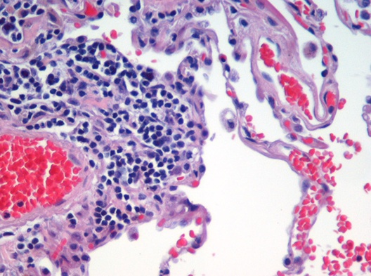
The study of tissues is known as histology or, in connection with disease, as histopathology. Xavier Bichat is considered as the "Father of Histology".[4] Plant histology is studied in both plant anatomy and physiology. The classical tools for studying tissues are the paraffin block in which tissue is embedded and then sectioned, the histological stain, and the optical microscope. Developments in electron microscopy, immunofluorescence, and the use of frozen tissue-sections have enhanced the detail that can be observed in tissues. With these tools, the classical appearances of tissues can be examined in health and disease, enabling considerable refinement of medical diagnosis and prognosis.

Plant tissue

[edit]
Cross-section of a flax plant stem with several layers of different tissue types:

In plant anatomy, tissues are categorized broadly into three tissue systems: the epidermis, the ground tissue, and the vascular tissue.[5]

  • Epidermis – Cells forming the outer surface of the leaves and of the young plant body.
  • Vascular tissue – The primary components of vascular tissue are the xylem and phloem. These transport fluids and nutrients internally.
  • Ground tissue – Ground tissue is less differentiated than other tissues. Ground tissue manufactures nutrients by photosynthesis and stores reserve nutrients.

Plant tissues can also be divided differently into two types:

  1. Meristematic tissues
  2. Permanent tissues.

Meristematic tissue

[edit]

Meristematic tissue consists of actively dividing cells and leads to an increase in length and thickness of the plant. The primary growth of a plant occurs only in certain specific regions, such as in the tips of stems or roots. It is in these regions that meristematic tissue is present. Cells of this type of tissue are roughly spherical or polyhedral to rectangular in shape, with thin cell walls. New cells produced by meristem are initially those of meristem itself, but as the new cells grow and mature, their characteristics slowly change and they become differentiated as components of meristematic tissue, being classified as:

1.Primary meristem.

  • Apical meristem : Present at the growing tips of stems and roots, they increase the length of the stem and root. They form growing parts at the apices of roots and stems and are responsible for the increase in length, also called primary growth. This meristem is responsible for the linear growth of an organ.

2.Secondary meristem.

  • Lateral meristem: Cells which mainly divide in one plane and cause the organ to increase in diameter and girth. Lateral meristem usually occurs beneath the bark of the tree as cork cambium and in vascular bundles of dicotyledons as vascular cambium. The activity of this cambium forms secondary growth.
  • Intercalary meristem: Located between permanent tissues, it is usually present at the base of the node, internode, and on leaf base. They are responsible for growth in length of the plant and increasing the size of the internode. They result in branch formation and growth.

The cells of meristematic tissue are similar in structure and have a thin and elastic primary cell wall made of cellulose. They are compactly arranged without intercellular spaces between them. Each cell contains a dense cytoplasm and a prominent cell nucleus. The dense protoplasm of meristematic cells contains very few vacuoles. Normally the meristematic cells are oval, polygonal, or rectangular in shape.

Meristematic tissue cells have a large nucleus with small or no vacuoles because they have no need to store anything. Their basic function is to multiply and increase the girth and length of the plant, with no intercellular spaces.

Permanent tissues

[edit]

Permanent tissues may be defined as a group of living or dead cells formed by meristematic tissue and have lost their ability to divide and have permanently placed at fixed positions in the plant body. Meristematic tissues that take up a specific role lose the ability to divide. This process of taking up a permanent shape, size and a function is called cellular differentiation. Cells of meristematic tissue differentiate to form different types of permanent tissues. There are 2 types of permanent tissues:

  1. simple permanent tissues
  2. complex permanent tissues

Simple permanent tissue

[edit]

Simple permanent tissue is a group of cells that are similar in origin, structure, and function. They are of three types:

  1. Parenchyma
  2. Collenchyma
  3. Sclerenchyma
Parenchyma
[edit]

Parenchyma (Greek, para – 'beside'; enchyma– infusion – 'tissue') is the bulk of a substance. In plants, it consists of relatively unspecialized living cells with thin cell walls that are usually loosely packed so that intercellular spaces are found between cells of this tissue. These are generally isodiametric in shape. They contain small number of vacuoles or sometimes they even may not contain any vacuole. Even if they do so the vacuole is of much smaller size than of normal animal cells. This tissue provides support to plants and also stores food. Chlorenchyma is a special type of parenchyma that contains chlorophyll and performs photosynthesis. In aquatic plants, aerenchyma tissues, or large air cavities, give support to float on water by making them buoyant. Parenchyma cells called idioblasts have metabolic waste. Spindle shaped fibers are also present in this cell to support them and known as prosenchyma, succulent parenchyma also noted. In xerophytes, parenchyma tissues store water.

Collenchyma
[edit]
Cross section of collenchyma cells

Collenchyma (Greek, 'Colla' means gum and 'enchyma' means infusion) is a living tissue of primary body like Parenchyma. Cells are thin-walled but possess thickening of cellulose, water and pectin substances (pectocellulose) at the corners where a number of cells join. This tissue gives tensile strength to the plant and the cells are compactly arranged and have very little inter-cellular spaces. It occurs chiefly in hypodermis of stems and leaves. It is absent in monocots and in roots.

Collenchymatous tissue acts as a supporting tissue in stems of young plants. It provides mechanical support, elasticity, and tensile strength to the plant body. It helps in manufacturing sugar and storing it as starch. It is present in the margin of leaves and resists tearing effect of the wind.

Sclerenchyma
[edit]

Sclerenchyma (Greek, Sclerous means hard and enchyma means infusion) consists of thick-walled, dead cells and protoplasm is negligible. These cells have hard and extremely thick secondary walls due to uniform distribution and high secretion of lignin and have a function of providing mechanical support. They do not have inter-cellular spaces between them. Lignin deposition is so thick that the cell walls become stronger, rigid and impermeable to water, which are also known as a stone cells or sclereids. These tissues are mainly of two types: sclerenchyma fiber and sclereids. Sclerenchyma fiber cells have a narrow lumen and are long, narrow and unicellular. Fibers are elongated cells that are strong and flexible, often used in ropes. Sclereids have extremely thick cell walls and are brittle, and are found in nutshells and legumes.

Epidermis
[edit]

The entire surface of the plant consists of a single layer of cells called epidermis or surface tissue. The entire surface of the plant has this outer layer of the epidermis. Hence, it is also called surface tissue. Most of the epidermal cells are relatively flat. The outer and lateral walls of the cell are often thicker than the inner walls. The cells form a continuous sheet without intercellular spaces. It protects all parts of the plant. The outer epidermis is coated with a waxy thick layer called cutin, which prevents loss of water. The epidermis also consists of stomata (singular:stoma), which helps in transpiration.

Complex permanent tissue

[edit]

The complex permanent tissue consists of more than one type of cells having a common origin which work together as a unit. Complex tissues are mainly concerned with the transportation of mineral nutrients, organic solutes (food materials), and water. That's why it is also known as conducting and vascular tissue. The common types of complex permanent tissue are:

Xylem and phloem together form vascular bundles.

Xylem
[edit]

Xylem (Greek, xylos = wood) serves as a chief conducting tissue of vascular plants. It is responsible for the conduction of water and inorganic solutes. Xylem consists of four kinds of cells:

  • Tracheids
  • Vessels (or tracheae)
  • Xylem fibers or Xylem sclerenchyma
  • Xylem parenchyma
Cross section of 2-year-old Tilia americana, highlighting xylem ray shape and orientation

Xylem tissue is organised in a tube-like fashion along the main axes of stems and roots. It consists of a combination of parenchyma cells, fibers, vessels, tracheids, and ray cells. Longer tubes made up of individual cells are vessels, while vessel members are open at each end. Internally, there may be bars of wall material extending across the open space. These cells are joined end-to-end to form long tubes. Vessel members and tracheids are dead at maturity. Tracheids have thick secondary cell walls and are tapered at the ends. They do not have end openings such as the vessels. The end overlap with each other, with pairs of pits present. The pit pairs allow water to pass from cell to cell.

Though most conduction in xylem tissue is vertical, lateral conduction along the diameter of a stem is facilitated via rays.[citation needed] Rays are horizontal rows of long-living parenchyma cells that arise out of the vascular cambium.

Phloem
[edit]

Phloem consists of:

Phloem is an equally important plant tissue as it is also part of the 'plumbing system' of a plant. Primarily, phloem carries dissolved food substances throughout the plant. This conduction system is composed of sieve-tube member and companion cells, which are without secondary walls. The parent cells of the vascular cambium produce both xylem and phloem. This usually also includes fibers, parenchyma, and ray cells. Sieve tubes are formed from sieve-tube members laid end to end. The end walls, unlike vessel members in xylem, do not have openings. The end walls, however, are full of small pores where cytoplasm extends from cell to cell. These porous connections are called sieve plates. In spite of the fact that their cytoplasm is actively involved in the conduction of food materials, sieve-tube members do not have nuclei at maturity. It is the companion cells that are nestled between sieve-tube members that function in some manner bringing about the conduction of food. Sieve-tube members that are alive contain a polymer called callose, a carbohydrate polymer, forming the callus pad/callus, the colourless substance that covers the sieve plate. Callose stays in solution as long as the cell contents are under pressure. Phloem transports food and materials in plants upwards and downwards as required.

Animal tissue

[edit]

Animal tissues are grouped into four basic types: connective, muscle, nervous, and epithelial.[6] Collections of tissues joined in units to serve a common function compose organs. While most animals can generally be considered to contain the four tissue types, the manifestation of these tissues can differ depending on the type of organism. For example, the origin of the cells comprising a particular tissue type may differ developmentally for different classifications of animals. Tissue appeared for the first time in the diploblasts, but modern forms only appeared in triploblasts.

The epithelium in all animals is derived from the ectoderm and endoderm (or their precursor in sponges), with a small contribution from the mesoderm, forming the endothelium, a specialized type of epithelium that composes the vasculature. By contrast, a true epithelial tissue is present only in a single layer of cells held together via occluding junctions called tight junctions, to create a selectively permeable barrier. This tissue covers all organismal surfaces that come in contact with the external environment such as the skin, the airways, and the digestive tract. It serves functions of protection, secretion, and absorption, and is separated from other tissues below by a basal lamina.

The connective tissue and the muscular are derived from the mesoderm. The nervous tissue is derived from the ectoderm.

Epithelial tissues

[edit]

The epithelial tissues are formed by cells that cover the organ surfaces, such as the surface of skin, the airways, surfaces of soft organs, the reproductive tract, and the inner lining of the digestive tract. The cells comprising an epithelial layer are linked via semi-permeable, tight junctions; hence, this tissue provides a barrier between the external environment and the organ it covers. In addition to this protective function, epithelial tissue may also be specialized to function in secretion, excretion and absorption. Epithelial tissue helps to protect organs from microorganisms, injury, and fluid loss.

Functions of epithelial tissue:

  • The principle function of epithelial tissues are covering and lining of free surface
  • The cells of the body's surface form the outer layer of skin.
  • Inside the body, epithelial cells form the lining of the mouth and alimentary canal and protect these organs.
  • Epithelial tissues help in the elimination of waste.
  • Epithelial tissues secrete enzymes and/or hormones in the form of glands.
  • Some epithelial tissue perform secretory functions. They secrete a variety of substances including sweat, saliva, mucus, enzymes.

There are many kinds of epithelium, and nomenclature is somewhat variable. Most classification schemes combine a description of the cell-shape in the upper layer of the epithelium with a word denoting the number of layers: either simple (one layer of cells) or stratified (multiple layers of cells). However, other cellular features such as cilia may also be described in the classification system. Some common kinds of epithelium are listed below:

  • Simple squamous (pavement) epithelium
  • Simple cuboidal epithelium
  • Simple columnar epithelium
  • Simple ciliated (pseudostratified) columnar epithelium
  • Simple glandular columnar epithelium
  • Stratified non-keratinized squamous epithelium
  • Stratified keratinized epithelium
  • Stratified transitional epithelium

Connective tissue

[edit]

Connective tissues are made up of cells separated by non-living material, which is called an extracellular matrix. This matrix can be liquid or rigid. For example, blood contains plasma as its matrix and bone's matrix is rigid. Connective tissue gives shape to organs and holds them in place. Blood, bone, tendon, ligament, adipose, and areolar tissues are examples of connective tissues. One method of classifying connective tissues is to divide them into three types: fibrous connective tissue, skeletal connective tissue, and fluid connective tissue.

Muscle tissue

[edit]
Cross section through skeletal muscle and a small nerve at high magnification (H&E stain)

Muscle cells (myocytes) form the active contractile tissue of the body. Muscle tissue functions to produce force and cause motion, either locomotion or movement within internal organs. Muscle is formed of contractile filaments and is separated into three main types; smooth muscle, skeletal muscle and cardiac muscle. Smooth muscle has no striations when examined microscopically. It contracts slowly but maintains contractibility over a wide range of stretch lengths. It is found in such organs as sea anemone tentacles and the body wall of sea cucumbers. Skeletal muscle contracts rapidly but has a limited range of extension. It is found in the movement of appendages and jaws. Obliquely striated muscle is intermediate between the other two. The filaments are staggered and this is the type of muscle found in earthworms that can extend slowly or make rapid contractions.[7] In higher animals striated muscles occur in bundles attached to bone to provide movement and are often arranged in antagonistic sets. Smooth muscle is found in the walls of the uterus, bladder, intestines, stomach, oesophagus, respiratory airways, and blood vessels. Cardiac muscle is found only in the heart, allowing it to contract and pump blood through the body.

Nervous tissue

[edit]

Cells comprising the central nervous system and peripheral nervous system are classified as nervous (or neural) tissue. In the central nervous system, neural tissues form the brain and spinal cord. In the peripheral nervous system, neural tissues form the cranial nerves and spinal nerves, inclusive of the motor neurons.

Mineralized tissues

[edit]

Mineralized tissues are biological tissues that incorporate minerals into soft matrices. Such tissues may be found in both plants and animals.

History

[edit]
Xavier Bichat (1771–1802)

Xavier Bichat introduced the word tissue into the study of anatomy by 1801.[8] He was "the first to propose that tissue is a central element in human anatomy, and he considered organs as collections of often disparate tissues, rather than as entities in themselves".[9] Although he worked without a microscope, Bichat distinguished 21 types of elementary tissues from which the organs of the human body are composed,[10] a number later reduced by other authors.

In 2013, the work of de Bono et al introduced the concept of the Functional Tissue Unit (FTU) as a biophysical definition of spatial tissue domains that satisfy both long-range and short range (i.e., local) communication constraints for cellular maintenance and supracellular organization (i.e., architecture).[11] A FTU consists of a cylindrical diffusive field of parenchyma centered around a tube. This central tube conveys long-range flow of a body fluid (e.g., blood, bile, air, urinary ultra-filtrate). It is compelling to draw parallels between the biophysical constraints that act upon a tissue domain and those acting on a protein domain. In this analogy, the FTU’s central tube is akin to the peptide backbone in a protein domain, and the cells in the surrounding diffusive cuff are analogous to interacting amino acid side chains.

Graphical examples of Functional Tissue Units


See also

[edit]

References

[edit]

Sources

[edit]
[edit]
Revisions and contributorsEdit on WikipediaRead on Wikipedia
from Grokipedia
In biology, a tissue is a group of structurally and functionally similar cells and their associated substances that together perform specific tasks in multicellular organisms. This level of organization enables specialization and division of labor among cells, supporting complex physiological processes unattainable by individual cells. Tissues serve as building blocks of organs, where diverse types integrate to facilitate functions such as movement, protection, and communication. Tissue classification varies between plants and animals. In plants, tissues are grouped into meristematic (actively dividing cells for growth) and permanent (specialized, non-dividing cells), with permanent tissues further divided into simple (e.g., , collenchyma, sclerenchyma) and complex (e.g., , ). In animals, including humans, there are four primary tissue types: epithelial, connective, muscle, and nervous. Epithelial tissue forms sheets covering body surfaces, lining cavities, and glands, providing protection, absorption, and secretion. offers support, binds tissues, and includes subtypes like , , blood, and for structure, transport, and . Muscle tissue enables contraction for movement, with subtypes skeletal (voluntary), cardiac (involuntary heart), and smooth (involuntary organs). , made of neurons and glial cells, conducts electrical and chemical signals for coordination and . These tissues interact dynamically, with development, , and repair influenced by genetic, environmental, and physiological factors, highlighting their in and .

General Concepts

Definition and Characteristics

In , a tissue is defined as a group of cells, along with their associated intercellular substances, that share a common structure and function to perform a specific within a . The , a nonliving material surrounding the cells, provides , facilitates , and serves as a medium for biochemical exchange between cells. Key characteristics of tissues include their highly organized cellular arrangement, which allows for efficient division of labor, and the presence of specialized intercellular connections that enable communication and maintain tissue integrity. In , these connections often take the form of plasmodesmata, cytoplasmic channels that link adjacent cells for symplastic transport of nutrients and signals. In animals, structures such as tight junctions seal intercellular spaces to regulate permeability and prevent leakage across tissue barriers. The composition of the extracellular matrix varies, ranging from fibrous proteins and in animals to cellulosic walls in , influencing tissue rigidity and flexibility. Tissues represent an intermediate level of biological organization, distinct from individual cells—the fundamental units of life capable of independent metabolism—and from organs, which integrate multiple tissue types to achieve complex functions. For instance, while a single cell operates autonomously, a tissue amplifies this capacity through collective action, and an organ coordinates tissues for systemic roles. In multicellular organisms, this tissue level enables specialization and efficiency, forming the foundation for higher organizational structures like organs and organ systems. The term "tissue" originates from the Middle English "tissu," borrowed from "tissu" meaning "woven" or "fabric," derived from Latin "texere" (to weave), evoking the interlaced arrangement of cells observed under .

Functions and Importance

Biological tissues perform a diverse array of primary functions critical to the of multicellular organisms, encompassing support, , , , absorption, contraction, conduction, and coordination. These roles are specialized across tissue types, with epithelial tissues primarily handling , absorption, and to form barriers and regulate exchanges; connective tissues providing , binding, and nutrient ; muscle tissues enabling contraction for movement and force generation; and nervous tissues supporting conduction of signals and coordination of responses. Collectively, these functions ensure the organism's ability to maintain integrity, respond to stimuli, and sustain vital processes. The significance of tissues extends to their foundational role in multicellularity, where they enable division of labor among cell populations, allowing specialized groups to perform distinct tasks more efficiently than in unicellular forms. This specialization facilitates by coordinating internal conditions, such as distribution and waste removal, and supports to environmental challenges through enhanced functional integration. In evolutionary history, tissues represent a transformative that propelled the advancement from simple unicellular progenitors to complex multicellular architectures, increasing organismal size, longevity, and ecological versatility over billions of years. Tissues as a key evolutionary milestone are evident in clades like , where the development of organized tissues permitted greater structural complexity and physiological specialization beyond diploblastic forms, and in Embryophyta, where tissue differentiation from algal ancestors enabled vascular support, protection against , and efficient resource transport essential for terrestrial life. Through inter-tissue interactions, such as signaling via extracellular matrices and junctions, tissues integrate to form organs and organ systems that execute higher-level functions; for instance, epithelial tissues collaborate with connective tissues to create selective barriers that prevent entry while permitting necessary exchanges. This underscores tissues' indispensable contribution to organismal unity and survival.

Plant Tissues

Meristematic Tissues

Meristematic tissues consist of undifferentiated, actively dividing cells that serve as the primary sites of growth in plants, enabling continuous development throughout the plant's life. These tissues are composed of totipotent cells, meaning they have the potential to differentiate into any cell type, a unique feature allowing plants to regenerate entire organisms from single cells under appropriate conditions. The cells exhibit characteristic features such as thin primary cell walls for flexibility during division, dense cytoplasm rich in organelles, and prominent nuclei indicating high metabolic and synthetic activity. Meristematic tissues are classified by their position and function into three main types: apical, lateral, and intercalary meristems. Apical meristems are located at the tips of and shoots, consisting of the promeristem—an initial cluster of embryonic-like cells—and primary meristems that give rise to protoderm, ground meristem, and procambium./04:_Plant_Physiology_and_Regulation/4.06:_Development/4.6.02:_Meristems) Lateral meristems, such as the and , occur along the sides of stems and and are considered secondary meristems derived from primary tissues. Intercalary meristems are found at the bases of leaves or internodes, particularly in grasses, facilitating localized growth. The primary function of meristematic tissues is to produce new cells through division, supporting unique to , where no fixed size limit exists. Apical meristems drive primary growth by elongating and shoots, while lateral meristems promote by increasing girth through the addition of vascular and protective layers. Intercalary meristems enable regrowth after damage, such as in grazed grasses. These tissues generate daughter cells that eventually differentiate into permanent tissues, contributing to the plant's overall structure and adaptability. At the cellular level, meristematic activity involves frequent to duplicate genetic material and to partition the , with forming a during the latter process. The arises from Golgi-derived vesicles that fuse at the cell's equator, depositing and enzymes to construct the new between daughter cells. This mechanism ensures precise separation in the absence of a centrosome-based apparatus, supporting the organized expansion of tissues.

Simple Permanent Tissues

Simple permanent tissues consist of mature, non-dividing cells in that are structurally and functionally similar, derived from meristematic tissues through differentiation. These tissues primarily form the system, providing mechanical support, storage, and metabolic functions without involvement in transport. They are characterized by cells connected via plasmodesmata, which facilitate intercellular communication and material exchange. Parenchyma is the most abundant type of simple permanent tissue, composed of living cells with thin, flexible primary cell walls made primarily of and . These cells are typically isodiametric or elongated, retain the ability to divide under certain conditions, and often contain chloroplasts for in leaves or serve as storage sites for and other reserves in and stems. For example, parenchyma forms the in stems and the mesophyll in leaves, enabling and wound healing through callus formation. Collenchyma provides flexible support in growing parts, consisting of living cells with unevenly thickened primary cell walls, particularly at the corners, due to additional and deposition. These elongated cells lack secondary walls and , allowing them to stretch as the plant grows, and they often occur in strands beneath the . Found in petioles, young stems, and leaf veins—for instance, the stringy fibers in stalks—collenchyma responds to mechanical stimuli like by further wall thickening for tensile strength. Sclerenchyma offers rigid mechanical support in mature regions, featuring cells with thick secondary cell walls impregnated with , rendering them dead at maturity and impermeable to . This tissue includes two subtypes: fibers, which are long and slender (e.g., in for production), and sclereids, which are irregular and branched (e.g., in nutshells or grit for hardness). Distributed throughout stems, leaves, , and coats, sclerenchyma provides compressive and tensile strength, with its lignified walls contrasting the pectin-rich compositions of other simple tissues. Overall, simple permanent tissues are distributed in the cortex, , and mesophyll of stems, leaves, and , adapting to roles in flexibility (collenchyma), storage and (), and permanence (sclerenchyma) to maintain plant integrity during growth and environmental stress.

Complex Permanent Tissues

Complex permanent tissues in are specialized, non-dividing structures composed of multiple distinct cell types that collaborate to perform specific functions, primarily long-distance and mechanical support. Unlike simple permanent tissues, which consist of a single , complex tissues exhibit heterogeneity to facilitate efficient conduction and structural integrity. These tissues originate from meristematic cells during primary and . The primary types of complex permanent tissues are and , which together form the vascular system. is responsible for the unidirectional transport of water and dissolved minerals from to aerial parts, while also providing rigidity. It comprises four main cell types: tracheids, present in all vascular plants and featuring tapered ends with pits for lateral water movement; vessel elements, unique to angiosperms and stacked end-to-end to form continuous vessels; fibers, elongated sclerenchyma cells that enhance tensile strength; and cells, which store nutrients and facilitate radial transport. Most cells, except , are dead at maturity, with lignified secondary walls that contribute to support. Phloem, in contrast, enables bidirectional transport of sugars, , and other organic compounds produced by , distributing them to non-photosynthetic tissues. Its key components include sieve tube elements, which form sieve tubes connected by sieve plates; companion cells, nucleated cells that provide metabolic support to the enucleate sieve elements via plasmodesmata; fibers for mechanical reinforcement; and cells for storage and short-distance transport. Unlike , phloem cells remain alive at maturity, though sieve elements lose nuclei and most organelles. in woody plants involves the , a lateral that produces additional and layers, increasing girth. Structural adaptations optimize transport efficiency and resilience in these tissues. In xylem, perforation plates at vessel ends—simple openings in angiosperms or scalariform/barred in some —minimize resistance to flow, while pit membranes in tracheids and vessels allow selective passage and prevent air bubbles from spreading during , a where tension causes columns to break; lignification further resists collapse under negative pressure. Phloem adaptations include callose deposition on sieve plates, a that plugs pores in response to or to prevent leakage, and the symplastic continuity between sieve elements and companion cells for loading/unloading of solutes. These features ensure reliable conduction under varying environmental stresses.

Animal Tissues

Epithelial Tissue

Epithelial tissue, also known as epithelium, consists of closely packed sheets of cells that cover external body surfaces, line internal cavities and organs, and form glands. These cells are derived from all three primary germ layers—ectoderm, mesoderm, and endoderm—and exhibit little intercellular material, distinguishing them from other tissue types. Epithelial tissues are avascular, relying on diffusion from underlying connective tissue for nourishment, and are characterized by a high regenerative capacity due to frequent exposure to environmental stresses. Epithelial tissues are classified based on the number of cell layers and the shape of the cells. Simple epithelia consist of a single layer of cells, facilitating rapid , , or absorption, while stratified epithelia feature two or more layers, providing greater against abrasion and penetration. Cell shapes include squamous (flat and scale-like, ideal for ), cuboidal (cube-shaped, suited for and absorption), and columnar (tall and column-like, optimized for absorption and ). Pseudostratified epithelia appear multilayered but are actually single-layered with nuclei at varying heights, often ciliated for transport functions. Epithelial cells exhibit distinct polarity, with an apical surface facing the lumen or external environment, a basal surface anchored to a , and lateral surfaces connecting adjacent cells. The apical surface may bear modifications such as microvilli to increase surface area for absorption or cilia for , while the basal surface adheres to the underlying . Locations include external coverings like the of the skin and internal linings such as the , respiratory airways, blood vessels (), and body cavities (). The primary functions of epithelial tissue encompass , absorption, , and . Protective roles are prominent in stratified squamous epithelia of the skin and oral cavity, shielding against mechanical injury and pathogens. Absorption occurs via simple columnar epithelia in the intestines, enhanced by microvilli on enterocytes, while is key in simple squamous epithelia of glomeruli and alveoli. is mediated by glandular epithelia, which form exocrine glands (e.g., salivary glands releasing enzymes via ducts) or endocrine glands (e.g., releasing hormones directly into blood). Cell cohesion is maintained by specialized junctions, including tight junctions that seal intercellular spaces to prevent leakage and desmosomes that provide mechanical strength through cadherin-mediated attachments. Specializations adapt epithelial tissues to specific needs, such as ciliated epithelium in the , where coordinated ciliary beating propels and trapped particles toward the . Microvilli, forming the in intestinal epithelia, dramatically expand surface area—up to 600-fold in some cases—for nutrient uptake without increasing overall tissue volume. These features underscore the tissue's role as a dynamic interface between the body and its environment, often integrating with underlying via the for structural support.

Connective Tissue

Connective tissue is a class of biological tissue in animals that primarily provides , binding, and protection to other tissues and organs, distinguished by its abundant relative to cellular content. It consists of three main components: cells suspended within an composed of protein fibers and . The cells include resident types such as fibroblasts, which produce the matrix; adipocytes, specialized for storage; and macrophages, involved in and immune surveillance. Fibers in the matrix are predominantly for tensile strength, for elasticity, and reticular fibers for fine support networks. The is an amorphous gel-like material that hydrates the tissue and facilitates nutrient . Connective tissues are classified into loose, dense, and specialized types based on the arrangement and composition of their matrix. Loose connective tissues, such as , feature loosely arranged and elastic fibers with abundant , providing flexibility and cushioning between organs. , a subtype of loose connective tissue, consists mainly of adipocytes and serves for and . Dense connective tissues are characterized by tightly packed fibers; regular dense connective tissue, like tendons and ligaments, has parallel bundles for unidirectional strength, while irregular dense connective tissue, found in the , has fibers in multiple directions for multidirectional resistance. Specialized connective tissues include , , and osseous tissue (detailed separately as mineralized tissues). provides flexible support without vascularization; , with and minimal fibers, occurs in articular surfaces and the for smooth, resilient surfaces; , incorporating elastic fibers alongside , is present in the and for shape maintenance with flexibility; and , blending type I and II collagens with dense fiber bundles, supports high-stress areas like intervertebral discs. , a fluid connective tissue, consists of plasma (the ) with suspended cells including erythrocytes for oxygen , leukocytes for immune defense, and platelets for clotting. The primary functions of connective tissue encompass structural support to maintain organ shape, nutrient and waste transport via the matrix, energy storage in adipose forms, immune defense through macrophages and mast cells, and tissue repair by fibroblast-mediated matrix remodeling. Vascularity varies among types, with loose and dense tissues being well-vascularized for nutrient delivery, while cartilage relies on diffusion from surrounding perichondrium due to its avascular nature. The extracellular matrix's properties are critical to connective tissue function; collagen fibers impart high tensile strength to withstand mechanical stress, while the ground substance, rich in glycosaminoglycans such as and , binds water to maintain hydration, , and resilience against compression. These components interact dynamically, with proteoglycans in the linking to to form hydrated networks that resist deformation.

Muscle Tissue

Muscle tissue is a specialized type of animal tissue composed of elongated cells capable of contraction, enabling movement, force generation, and maintenance of posture. It is one of the four primary types of animal tissues, alongside epithelial, connective, and nervous tissues. There are three main types of muscle tissue: , , and , each adapted to specific physiological roles. is striated, voluntary, and consists of multinucleated fibers that attach to bones via tendons, facilitating locomotion and voluntary movements. is also striated but involuntary, forming the myocardium of the heart with branched fibers connected by intercalated discs that include gap junctions for synchronized contractions. is non-striated and involuntary, found in the walls of hollow organs such as blood vessels, the digestive tract, and , where it regulates visceral movements like . The microscopic structure of muscle tissue revolves around contractile proteins and , which form filaments that interact to produce shortening. In skeletal and cardiac muscle, these filaments are organized into repeating units called sarcomeres, giving the tissue its striated appearance and allowing precise control of contraction length. Intercalated discs in not only contain gap junctions for electrical coupling but also desmosomes and adherens junctions for mechanical stability during rhythmic pumping. Smooth muscle lacks sarcomeres, with actin-myosin filaments arranged in a more irregular lattice, and features gap junctions in some tissues to coordinate contractions across cells. These structural adaptations support the tissue's role in generating force without fatigue in diverse contexts. Muscle contraction occurs through the sliding filament mechanism, where myosin heads bind to filaments, powered by , causing filaments to slide past each other and shorten the in striated types or the cell in . This process generates force for functions such as heartbeat in , in for nutrient propulsion, and posture maintenance in . Energy for contraction is derived from ATP produced via cellular , with calcium ions playing a key regulatory role by exposing binding sites on . Under nervous control from the somatic or autonomic systems, muscle tissue responds to stimuli to perform these essential roles. Regeneration of muscle tissue is limited in adults compared to development, relying on resident stem cells to repair minor damage. In , cells—quiescent stem cells located between the and —activate upon injury, proliferate, and fuse with damaged fibers to restore function, though large-scale regeneration is inefficient without external support. Cardiac muscle has minimal regenerative capacity, with cardiomyocytes largely post-mitotic, leading to scar formation after injury; recent studies highlight limited contributions from cells. Smooth muscle exhibits some regenerative potential through proliferation of existing cells or recruitment from progenitors, but it is constrained in adults, emphasizing the tissue's vulnerability to chronic damage.

Nervous Tissue

Nervous tissue is the primary component of the in animals, consisting of excitable cells specialized for rapid communication and their supporting elements. The functional units are neurons, which include a cell body (soma) containing the nucleus, dendrites that receive incoming signals, and a long that conducts outgoing impulses away from the soma. Synapses, specialized junctions between neurons or between neurons and target cells, facilitate signal transmission through chemical or electrical means. Supporting neuroglia, or glial cells, outnumber neurons and provide structural, metabolic, and protective roles; key types include , which maintain the blood-brain barrier and regulate extracellular balance; , which insulate axons in the ; and , which act as immune defenders by phagocytosing debris and pathogens. The core functions of nervous tissue involve sensing environmental changes, processing information, and coordinating responses to maintain . Neurons detect sensory input via specialized receptors that convert stimuli into electrical signals, which are then integrated in neural circuits to evaluate relevance and generate appropriate outputs. Motor output occurs when integrated signals propagate to effectors like muscles or glands, enabling actions such as contraction or secretion. This signaling relies on action potentials, self-propagating electrochemical waves along axons, driven by voltage-gated channels that exploit sodium (Na⁺) and (K⁺) gradients across the ; opens Na⁺ channels, allowing influx that triggers further propagation, followed by K⁺ efflux for repolarization. Nervous tissue is organized into the (CNS), comprising the and for integration, and the peripheral nervous system (PNS), consisting of that connect the CNS to the body for sensory and motor relay. Myelination, the wrapping of axons in lipid-rich sheaths, enhances conduction efficiency; in the CNS, form myelin segments around multiple axons, while in the PNS, Schwann cells myelinate single axons. This insulation enables , where action potentials "jump" between unmyelinated nodes of Ranvier, increasing speed from about 0.5–2 m/s in unmyelinated fibers to 70–120 m/s in large myelinated ones, crucial for rapid responses. Adaptations in nervous tissue allow dynamic responses to experience and injury, exemplified by , the ability of neural circuits to reorganize through changes in synaptic strength, such as that strengthens connections for learning and . Signal transmission at synapses involves neurotransmitter release from presynaptic vesicles into the synaptic cleft, where molecules like bind receptors on postsynaptic cells to propagate or modulate signals—, for instance, excites at neuromuscular junctions and influences plasticity in the brain. , released in reward-related pathways, modulates by enhancing or depressing transmission, supporting adaptive behaviors like and formation.

Mineralized Tissues

Mineralized tissues in vertebrates are specialized connective tissues characterized by the deposition of crystals, a mineral with the formula Ca₁₀(PO₄)₆(OH)₂, within an organic matrix to provide exceptional hardness and rigidity. This mineralization process, known as , integrates the inorganic phase (comprising about 65-70% of by weight) with an organic framework primarily of type I, enabling these tissues to withstand mechanical stresses while maintaining metabolic activity. Unlike soft connective tissues, mineralized tissues are unique to vertebrates and serve as the primary structural components of the and . Bone is the most abundant mineralized tissue, consisting of two main types: compact (cortical) bone, which forms the dense outer layer offering high compressive strength, and spongy (cancellous or trabecular) bone, which fills the interior with a porous network of trabeculae for lightweight support and metabolic functions. 's dynamic structure is maintained by three key cell types: osteoblasts, which synthesize and mineralize the ; osteocytes, mature cells embedded within the matrix that sense mechanical loads and coordinate remodeling; and osteoclasts, multinucleated cells that resorb bone through acidification and enzymatic degradation. This cellular interplay allows continuous remodeling, adapting bone architecture to physiological demands. In addition to bone, mineralized tissues include dental structures such as enamel, , and , each with distinct compositions tailored to their roles in mastication. Enamel, the hardest substance in the (approximately 96% by weight), is an acellular, highly mineralized epithelial-derived tissue covering the crown of , providing a protective barrier against and acids. , forming the bulk of the beneath enamel, is about 70% mineralized and features a tubular structure containing odontoblastic processes that transmit sensory signals. , a thinner acellular or cellular layer (around 50% ) covering the , facilitates anchorage to the periodontal ligament. These tissues fulfill critical functions beyond structural support, including the storage and of essential minerals like calcium and , which are mobilized from reservoirs during metabolic needs. Spongy , in particular, houses red where hematopoiesis occurs, producing blood cells throughout life. 's adaptability is exemplified by , which posits that bone remodels in response to mechanical loading, increasing density in high-stress areas and resorbing in low-load regions to optimize strength and efficiency. Bone formation, or , occurs through two primary mechanisms unique to vertebrates: , where mesenchymal cells directly differentiate into osteoblasts to form without a cartilage intermediate (as in flat bones like the ); and , involving a model that is gradually replaced by through vascular invasion and ossification centers (typical for long bones). Dental mineralized tissues form via specialized processes: ameloblasts secrete enamel matrix for mineralization before their , leaving it acellular; odontoblasts produce throughout life; and cementoblasts deposit incrementally. These processes ensure the integrated development of rigid, functional structures from precursors.

Tissue Development

Histogenesis in Plants

Histogenesis in plants encompasses the developmental processes by which undifferentiated cells in meristems undergo patterned division, expansion, and specialization to form distinct tissue types, contrasting with animal histogenesis by enabling indeterminate, modular growth throughout the plant's life. This formation establishes the primary body plan, including dermal, ground, and vascular tissues, through coordinated cellular activities in shoot and apical meristems. The stages of histogenesis initiate with cell proliferation in meristematic zones, where rapid mitotic divisions produce daughter cells that maintain totipotency—the inherent ability of plant cells to regenerate entire organisms under appropriate conditions. Following proliferation, cells enter an elongation phase, increasing in size primarily along the growth axis to contribute to organ extension, as observed in root-tip meristems. Differentiation then ensues, transforming these cells into specialized types such as tracheids or parenchyma, often guided by positional cues within the organized layers of shoot apical meristems (tunica-corpus structure) or root apical meristems (quiescent center and surrounding initials). For instance, auxin gradients establish polarity, with higher concentrations promoting vascular cell fate in procambial strands. Key mechanisms underlying these stages involve regulated and hormonal signaling. Homeobox genes, such as those in the KNOX family, play pivotal roles in maintaining indeterminacy and directing tissue patterning during and organ development. Hormonally, s drive directional transport to form concentration gradients that specify vascular differentiation, while cytokinins antagonize effects to refine patterning, ensuring bisymmetric vascular bundles in roots. This interplay, for example, translates cotyledonary responses into embryonic root vascular symmetry via cytokinin-mediated inhibition. Environmental factors further modulate histogenesis by influencing distribution and activity. Light signals, through phototropins, interact with transport to shape tissue orientation and elongation in shoots, counteracting unilateral growth biases. , sensed via statoliths in root cells, redirects fluxes to promote asymmetric elongation and root curvature, thereby patterning geotropic tissue responses. These cues integrate with totipotency to facilitate regeneration, allowing wounded tissues to redifferentiate meristematic cells into functional structures, as seen in formation during repair.

Histogenesis in Animals

Histogenesis in animals refers to the developmental processes by which specialized tissues form from undifferentiated precursor cells during embryogenesis and regeneration. This occurs primarily through the differentiation of cells derived from the three primary germ layers—, , and —established during , a pivotal stage where the reorganizes into a trilaminar structure. involves , ingression, and of cells, leading to the formation of these layers, which serve as the foundational blueprint for all subsequent tissue development. Key processes in histogenesis include embryonic induction, , and cytodifferentiation. Embryonic induction entails signaling between tissues that directs cell fate, such as the underlying inducing the overlying to form neural tissue via diffusible factors like BMP inhibitors. encompasses the physical shaping of tissues through cell movements, changes, and mechanical forces, resulting in organ architecture. Cytodifferentiation follows, where cells acquire specific functions and structures; for instance, cells, derived from at the border, migrate and differentiate into neurons, , and melanocytes under the influence of transcription factors like Sox10. , or , plays a crucial role in shaping tissues by eliminating excess cells, as seen in the interdigital regions during limb formation, where it sculpts digits through caspase-mediated execution. The contributes to external epithelia, such as and oral lining, as well as the entire , including the central and peripheral components. The gives rise to connective tissues like and , muscle types (skeletal, cardiac, smooth), and the cardiovascular system, with paraxial mesoderm forming somites that segment into sclerotome for skeleton and for muscles. The forms internal epithelia of the digestive and respiratory tracts, along with associated glands like the liver and . Beyond embryogenesis, histogenesis continues in adult animals through regeneration, mediated by stem cell niches that maintain tissue and repair damage. The hematopoietic stem cell niche in , comprising stromal cells and endothelial components, supports production and regeneration by providing cytokines like SCF and , enabling s to self-renew and differentiate into lineages. exemplifies regenerative histogenesis, progressing through four overlapping phases: (clot formation), (neutrophil and recruitment), proliferation ( formation with fibroblasts and ), and remodeling ( reorganization for maturation). These phases restore tissue integrity, though full regeneration varies by and site, often involving epithelial and reformation.

Historical Perspectives

Early Observations

The advent of in the enabled the first detailed observations of biological structures, laying the groundwork for understanding tissues as organized materials, though without recognition of their cellular composition. The , improved upon by scientists like around 1660, combined multiple lenses to achieve magnifications sufficient for viewing minute details, marking a pivotal technological advancement that shifted biological inquiry from to finer scales. Earlier prototypes dated to the late , but Hooke's refinements, including stable mounting and illumination, made systematic observation practical for natural philosophers. Marcello Malpighi, an Italian physician and microscopist, conducted pioneering examinations of plant and animal structures in 1661, producing detailed drawings that revealed layered organizations resembling fibrous networks. His observations of , including vascular bundles and epidermal layers, depicted tissues as interconnected meshes, influencing early botanical studies. Building on this, Robert Hooke published in 1665, where he described thin slices of cork under his compound microscope as comprising uniform, box-like "cells"—empty chambers akin to honeycomb partitions—providing the first visual evidence of plant tissue architecture and coining the term "cell" for these compartments. These findings portrayed plant tissues as rigid, porous frameworks rather than dynamic living units. In the realm of animal tissues, Antonie van Leeuwenhoek's single-lens microscopes in the 1670s yielded groundbreaking insights into fluid and contractile elements. By 1674, he observed red cells as disc-shaped corpuscles circulating in capillaries, illustrating as a tissue composed of suspended particles within a fluid matrix. Leeuwenhoek also examined muscle fibers, noting their striated, fibrillar arrangement and contractile behavior under magnification, which suggested tissues as interwoven bundles capable of motion. These early observations were constrained by the absence of cellular theory, viewing tissues primarily as woven fabrics or fibrous aggregates without discerning individual living cells as building blocks. Seventeenth- and eighteenth-century naturalists conceptualized the body as a mechanical assembly of threads and membranes, with "tissue" deriving from the Latin textum meaning "woven thing," emphasizing structural continuity over discrete units. This perspective, rooted in iatromechanical ideas, treated tissues as passive, interlaced materials formed during embryogenesis, limiting interpretations to gross textures rather than regenerative or modular processes.

Key Developments and Classifications

The foundational classification of animal tissues was established by French anatomist in 1801, who identified four primary types—epithelial, connective, muscular, and nervous—based on their functional and structural similarities across organs, without the aid of . Bichat introduced the term "tissue" (from the French tissu, meaning "woven") to denote these fundamental components of organs. This system marked a shift from organ-centric views to tissue-level analysis, emphasizing that organs are composites of these tissue types, and laid the groundwork for modern . In parallel, for plants, German botanist Gottlieb Haberlandt proposed an anatomico-physiological classification in his 1884 book Physiological Plant Anatomy, delineating three main tissue systems—dermal (protective outer layer), ground (storage and support), and vascular (transport)—along with a fourth meristematic system responsible for growth and differentiation. Haberlandt's framework addressed earlier gaps by incorporating meristematic tissues as dynamic, undifferentiated regions essential for , integrating physiological roles with anatomical structure. The advent of cell theory profoundly influenced tissue understanding in the mid-19th century. In 1838, botanist Matthias Schleiden asserted that plants are aggregates of cells, each forming the basic unit of structure and function, as detailed in his publication Beiträge zur Phytogenesis. Theodor Schwann extended this to animals in 1839, concluding in Mikroskopische Untersuchungen über die Übereinstimmung in der Struktur und dem Wachstum der Tiere und Pflanzen that all tissues arise from cells, unifying plant and animal biology under a cellular paradigm. This theory reframed tissues as multicellular assemblies, challenging prior notions of spontaneous generation. Rudolf Virchow further refined it in 1858 with his principle omnis cellula e cellula ("every cell from a cell"), articulated in Cellular Pathology, which emphasized cellular origins in disease and tissue repair, solidifying the cellular basis of all tissues. Twentieth-century advancements revealed finer tissue details through technological and molecular innovations. The introduction of electron microscopy in the 1940s enabled visualization of tissue , such as arrangements and intercellular junctions, far beyond light microscopy's limits, as demonstrated in early biological applications that uncovered subcellular components in epithelial and muscle tissues. By the 1980s, identified tissue-specific genes, notably the family, whose discovery and cloning revealed regulatory roles in patterning animal tissues along body axes during development. These genes, conserved across species, control differential in tissues like neural and connective types.

References

Add your contribution
Related Hubs
User Avatar
No comments yet.