Hubbry Logo
Radiation burnRadiation burnMain
Open search
Radiation burn
Community hub
Radiation burn
logo
8 pages, 0 posts
0 subscribers
Be the first to start a discussion here.
Radiation burn
Radiation burn
from Wikipedia
Radiation burn
Other namesRadiodermatitis
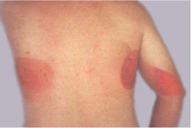
Ionizing radiation burn: large red patches of skin on the back and arm from multiple prolonged fluoroscopy procedures
SpecialtyDermatology Edit this on Wikidata

A radiation burn is a damage to the skin or other biological tissue and organs as an effect of radiation. The radiation types of greatest concern are thermal radiation, radio frequency energy, ultraviolet light and ionizing radiation.

The most common type of radiation burn is a sunburn caused by UV radiation. High exposure to X-rays during diagnostic medical imaging or radiotherapy can also result in radiation burns. As the ionizing radiation interacts with cells within the body—damaging them—the body responds to this damage, typically resulting in erythema—that is, redness around the damaged area. Radiation burns are often discussed in the same context as radiation-induced cancer due to the ability of ionizing radiation to interact with and damage DNA, occasionally inducing a cell to become cancerous. Cavity magnetrons can be improperly used to create surface and internal burning. Depending on the photon energy, gamma radiation can cause deep gamma burns, with 60Co internal burns common. Beta burns tend to be shallow as beta particles are not able to penetrate deeply into a body; these burns can be similar to sunburn. Alpha particles can cause internal alpha burns if inhaled, with external damage (if any) being limited to minor erythema.

Radiation burns can also occur with high power radio transmitters at any frequency where the body absorbs radio frequency energy and converts it to heat.[1] The U.S. Federal Communications Commission (FCC) considers 50 watts to be the lowest power above which radio stations must evaluate emission safety. Frequencies considered especially dangerous occur where the human body can become resonant, at 35 MHz, 70 MHz, 80-100 MHz, 400 MHz, and 1 GHz.[2] Exposure to microwaves of too high intensity can cause microwave burns.

Types

[edit]

Radiation dermatitis (also known as radiodermatitis) is a skin disease associated with prolonged exposure to ionizing radiation.[3]: 131–2  Radiation dermatitis occurs to some degree in most patients receiving radiation therapy, with or without chemotherapy.[4]

There are three specific types of radiodermatitis: acute radiodermatitis, chronic radiodermatitis, and eosinophilic, polymorphic, and pruritic eruption associated with radiotherapy.[3]: 39–40  Radiation therapy can also cause radiation cancer.[3]: 40 

With interventional fluoroscopy, because of the high skin doses that can be generated in the course of the intervention, some procedures have resulted in early (less than two months after exposure) and/or late (two months or more after exposure) skin reactions, including necrosis in some cases.[5]: 773 

Radiation dermatitis, in the form of intense erythema and vesiculation of the skin, may be observed in radiation ports.[3]: 131 

As many as 95% of patients treated with radiation therapy for cancer will experience a skin reaction. Some reactions are immediate, while others may be later (e.g., months after treatment).[6]

Acute

[edit]

Acute radiodermatitis occurs when an "erythema dose" of ionizing radiation is given to the skin, after which visible erythema appears up to 24 hours after.[3]: 39  Radiation dermatitis generally manifests within a few weeks after the start of radiotherapy.[4]: 143  Acute radiodermatitis, while presenting as red patches, may sometimes also present with desquamation or blistering.[7] Erythema may occur at a dose of 2 Gy radiation or greater.[8]

Chronic

[edit]
Chronic radiodermatitis on the neck and jaw from X-ray exposure

Chronic radiodermatitis occurs with chronic exposure to "sub-erythema" doses of ionizing radiation over a prolonged period, producing varying degrees of damage to the skin and its underlying parts after a variable latent period of several months to several decades.[3]: 40  In the past this type of radiation reaction occurred most frequently in radiologists and radiographers who were constantly exposed to ionizing radiation, especially before the use of X-ray filters.[3]: 40  Chronic radiodermatitis, squamous and basal cell carcinomas may develop months to years after radiation exposure.[7]: 130 [9] Chronic radiodermatitis presents as atrophic indurated plaques, often whitish or yellowish, with telangiectasia, sometimes with hyperkeratosis.[7]: 130 

Other

[edit]

Eosinophilic, polymorphic, and pruritic eruption associated with radiotherapy is a skin condition that occurs most often in women receiving cobalt radiotherapy for internal cancer.[3]: 39–40 

Radiation-induced erythema multiforme may occur when phenytoin is given prophylactically to neurosurgical patients who are receiving whole-brain therapy and systemic steroids.[3]: 130 

Delayed effects

[edit]

Radiation acne is a cutaneous condition characterized by comedo-like papules occurring at sites of previous exposure to therapeutic ionizing radiation, skin lesions that begin to appear as the acute phase of radiation dermatitis begins to resolve.[10]: 501 

Radiation recall reactions occur months to years after radiation treatment, a reaction that follows recent administration of a chemotherapeutic agent and occurs with the prior radiation port, characterized by features of radiation dermatitis.[3][11] Restated, radiation recall dermatitis is an inflammatory skin reaction that occurs in a previously irradiated body part following drug administration.[12] There does not appear to be a minimum dose, nor an established radiotherapy dose relationship.[12]

Alpha burns

[edit]

"Alpha burns" are caused by alpha particles, which can cause extensive tissue damage if emitted within the body by inhaled or ingested sources.[13] Due to the keratin in the epidermal layer of the skin, external alpha burns are limited to only mild reddening of the outermost layer of skin.[14]

Beta burns

[edit]

"Beta burns"—caused by beta particles—are shallow surface burns, usually of skin and less often of lungs or gastrointestinal tract, caused by beta particles, typically from hot particles or dissolved radionuclides that came to direct contact with or close proximity to the body. They can appear similar to sunburn. Unlike gamma rays, beta emissions are stopped much more effectively by materials and therefore deposit all their energy in only a shallow layer of tissue, causing more intense but more localized damage. On cellular level, the changes in skin are similar to radiodermatitis.

The dose is influenced by relatively low penetration of beta emissions through materials. The cornified keratine layer of epidermis has enough stopping power to absorb beta radiation with energies lower than 70 keV. Further protection is provided by clothing, especially shoes. The dose is further reduced by limited retention of radioactive particles on skin; a 1 millimeter particle is typically released in 2 hours, while a 50 micrometer particle usually does not adhere for more than 7 hours. Beta emissions are also severely attenuated by air; their range generally does not exceed 6 feet (1.8 m) and intensity rapidly diminishes with distance.[15]

The eye lens seems to be the most sensitive organ to beta radiation,[16] even in doses far below maximum permissible dose. Safety goggles are recommended to attenuate strong beta.[17]

Careful washing of exposed body surface, removing the radioactive particles, may provide significant dose reduction. Exchanging or at least brushing off clothes also provides a degree of protection.

If the exposure to beta radiation is intense, the beta burns may first manifest in 24–48 hours by itching and/or burning sensation that last for one or two days, sometimes accompanied by hyperaemia. After 1–3 weeks burn symptoms appear; erythema, increased skin pigmentation (dark colored patches and raised areas), followed by epilation and skin lesions. Erythema occurs after 5–15 Gy, dry desquamation after 17 Gy, and bullous epidermitis after 72 Gy.[15] Chronic radiation keratosis may develop after higher doses. Primary erythema lasting more than 72 hours is an indication of injury severe enough to cause chronic radiation dermatitis. Edema of dermal papillae, if present within 48 hours since the exposition, is followed by transepidermal necrosis. After higher doses, the malpighian layer cells die within 24 hours; lower doses may take 10–14 days to show dead cells.[18] Inhalation of beta radioactive isotopes may cause beta burns of lungs and nasopharyngeal region, ingestion may lead to burns of gastrointestinal tract; the latter being a risk especially for grazing animals.

  • In first degree beta burns the damage is largely limited to epidermis. Dry or wet desquamation occurs; dry scabs are formed, then heal rapidly, leaving a depigmented area surrounded with irregular area of increased pigmentation. The skin pigmentation returns to normal within several weeks.
  • Second degree beta burns lead to formation of blisters.
  • Third and fourth degree beta burns result in deeper, wet ulcerated lesions, which heal with routine medical care after covering themselves with dry scab. In case of heavy tissue damage, ulcerated necrotic dermatitis may occur. Pigmentation may return to normal within several months after wound healing.[15]

Lost hair begins regrowing in nine weeks and is completely restored in about half a year.[19]

The acute dose-dependent effects of beta radiation on skin are as follows:[20]

0–6 Gy no acute effect
6–20 Gy moderate early erythema
20–40 Gy early erythema in 24 hours, skin breakdown in 2 weeks
40–100 Gy severe erythema in less than 24 hours
100–150 Gy severe erythema in less than 4 hours, skin breakdown in 1–2 weeks
150–1000 Gy blistering immediate or up to 1 day

According to other source:[21]

2–6 Gy transient erythema 2–24 h
3–5 Gy dry desquamation in 3–6 weeks
3–4 Gy temporary epilation in 3 weeks
10–15 Gy erythema 18–20 days
15–20 Gy moist desquamation
25 Gy ulceration with slow healing
30–50 Gy blistering, necrosis in 3 weeks
100 Gy blistering, necrosis in 1–3 weeks

As shown, the dose thresholds for symptoms vary by source and even individually. In practice, determining the exact dose tends to be difficult.

Similar effects apply to animals, with fur acting as additional factor for both increased particle retention and partial skin shielding. Unshorn thickly wooled sheep are well protected; while the epilation threshold for sheared sheep is between 23 and 47 Gy (2500–5000 rep) and the threshold for normally wooled face is 47–93 Gy (5000–10000 rep), for thickly wooled (33 mm hair length) sheep it is 93–140 Gy (10000–15000 rep). To produce skin lesions comparable with contagious pustular dermatitis, the estimated dose is between 465 and 1395 Gy.[22]

Energy vs penetration depth

[edit]
Nuclide t12 Yield Q[a 1] βγ
(a) (%)[a 2] (keV)
155Eu 4.74   0.0803[a 3] 252 βγ
85Kr 10.73   0.2180[a 4] 687 βγ
113mCd 13.9   0.0008[a 3] 316 β
90Sr 28.91 4.505     2826[a 5] β
137Cs 30.04 6.337     1176 βγ
121mSn 43.9 0.00005   390 βγ
151Sm 94.6 0.5314[a 3] 77 β
  1. ^ Decay energy is split among β, neutrino, and γ if any.
  2. ^ Per 65 thermal neutron fissions of 235U and 35 of 239Pu.
  3. ^ a b c Neutron poison; in thermal reactors, most is destroyed by further neutron capture.
  4. ^ Less than 1/4 of mass-85 fission products as most bypass ground state: Br-85 → Kr-85m → Rb-85.
  5. ^ Has decay energy 546 keV; its decay product Y-90 has decay energy 2.28 MeV with weak gamma branching.

The effects depend on both the intensity and the energy of the radiation. Low-energy beta (sulfur-35, 170 keV) produces shallow ulcers with little damage to dermis, while cobalt-60 (310 keV), caesium-137 (550 keV), phosphorus-32 (1.71 MeV), strontium-90 (650 keV) and its daughter product yttrium-90 (2.3 MeV) damage deeper levels of the dermis and can result in chronic radiation dermatitis. Very high energies from electron beams from particle accelerators, reaching tens of megaelectronvolts, can be deeply penetrating. Conversely, megavolt-scale beams can deposit their energy deeper with less damage to the dermis; modern radiotherapy electron beam accelerators take advantage of this. At yet higher energies, above 16 MeV, the effect does not show significantly anymore, limiting the usefulness of higher energies for radiotherapy. As a convention, surface is defined as the topmost 0.5 mm of skin.[23] High-energy beta emissions should be shielded with plastic instead of lead, as high-Z elements generate deeply penetrating gamma bremsstrahlung.

The electron energies from beta decay are not discrete but form a continuous spectrum with a cutoff at maximum energy. The rest of the energy of each decay is carried off by an antineutrino which does not significantly interact and therefore does not contribute to the dose. Most energies of beta emissions are at about a third of the maximum energy.[17] Beta emissions have much lower energies than what is achievable from particle accelerators, no more than few megaelectronvolts.

The energy-depth-dose profile is a curve starting with a surface dose, ascending to the maximum dose in a certain depth dm (usually normalized as 100% dose), then descends slowly through depths of 90% dose (d90) and 80% dose (d80), then falls off linearly and relatively sharply though depth of 50% dose (d50). The extrapolation of this linear part of the curve to zero defines the maximum electron range, Rp. In practice, there is a long tail of weaker but deep dose, called "bremsstrahlung tail", attributable to bremsstrahlung. The penetration depth depends also on beam shape, narrower beam tend to have less penetration. In water, broad electron beams, as is the case in homogeneous surface contamination of skin, have d80 about E/3 cm and Rp about E/2 cm, where E is the beta particle energy in MeV.[24]

The penetration depth of lower-energy beta in water (and soft tissues) is about 2 mm/MeV. For a 2.3 MeV beta the maximum depth in water is 11 mm, for 1.1 MeV it is 4.6 mm. The depth where maximum of the energy is deposited is significantly lower.[25]

The energy and penetration depth of several isotopes is as follows:[26]

Isotope Half-life Specific activity
(TBq/g)
Avg.
(keV)
Max.
(keV)
In air
(mm)
In tissue
(mm)
Comments
Tritium 12.3 years 357 5.7 18.6 6 0.006 no beta passes the dead layer of skin; however, tritium and its compounds may diffuse through skin
Carbon-14 5730 years 0.165 49 156 240 0.28 about 1% of beta passes through the dead layer of skin
Sulfur-35 87.44 days 1580 48.8 167.47 260 0.32
Phosphorus-33 25.3 days 5780 76.4 248.5 500 0.6
Phosphorus-32 14.29 days 10600 695 1710 6100 7.6 risk of Bremsstrahlung if improperly shielded

For a wide beam, the depth-energy relation for dose ranges is as follows, for energies in megaelectronvolts and depths in millimeters. The dependence of surface dose and penetration depth on beam energy is clearly visible.[24]

MeV Surface
dose %
Max.
depth
90% 80% 50% 10% Rp
5 74% 9 12 14 17 22 23
7 76% 16 20 22 27 33 34
10 82% 24 31 34 39 48 49
13 88% 32 40 43 51 61 64
16 93% 34 51 56 65 80 80
19 94% 26–36 59 67 78 95 95
22 96% 26–36 65 76 93 113 114
25 96% 26–36 65 80 101 124 124

Causes

[edit]

Radiation burns are caused by exposure to high levels of radiation. Levels high enough to cause burn are generally lethal if received as a whole-body dose, whereas they may be treatable if received as a shallow or local dose.

Medical imaging

[edit]

Fluoroscopy may cause burns if performed repeatedly or for too long.[10]

Similarly, X-ray computed tomography and traditional projectional radiography have the potential to cause radiation burns if the exposure factors and exposure time are not appropriately controlled by the operator.

A study of radiation-induced skin injuries[27][28] has been performed by the Food and Drug Administration (FDA) based on results from 1994,[29] followed by an advisory to minimize further fluoroscopy-induced injuries.[30] The problem of radiation injuries due to fluoroscopy has been further investigated in review articles in 2000,[31] 2001,[32][33] 2009[34] and 2010.[35][36][37]

Radioactive fallout

[edit]

Beta burns are frequently the result of exposure to radioactive fallout after nuclear explosions or nuclear accidents. Shortly after the explosion, the fission products have very high beta activity, with about two beta emissions per each gamma photon.

After the Trinity test, the fallout caused localized burns on the backs of cattle in the area downwind.[38] The fallout had the appearance of small flaky dust particles. The cattle showed temporary burns, bleeding, and loss of hair. Dogs were also affected; in addition to localized burns on their backs, they also had burned paws, likely from the particles lodged between their toes as hoofed animals did not show problems with feet. About 350–600 cattle were affected by superficial burns and localized temporary loss of dorsal hair; the army later bought 75 most affected cows as the discolored regrown hair lowered their market value.[39] The cows were shipped to Los Alamos and Oak Ridge, where they were observed. They healed, now sporting large patches of white fur; some looked as if they had been scalded.[40]

The fallout produced by the Castle Bravo test was unexpectedly strong. A white snow-like dust, nicknamed by the scientists "Bikini snow" and consisting of contaminated crushed calcined coral, fell for about 12 hours upon the Rongelap Atoll, depositing a layer of up to 2 cm. Residents developed beta burns, mostly on the backs of their necks and on their feet,[38] and were resettled after three days. After 24–48 hours their skin was itching and burning; in a day or two the sensations subsided, to be followed after 2–3 weeks by epilation and ulcers. Darker-colored patches and raised areas appeared on their skin, blistering was uncommon. Ulcers formed dry scabs and healed. Deeper lesions, painful, weeping and ulcerated, formed on more contaminated residents; the majority healed with simple treatment. In general, the beta burns healed with some cutaneous scarring and depigmentation. Individuals who bathed and washed the fallout particles from their skin did not develop skin lesions.[20] The fishing ship Daigo Fukuryu Maru was affected by the fallout as well; the crew suffered skin doses between 1.7 and 6.0 Gy, with beta burns manifesting as severe skin lesions, erythema, erosions, sometimes necrosis, and skin atrophy. Twenty-three U.S. radar servicemen of the 28-member weather station on Rongerik[41] were affected, experiencing discrete 1–4 mm skin lesions which healed quickly, and ridging of fingernails several months later. Sixteen crew members of the aircraft carrier USS Bairoko received beta burns, and there was an increased cancer rate.[15]

During the Zebra test of the Operation Sandstone in 1948, three men had beta burns on their hands when removing sample collection filters from drones flying through the mushroom cloud; their estimated skin surface dose was 28 to 149 Gy, and their disfigured hands required skin grafts. A fourth man showed weaker burns after the earlier Yoke test.[42]

The Upshot–Knothole Harry test at the Frenchman Flat site released a large amount of fallout. A significant number of sheep died after grazing on contaminated areas. The AEC however had a policy to compensate farmers only for animals showing external beta burns, so many claims were denied. Other tests on the Nevada Test Site also caused fallout and corresponding beta burns to sheep, horses and cattle.[43] During the Operation Upshot–Knothole, sheep as far as 50 miles (80 km) from the test site developed beta burns to their backs and nostrils.[42]

During underground nuclear testing in Nevada, several workers developed burns and skin ulcers, in part attributed to exposure to tritium.[44]

Nuclear accidents

[edit]

Beta burns were a serious medical issue for some victims of the Chernobyl disaster; from 115 patients treated in Moscow, 30% had burns covering 10–50% of body surface, 11% were affected on 50–100% of skin; the massive exposure was often caused by clothes drenched with radioactive water. Some firefighters developed beta burns of lungs and nasopharyngeal region after inhalation of massive amounts of radioactive smoke. Out of 28 deaths, 16 had skin injuries listed among the causes. The beta activity was extremely high, with beta/gamma ratio reaching 10–30 [clarification needed] and beta energy high enough to damage basal layer of the skin, resulting in large area portals for infections, exacerbated by damage to bone marrow and weakened immune system. Some patients received skin dose of 400–500 Gy. The infections caused more than half of the acute deaths. Several died of fourth degree beta burns between 9–28 days after dose of 6–16 Gy. Seven died after dose of 4–6 Gy and third degree beta burns in 4–6 weeks. One died later from second degree beta burns and dose 1-4 Gy.[44] The survivors have atrophied skin which is spider veined and with underlying fibrosis.[15]

The burns may manifest at different times at different body areas. The Chernobyl liquidators' burns first appeared on wrists, face, neck and feet, followed by chest and back, then by knees, hips and buttocks.[45]

Industrial radiography sources are a common source of beta burns in workers.

Radiation therapy sources can cause beta burns during exposure of the patients. The sources can be also lost and mishandled, as in the Goiânia accident, during which several people had external beta burns and more serious gamma burns, and several died. Numerous accidents also occur during radiotherapy due to equipment failures, operator errors, or wrong dosage.

Electron beam sources and particle accelerators can be also sources of beta burns.[46] The burns may be fairly deep and require skin grafts, tissue resection or even amputation of fingers or limbs.[47]

Treatment

[edit]

Radiation burns should be covered by a clean, dry dressing as soon as possible to prevent infection. Wet dressings are not recommended.[48] The presence of combined injury (exposure to radiation plus trauma or radiation burn) increases the likelihood of generalized sepsis.[49] This requires administration of systemic antimicrobial therapy.[50]

See also

[edit]

References

[edit]
[edit]
Revisions and contributorsEdit on WikipediaRead on Wikipedia
from Grokipedia
A radiation burn, also termed cutaneous radiation injury or acute radiation dermatitis, is tissue damage primarily to the skin caused by exposure to ionizing radiation, which ionizes atoms in cells leading to free radical production, DNA strand breaks, and subsequent cell death. Unlike thermal or chemical burns, it arises not from macroscopic heat or caustic agents but from microscopic ionization events that disrupt cellular function and trigger inflammatory responses. The injury's severity correlates directly with absorbed dose, typically measured in Gray (Gy), with early erythema appearing at 2-6 Gy, progressing to dry desquamation at 10-15 Gy, moist desquamation above 20 Gy, and dermal necrosis beyond 30-35 Gy. Such burns commonly occur in medical settings during radiotherapy, affecting over 90% of patients to varying degrees due to targeted high-dose exposure for , or from accidental overexposures in fluoroscopic procedures, , or nuclear accidents. Symptoms manifest days to weeks post-exposure, including pruritus, , hyperpigmentation or , blistering, and epilation, with latent periods shortening as dose increases; high-dose cases (>100 Gy localized) can cause immediate vascular damage and dry . In therapeutic radiotherapy, particularly for head and neck cancers, late or chronic skin reactions—such as ulceration or necrosis in the neck area—can develop months to years after treatment completion. Management focuses on conservative care, prevention, and control, though severe injuries may necessitate , , or advanced therapies like application in research contexts. Prevention relies on precise , shielding, and exposure limits, underscoring the deterministic nature of effects where exceeding thresholds predictably induces harm.

Pathophysiology

Mechanisms of Tissue Damage

Ionizing radiation deposits energy in tissue primarily through interactions such as the , , and , which eject electrons that ionize atoms along their paths, creating ion pairs and excited states. These processes result in localized energy absorption measured in grays (Gy), where 1 Gy equals 1 joule per , leading to the formation of free radicals and reactive species that overwhelm cellular repair mechanisms. In skin tissue, this energy deposition predominantly affects the basal layer of the and underlying , initiating biophysical damage through high (LET) events that cluster ionizations within nanometers. Damage occurs via direct ionization of critical biomolecules, such as DNA in keratinocytes and endothelial cells, causing double-strand breaks (DSBs) that are difficult to repair accurately, or indirectly through radiolysis of cellular water, producing reactive oxygen species (ROS) like hydroxyl radicals that oxidize DNA bases, lipids, and proteins. Direct effects account for about one-third of DNA lesions at typical doses, with indirect effects amplified in aqueous environments, leading to clustered oxidative damage that propagates to vascular endothelium, impairing perfusion and exacerbating hypoxic injury. DSBs, in particular, trigger cell cycle arrest or apoptosis if unrepaired, depleting proliferative cells in the germinal layer and compromising tissue regeneration. Penetration and damage profiles vary by radiation type: alpha particles, with high LET but minimal skin penetration (stopped by stratum corneum), cause dense ionization if internalized but rarely external burns; beta particles penetrate several millimeters into skin, depositing dose superficially and inducing erythema at 2-6 Gy; gamma rays penetrate deeply with lower LET, requiring higher doses (>20 Gy) for moist desquamation via uniform energy spread; neutrons indirectly damage via nuclear recoils, enhancing ROS production akin to high-LET effects. These thresholds reflect empirical absorbed doses where cellular lethality exceeds mitotic replacement, with skin erythema onset at 2-4 Gy for transient effects and escalation beyond 10 Gy for deeper endothelial compromise.

Dose-Response Relationships

Radiation burns manifest as deterministic effects with well-defined threshold doses, below which no clinically observable skin injury occurs, contrasting with the linear no-threshold (LNT) model's assumptions for stochastic risks. Empirical thresholds derived from human interventional radiology incidents, radiotherapy data, and animal irradiation studies establish transient erythema—the initial indicator of epidermal damage—at 2-6 Gy for single acute exposures to low-linear energy transfer (LET) radiation like X-rays or gamma rays, with severity scaling nonlinearly thereafter due to progressive depletion of proliferating basal cells. No deterministic skin effects have been documented below 2 Gy in these datasets, underscoring a clear biological threshold governed by cellular repair capacity and mitotic inhibition rather than cumulative probabilistic damage. Dose escalation yields predictable escalations: dry emerges at 10-15 Gy, moist at 15-25 Gy, and dermal or ulceration above 25-30 Gy, with high dose rates (e.g., >1 Gy/min) exacerbating outcomes by outpacing and inducing vascular endothelial . These relationships hold for beta-particle or exposures but vary with particle type, as high-LET alpha particles cause denser tracks, lowering thresholds by factors of 2-5 compared to electrons. Observations from controlled models and accidental overexposures confirm this steep sigmoidal response curve, where effect probability transitions from 0% to near 100% over a narrow 5-10 Gy band above threshold. Protraction or —delivering total dose over extended time or multiple sessions—attenuates severity through sublethal damage repair and repopulation, as demonstrated in assays where equivalent single doses of 16-22 Gy induce moist , but fractionated regimens (e.g., 2 Gy per fraction) tolerate 50-70 Gy total without due to interfraction recovery of clonogenic . This sparing is quantified by the linear-quadratic model, where the beta term (interaction of lesions) diminishes with lower dose rates, reducing biologically effective dose by 20-50% for skin endpoints in and trials. For high-dose regimes producing burns, the LNT paradigm—positing proportional risk from zero dose without threshold—is mechanistically inapplicable, as deterministic thresholds reflect saturation of repair pathways rather than incremental hits; critiques emphasize that empirical data from nonuniform exposures (e.g., reactor workers or survivors of high-local-dose events) show burns confined to >10 Gy loci without low-dose propagation, invalidating LNT extrapolation for tissue reactions and favoring threshold models for causal prediction of acute injury.

Classification

Acute Radiation Burns

Acute radiation burns arise from high-dose exposure to the skin, typically exceeding 2-10 Gy, leading to deterministic effects such as and in the and . These injuries manifest within hours to weeks, distinguishing them from chronic dermatitis by their rapid onset and potential for severe tissue necrosis without intervening latency periods characteristic of lower-dose exposures. The progression of acute radiation burns follows phases akin to those in , beginning with a prodromal stage of transient appearing 1-24 hours post-exposure at doses above 2 Gy, often resolving temporarily before recurring. In the manifest illness phase, occurring 2-5 weeks after exposure, symptoms escalate to include epilation at thresholds around 7 Gy, dry at 10-15 Gy, and wet or blistering at 15-25 Gy, reflecting basal cell layer depletion and impaired regeneration. For beta radiation, which deposits energy superficially, empirical thresholds are lower for effects; doses of 5-10 Gy can induce first-degree , progressing to second-degree burns with moist at 10-15 Gy superficial dose, as observed in fallout scenarios where contaminated particles cause localized hot spots. Ulceration and emerge at >20 Gy, with severity correlating to and contamination duration, as evidenced in historical industrial accidents. Examples from criticality accidents illustrate acute burn severity; in the 1999 Tokaimura incident, worker Hisashi Ouchi received an estimated 17 Gy whole-body dose with extreme localized skin and blistering within days, leading to multi-organ failure including dermal . Similarly, the 1958 Y-12 criticality exposed workers to doses up to 10-20 Gy equivalents, resulting in prompt epilation and requiring medical intervention. These cases underscore the dose-dependent rapidity, with burns appearing hours post-exposure in high-flux /gamma fields.

Chronic Radiation Dermatitis

Chronic radiation dermatitis manifests as delayed alterations following cumulative exposure, typically emerging months to years after the initial insult, often defined as persisting or developing beyond 90 days post-treatment. In therapeutic contexts, particularly radiation therapy for head and neck cancers affecting the neck skin, late skin reactions including ulceration or skin breakdown typically occur months to years after treatment completion. Delayed changes such as ulcers often appear 4-6 months post-treatment, while chronic reactions including ulceration and necrosis commonly manifest after 6 months. Unlike acute reactions, which involve rapid inflammatory responses, chronic dermatitis arises from insidious vascular and remodeling, leading to permanent structural changes that can progressively impair integrity and function. Characteristic features include dermal atrophy, irregular pigmentation (hypo- or ), subcutaneous , telangiectasias, and potential ulceration or , resulting from endothelial cell depletion and subsequent tissue hypocellularity. These changes stem from obliterative endarteritis, which compromises vascular supply and promotes ischemic hypoxia, contrasting with the acute phase's predominant and exudative . Histopathologically, chronic lesions exhibit hyalinized bundles, loss of adnexal structures, and ectatic vessels without the acute epidermal or dense lymphocytic infiltrates. In fractionated regimens common to radiotherapy, chronic dermatitis correlates with cumulative skin doses of approximately 50-60 Gy, where biological equivalent doses exceed tissue tolerance thresholds, yielding Radiation Therapy Oncology Group (RTOG)/European Organization for Research and Treatment of Cancer (EORTC) late morbidity grades 2-3—manifesting as moderate , , or ulceration. Lower thresholds, such as 10-20 Gy in hypofractionated or repeated exposures, can precipitate grade 2 changes in susceptible individuals, particularly with concurrent vascular compromise. These late effects reflect non-repairable damage to slowly proliferating fibroblasts and , amplifying with total dose and fraction size beyond reparative capacity.

Particle-Specific Burns

Alpha particles, characterized by high linear energy transfer (LET) values of approximately 100 keV/μm, deposit their energy over very short ranges in biological tissue, typically 20–100 μm. This limited penetration prevents external alpha radiation from reaching viable epidermal cells beneath the stratum corneum, rendering it incapable of inducing cutaneous burns from whole-body or distant exposure. However, direct contamination of mucous membranes, inhalation, or ingestion of alpha-emitting radionuclides can result in highly localized tissue destruction due to dense ionization tracks, leading to mucosal necrosis or ulceration akin to superficial burns in affected areas. In the 2006 poisoning of Alexander Litvinenko by polonium-210, an alpha emitter with specific activity of 166 TBq/g, internalized particles caused rapid cellular disruption in the gastrointestinal tract and bone marrow via alpha emissions, manifesting as severe internal damage rather than external skin burns. Beta particles exhibit moderate LET (0.2–10 keV/μm) and greater penetration than alpha, with ranges in soft tissue varying by energy: for 1 MeV electrons, approximately 0.5 cm, and up to 1–2 cm for higher energies. This allows beta radiation to deposit maximum dose at or near the skin surface while affecting dermal layers, producing characteristic burns with erythema, dry or moist desquamation, and blistering that resemble second-degree thermal injuries. The energy-depth profile shows a Bragg-like peak near the surface for lower energies, with contamination from beta-emitting fallout—such as in nuclear reactor accidents—often resulting in irregular "hot particle" lesions due to uneven dose distribution. Unlike alpha, beta burns arise from external exposure but are mitigated by thin shielding like clothing. Gamma rays and neutrons differ markedly in burn etiology due to their low-to-high effective LET and deep penetration. Gamma radiation, with low LET (~0.2 keV/μm), induces uniform volumetric heating across tissues, rarely producing localized superficial burns unless surface fluence exceeds 10–20 Gy, at which point epilation and erythema may occur alongside deeper effects. Neutrons, uncharged and highly penetrating, generate secondary charged particles (protons, alphas) upon interaction with tissue nuclei, yielding variable LET (up to 100 keV/μm for recoil protons) that can cause skin erythema at fluences above 10^13 n/cm², though burns are less superficial and more associated with systemic neutron-induced damage than alpha or beta. High-LET effects from neutrons amplify relative biological effectiveness (RBE) for tissue necrosis compared to low-LET photons, but localized burns require proximity to unshielded sources.

Etiology and Exposure Sources

Medical and Therapeutic Exposures

Radiation burns in medical and therapeutic contexts primarily arise as iatrogenic complications from cancer radiotherapy and prolonged fluoroscopically guided interventional procedures, though severe cases remain relatively rare due to dose monitoring and technological advancements. In standard external beam radiotherapy, acute skin reactions occur in up to 95% of patients, predominantly as mild to moderate dermatitis with erythema and dry desquamation, while severe moist desquamation or ulceration—akin to burns—affects fewer than 5%, typically in high-dose sites like the breast or head and neck.00063-9/fulltext) Severe therapeutic radiation burns have historically resulted from equipment malfunctions, as in the linear accelerator incidents from June 1985 to January 1987, where software errors caused unintended megavoltage electron beam overdoses exceeding 100 Gy in six patients across North American clinics, leading to deep dermal , excruciating , and at least three fatalities from secondary infections or complications. These events highlighted vulnerabilities in early computerized systems but spurred regulatory improvements, rendering such overdoses exceptional today. In routine practice, the incidence of severe skin burns is estimated below 0.1%, further mitigated by intensity-modulated (IMRT), which reduces acute severity by optimizing dose distribution and sparing superficial tissues compared to conventional wedged fields.03685-7/fulltext) Fluoroscopy during interventional radiology procedures, such as or , poses risks when peak skin doses surpass 5 Gy, inducing transient at 2-6 Gy and potential ulceration or above 10-15 Gy due to prolonged exposure times often exceeding 60 minutes. Clinically apparent skin injuries manifest in under 1% of cases, with even rarer severe burns (<0.1%) linked to complex, high-dose interventions like cardiac ablations, where cumulative doses can reach 20-50 Gy without real-time tracking. Diagnostic computed tomography (CT) scans rarely cause burns, as absorbed skin doses typically remain below 1 Gy per procedure, though repeated or high-dose perfusion studies may approach erythematous thresholds in vulnerable patients. Overall, these exposures underscore the deterministic nature of radiation skin damage, where injury probability escalates sharply above threshold doses, but modern dosimetry limits iatrogenic burns to exceptional outliers.

Accidental and Industrial Exposures

Accidental and industrial exposures to ionizing radiation capable of producing burns primarily arise from mishandling high-activity sealed sources in radiography or criticality excursions in nuclear fuel processing facilities, where skin doses surpass 10-20 Gy from gamma or beta emitters. These events differ from diffuse environmental exposures by delivering localized, high-intensity doses due to proximity to unshielded sources or fission products. Industrial radiography, involving isotopes like or , accounts for many such incidents, as sources can become detached from shielding during welding inspections or transport, leading to direct contact burns manifesting as erythema within hours and ulceration within days at doses above 15 Gy. In the 1999 Gilan, Iran accident, a radiography worker unknowingly handled an unshielded 185 GBq iridium-192 source for several minutes, receiving a localized skin dose estimated at over 100 Gy, which caused severe beta and gamma burns to the hands requiring surgical debridement and grafting. Similarly, the Yanango, Peru incident in January 1999 involved a welder who pocketed a 1.4 TBq iridium-192 source, resulting in thigh burns from prolonged proximity (equivalent to 20-50 Gy skin dose) and necessitating amputation due to necrosis. Contaminated clothing exacerbates these injuries by trapping beta-emitting contaminants, creating hotspots; for instance, workers handling plutonium or uranium precipitates without proper decontamination have developed patchy dermal burns from self-absorption of particles emitting up to 10 Gy/hour locally. Criticality accidents in industrial settings, such as the September 30, 1999 Tokaimura event in Japan, expose workers to prompt gamma and neutron fluxes yielding skin doses exceeding 20 Gy amid a sustained chain reaction in uranium solution. Technician Hisashi Ouchi absorbed approximately 17 Sv total, with localized skin exposures causing rapid erythema, desquamation, and full-thickness burns across much of his body, compounded by beta contributions from fission products. Two other workers received 6-10 Gy skin doses, manifesting as moderate burns alongside acute radiation syndrome. Such mishaps stem from procedural violations, like exceeding safe precipitation volumes, highlighting vulnerabilities in fuel reprocessing. Radiation burns constitute less than 1% of documented industrial overexposures, per IAEA compilations of over 600 global incidents since 1980, as most occupational doses remain below deterministic thresholds due to shielding protocols, though underreporting in developing regions may skew figures. These events underscore the need for remote handling and dose monitoring, as burns often accompany higher systemic doses leading to multi-organ failure.

Environmental and Fallout Exposures

Environmental exposures to ionizing radiation from nuclear fallout primarily involve beta particles from fission products adhering to skin or clothing, causing localized burns where contamination occurs without adequate shielding. Such incidents are empirically confined to high-dose proximal zones near release points, with rapid dose attenuation limiting widespread acute effects due to inverse square law dilution, atmospheric dispersion, and ground deposition. In the 1945 atomic bombings of Hiroshima and Nagasaki, radioactive "black rain"—fallout-laden precipitation occurring 1-2 hours post-detonation—deposited fission products like cesium-137 and strontium-90, resulting in beta burns on exposed skin for individuals outdoors within 2-3 km of the hypocenters. These injuries affected thousands of survivors in contaminated areas, manifesting as erythema and ulceration from beta doses exceeding 10-20 Gy to the epidermis, though thermal burns from the initial flash predominated nearer ground zero. Beyond these radii, fallout doses were insufficient for acute cutaneous damage, as confirmed by survivor dosimetry. The 1986 Chernobyl reactor explosion released radionuclides via atmospheric plume and direct emission, exposing liquidators—including helicopter crews dropping boron and sand on the core—to intense gamma and beta fields, yielding acute radiation syndrome with severe skin burns in 134 cases among on-site personnel receiving 0.8-16 Gy whole-body equivalents. Population-wide, however, environmental fallout doses averaged below 30 mSv in affected regions, precluding acute burns due to dilution over distance and time; no off-site civilians exhibited such injuries, underscoring causal thresholds for deterministic effects. Following the 2011 Fukushima Daiichi meltdowns, airborne and deposited radionuclides like and cesium-137 dispersed via wind and rain, but public exposures remained subthreshold for burns, with maximum environmental doses around 10-20 mSv yielding no confirmed acute cutaneous cases among evacuees or residents. Isolated on-site beta burns occurred in three workers from direct immersion in highly contaminated water (exceeding 100 Gy local skin dose), but these stemmed from procedural lapses rather than broad fallout, reinforcing the rarity of such injuries absent concentrated proximal contact.

Clinical Manifestations

Symptoms and Staging

Radiation burns manifest with skin-specific symptoms that typically emerge after a latent period following exposure, distinguishing them from immediate-onset thermal injuries. Initial prodromal signs include transient itching, tingling, or mild erythema appearing within hours to days, often resolving temporarily before progression. Unlike thermal burns, which cause rapid blistering or charring due to direct heat coagulation, radiation burns exhibit delayed pain and develop a dry, leathery eschar in severe cases rather than fluid-filled vesicles. Clinical staging of radiation burns, particularly acute cutaneous radiation injury, follows the Common Terminology Criteria for Adverse Events (CTCAE) version 5.0, adapted for dermatitis radiation. Grade 1 involves faint erythema or dry desquamation, presenting as mild redness or flaking without significant discomfort. Grade 2 features moderate to brisk erythema with patchy moist desquamation confined to skin folds, accompanied by edema and increased tenderness. Higher grades indicate escalating severity: Grade 3 includes moist desquamation beyond skin folds, potential bleeding from minor trauma, and significant pain limiting daily activities. Grade 4 encompasses full-thickness dermal necrosis, ulceration, or spontaneous bleeding, often necessitating surgical intervention like skin grafting. Progression timelines vary by dose; erythema may peak 10-14 days post-exposure for doses around 10-20 Gy, with desquamation following 2-4 weeks later, and necrosis in doses exceeding 30 Gy potentially within weeks. Local radiation burns rarely induce acute radiation syndrome (ARS), which requires substantial whole-body exposure, though extensive skin involvement can contribute to secondary infection risks.

Histopathological Features

Histopathological examination of acute radiation burns demonstrates selective keratinocyte apoptosis, with pyknotic nuclei and eosinophilic cytoplasm evident within hours of exposure and peaking at 24-48 hours, reflecting DNA damage and mitotic arrest in radiosensitive basal cells. Basal cell vacuolization and hydropic degeneration of the basal layer occur prominently, accompanied by spongiosis, intracellular edema, and potential subepidermal vesiculation, leading to epidermal-dermal separation without widespread coagulative necrosis. Atypical keratinocytes, including dyskeratotic cells, may appear, alongside dermal edema, fibrin thrombi, and an inflammatory infiltrate of macrophages, eosinophils, lymphocytes, and plasma cells. Vascular changes are characteristic, featuring endothelial swelling, degeneration, and capillary occlusion, which contribute to early hyperemia, serum exudation, and ischemia, distinguishing radiation injury from thermal burns that exhibit immediate homogenization of tissue and carbonization residues. The absence of soot, charring, or full-thickness protein denaturation further aids differentiation, as radiation primarily induces nuclear atypia and targeted cell death while preserving overall tissue architecture initially. In later stages transitioning to chronic radiation dermatitis, the epidermis shows atrophy or acanthosis with cytologic atypia, while the dermis develops hypocellular fibrosis, dense eosinophilic collagen sclerosis, elastosis, and telangiectatic vessels with fibrointimal thickening, often with loss of adnexal structures and persistent microvascular damage. These features underscore the progressive, dose-dependent nature of ionizing radiation's effects on proliferative and vascular compartments.

Diagnosis

Clinical Assessment

Clinical assessment of suspected radiation burns prioritizes a thorough exposure history to estimate absorbed dose and predict injury severity. Clinicians elicit details on the radiation source (e.g., medical fluoroscopy, industrial radiography, or nuclear incident), duration of exposure, distance from the source, shielding materials used, and any available dosimetry data such as thermoluminescent dosimeter (TLD) readings or personnel monitoring records. Symptom chronology is critical, as acute cutaneous radiation injury often features a latent period of hours to weeks before erythema or epilation appears, contrasting with immediate onset in thermal or chemical burns. Physical examination evaluates skin within the presumed exposure field for dose-dependent manifestations, including transient erythema (at >2-6 Gy), dry (>10-15 Gy), moist desquamation with vesicles or (>18 Gy), and temporary epilation (>7 Gy). Lesions display sharply demarcated borders and geometric patterns mirroring the radiation field's shape, such as rectangular or circular distributions from beam ports, without the irregular feathering typical of burns. Initial findings lack signs of or from trauma, reflecting direct -induced mitotic arrest and in basal rather than vascular or microbial invasion. Differentiation from thermal burns relies on these features: radiation injuries exhibit progressive, multi-phasic over weeks to months due to ongoing vascular and fibrotic responses, absent in heat-induced protein denaturation. Doppler imaging, which assesses dermal to gauge depth in thermal burns, offers limited utility here, as radiation's stochastic damage to and stem cells disrupts microcirculation patterns unpredictably and delays observable hypoperfusion.

Imaging and Dosimetry

Thermoluminescent dosimeters (TLDs), if present during exposure, enable retrospective measurement of skin dose in radiation burn cases by quantifying trapped electrons in crystalline materials released as upon heating, with sensitivity to shallow depths of approximately 7 mg/cm² relevant to epidermal and dermal effects. These passive detectors provide estimates in grays (Gy), crucial for correlating exposure levels—typically exceeding 3-6 Gy for transient or 10-15 Gy for moist —with prognostic outcomes. In scenarios lacking direct dosimeters, simulations reconstruct nonuniform dose distributions by modeling particle interactions, transport, and energy deposition based on source parameters, shielding, and anatomical geometry, often applied in accidental overexposures to estimate peak skin doses and heterogeneity factors. Such computational approaches, validated against physical measurements, account for type (e.g., photons versus betas) and predict thresholds for deterministic effects like ulceration above 20-30 Gy. Cross-sectional imaging assesses damage depth beyond surface erythema. Computed tomography (CT) identifies subcutaneous fat stranding, , or vascular occlusion, while (MRI) offers superior soft-tissue contrast to detect early or in and subcutis, guiding decisions. (PET), often combined with CT or MRI, quantifies metabolic hyperactivity from inflammation or hypometabolism in devitalized tissue, aiding differentiation of reversible versus irreversible injury through uptake patterns of tracers like 18F-FDG. Biophysical dosimetry integrates relative biological effectiveness (RBE), the dose ratio of photons (reference, RBE=1) to other radiations yielding equivalent skin effects, remains near unity for photons and electrons but rises to 1.5-3 for high-linear energy transfer (LET) charged particles due to clustered DNA damage from dense tracks. Skin-specific models adjust gray-equivalent doses accordingly, enhancing accuracy for particle exposures where underestimation by physical dose alone could misinform prognosis.

Treatment

Immediate Interventions

The primary immediate intervention for radiation burns is to remove the patient from the radiation source to prevent additional exposure, which can exacerbate tissue damage. If external is suspected, such as in accidental exposures involving radioactive materials, gentle with soap and lukewarm water should be performed promptly to remove residual particles, followed by thorough rinsing; however, aggressive scrubbing must be avoided to prevent further trauma. Local wound care emphasizes supportive measures in a sterile environment, ideally a burn unit for severe cases. Affected should be cleansed gently with mild and patted dry, using non-adherent dressings to cover the area and minimize ; ice, heat, or adhesive materials are contraindicated as they may worsen or . , if present, should not be popped to reduce risk, as the intact blister fluid provides a during early healing. Prophylactic topical antibiotics combined with corticosteroids may be applied for open or moist lesions to control secondary bacterial , with systemic antibiotics initiated only upon clinical signs such as increased , pus, or fever. Pain management is critical and typically involves opioids for moderate to severe cases, titrated to effect while monitoring respiratory status. For burns exceeding 20% (BSA), intravenous fluid resuscitation is indicated using protocols adapted from guidelines, such as the (4 mL/kg/%BSA of lactated over 24 hours, half in the first 8 hours post-injury), to address from capillary leak. In deep or necrotic lesions, early surgical within days of injury, down to viable tissue, is recommended to limit spread and reduce long-term scarring, as supported by outcomes in radiation-injured patients.

Supportive and Reconstructive Care

Supportive care for radiation burns emphasizes wound management to mitigate ongoing tissue damage from hypoxia and impaired vascularity. Moist environments are facilitated using dressings, which maintain hydration, reduce , and promote epithelial cell migration while minimizing risk in desquamative lesions. (HBOT), delivered at pressures exceeding 2 atmospheres absolute, enhances oxygen delivery to hypoxic tissues, supporting neovascularization and synthesis in irradiated areas, with approximately one-third of U.S. HBOT cases addressing late radiation effects. Reconstructive interventions address healing deficits from avascular necrosis and . Skin grafting over irradiated beds exhibits near-100% failure rates due to inadequate vascular supply, necessitating vascularized tissue flaps for ulcer coverage to ensure viability and integration. These approaches are staged after initial , with flaps preferred for their blood supply to overcome -induced hypovascularity. Nutritional support counters the hypermetabolic catabolic state induced by tissue injury, similar to thermal burns. High-protein intake, targeting greater than 2 g/kg body weight per day, preserves and aids tissue repair by providing essential for protein synthesis amid elevated nitrogen losses. Enteral formulas with high caloric supplement oral intake when gastrointestinal tolerance is compromised.

Emerging Therapies

Multifunctional biomaterials have emerged as promising agents for mitigating radiation-induced skin injuries by targeting (ROS) and inflammation post-2020. These include antioxidant-loaded nanoparticles and hydrogels that deliver scavengers like mimics or polyphenols to neutralize oxidative damage, promoting epithelial regeneration in preclinical rodent models exposed to doses of 20-40 Gy. A 2025 review synthesizes evidence from studies showing these materials reduce fibrosis markers such as deposition by 30-50% compared to controls, attributing efficacy to sustained ROS quenching and anti-inflammatory modulation. Similarly, phycocyanin-based microspheres have demonstrated antibacterial and wound-healing effects in irradiated complicated by , accelerating closure in animal models via enhanced and reduced bacterial load. Photobiomodulation therapy (PBMT), involving low-level or LED light at 600-1000 nm wavelengths, has gained endorsement in the Multinational Association of Supportive Care in Cancer (MASCC) 2023 guidelines for preventing grade 2+ acute radiation in patients undergoing radiotherapy. Clinical trials report PBMT reduces incidence by 20-40% through mechanisms like mitochondrial and ROS downregulation, with sessions typically administered 2-3 times weekly during treatment. While silver dressings remain standard for managing exudative wounds due to antimicrobial properties, emerging formulations incorporate them into bioactive scaffolds for targeted delivery, though randomized data post-2023 is limited to small cohorts showing modest improvements in healing time. Regenerative approaches, including stem cell therapies, are in early investigative stages for reversing radiation fibrosis. In August 2025, the International Atomic Energy Agency initiated a project evaluating mesenchymal stem cells for severe radiation skin injuries, aiming to enhance vascularization and extracellular matrix remodeling in Phase I/II trials involving accidental exposure victims. Preclinical data from 2023-2024 models indicate adipose-derived stem cells reduce fibrotic scarring by 25-35% via paracrine factors like VEGF. Gene editing techniques, such as CRISPR-Cas9 targeting TGF-β pathways, show promise in vitro for fibrosis attenuation but lack human trials specific to radiation burns as of 2025, with ongoing rodent studies reporting partial reversal of dermal thickening. These therapies face challenges in scalability and radiation resistance of edited cells, necessitating further dosimetry-guided validation.

Prevention

Protective Measures

The ALARA principle—as low as reasonably achievable—guides protective measures against radiation burns by minimizing exposure through reduced time near sources, increased distance (following the ), and effective shielding. In high-risk scenarios like interventional , minimizing procedure duration and maximizing operator distance from the beam reduces peak skin doses that can exceed 10 Gy and cause burns. Shielding employs materials tailored to radiation type: dense lead aprons (0.25–0.5 mm thick) attenuate over 90–99% of scattered X-rays and gamma rays during , protecting personnel skin from deterministic effects like or burns when worn correctly. For beta particles prevalent in , lightweight shields such as plexiglass, plastic sheets (about 1 cm thick), or even standard clothing suffice to block penetration and prevent superficial burns, as beta range in tissue is limited to millimeters. In nuclear operations, (PPE) like suits forms a barrier against alpha and beta-emitting particulates, preventing direct contact and that could lead to localized burns; these suits filter particles down to 1 micron but offer negligible shielding against penetrating gamma rays. Pharmacological agents, administered pre-exposure, include , a compound that scavenges free radicals to protect normal tissues; clinical use in radiotherapy shows reduced acute reactions, though against high-dose burns remains mixed, with animal models demonstrating mitigation but limited for severe cutaneous injury. Topical antioxidants have been explored similarly, but evidence for preventing burns is inconclusive, lacking robust randomized trials.

Risk Mitigation Protocols

Following the Fukushima Daiichi accident in 2011, the (IAEA) revised its safety standards to strengthen defense-in-depth principles, mandating greater reliance on remote handling equipment and automated systems in nuclear facilities to minimize operator exposure to high fields that could cause burns. These protocols emphasize redundant barriers and procedural controls to prevent scenarios where personnel must manually intervene in irradiated zones, thereby reducing deterministic skin injuries from acute doses exceeding 2-6 Gy. In medical settings involving , regulatory frameworks such as those from the U.S. (FDA) and the American Association of Physicists in Medicine (AAPM) establish skin dose thresholds of approximately 2 Gy as the minimum for transient , requiring facilities to implement real-time monitoring systems like dose-area product meters and peak skin dose tracking to halt procedures before deterministic effects manifest. Procedures exceeding this limit necessitate documentation and follow-up, with institutional protocols integrating and collimation to enforce compliance and avert burns from prolonged beam entry. Radiation safety programs across industries incorporate simulation-based training to address human factors, which contribute to over 70% of incidents in fields like radiotherapy, by replicating high-risk scenarios to enhance and procedural adherence without actual exposure. These institutional requirements, often mandated by bodies like the IAEA and national regulators, prioritize and mock-up drills to institutionalize error-proof habits, thereby curtailing preventable overexposures in operational environments.

Epidemiology

Incidence and Prevalence

Approximately 95% of patients undergoing radiotherapy develop radiation dermatitis, the most common form of radiation burn, though over 90% of these cases manifest as mild reactions (grade 1 or 2 and dry ) rather than severe burns requiring specialized intervention. Severe radiation dermatitis (grade 3 or higher, involving moist or ulceration) occurs in fewer than 10% of radiotherapy cases overall, with rates varying by treatment site, , and patient factors such as skin type. Accidental radiation burns outside therapeutic contexts remain rare globally, constituting a minuscule proportion of total burn injuries; for instance, the (IAEA) documented around 3,000 radiation injuries across 405 accidents from 1944 to 1999, averaging fewer than 60 cases per year, with skin burns representing only a subset of these. In contrast to the millions of annual thermal and chemical burns reported worldwide (e.g., over 8.9 million burn incidents in 2019 per Global Burden of Disease data), severe accidental radiation burns number in the low dozens globally each year, primarily from industrial or medical procedure mishaps. Incidence trends for severe radiation burns have declined over decades due to technological advancements in , shielding, and safety protocols, as evidenced by IAEA and analyses of accident data showing reduced overexposures since the mid-20th century. Occupational cases, which account for a significant portion of non-therapeutic incidents, predominantly affect males (around 70-80% of exposed workers in radiation-handling fields) in the 30-50 age range, aligning with the demographics of nuclear, , and industrial personnel.

Mortality and Morbidity Statistics

Mortality from isolated cutaneous radiation injuries remains low, typically under 5% in managed cases without concurrent (ARS), as fatalities stem primarily from secondary bacterial infections or rather than the deterministic tissue itself. However, when radiation burns coincide with ARS from whole-body exposure exceeding 4 Gy or extensive beta , mortality surges due to compounded and wound ; in the 1986 Chernobyl accident, 28 of 134 diagnosed ARS cases among first responders—including firefighters with severe beta burns—died within months from multi-organ failure exacerbated by skin lesions covering up to 100% of . Morbidity in survivors of severe burns frequently involves chronic non-healing ulcers and , occurring in 10-60% of high-dose cases due to vascular damage and impaired function, often necessitating long-term care and increasing risks of . These complications contribute to prolonged , though disability-adjusted life years (DALYs) attributable to burns are comparatively lower than those from thermal burns of equivalent extent, reflecting rarer incidence and responsive supportive therapies that mitigate systemic inflammatory cascades absent in chemical exposures. In therapeutic contexts, up to 95% of patients experience some degree of persistent skin toxicity, underscoring the dose-dependent nature of late-onset morbidity.

Historical Incidents

Early Documented Cases

The earliest documented cases of radiation burns coincided with the rapid adoption of X-rays following Wilhelm Röntgen's 1895 discovery, with reports of skin dermatitis emerging by mid-1896 due to unprotected prolonged exposures during diagnostic and experimental imaging. A severe instance of X-ray-induced dermatitis was published in July 1896, detailing erythema, blistering, and ulceration on the hands of researchers handling unshielded tubes for extended periods, often exceeding hours without awareness of cumulative dose effects. Thomas Edison's fluoroscopy work in 1896 similarly yielded observations of skin injuries, as his assistant Clarence Dally developed chronic dermatitis from repeated hand exposures, progressing to multiple amputations by 1904 and highlighting the deterministic nature of high-dose skin damage. These cases empirically demonstrated that burns manifested in a dose-dependent manner—mild erythema at lower exposures versus deep ulceration and necrosis at higher ones—contrasting initial misconceptions of X-rays as harmless "rays of light." In the and , radium dial painters, primarily young women employed by firms like the , suffered from ingesting -226 via lip-pointing brushes to apply on watch dials, resulting in over 100 documented illnesses by the mid-1920s including jaw osteonecrosis ("") and bone fractures from alpha-particle internalization. Empirical autopsies and clinical follow-ups revealed these as localized tissue destruction from chronic high internal doses, akin to external burns but via systemic deposition, with thresholds tied to cumulative intake exceeding microgram levels rather than mysterious toxicity. Such observations debunked claims of spontaneous or non-dose-related harm, establishing causal links through measured radium burdens in affected bones. During the in the 1940s, criticality accidents in handling labs produced acute radiation burns from and gamma bursts. In August 1945, physicist received a lethal supracriticality exposure at Los Alamos, suffering severe hand blistering and systemic burns equivalent to a "three-dimensional sunburn" from unshielded fission products, dying 25 days later from doses estimated at 5-15 Gy. Similar empirical data from 1945 uranium enrichment incidents involving four workers confirmed skin and deeper as functions of proximity and exposure duration, reinforcing pre-nuclear findings that radiation tissue damage scaled predictably with rather than probabilistic thresholds.

Major Nuclear Events

The SL-1 reactor accident occurred on January 3, 1961, at the National Reactor Testing Station in , , when three operators were killed instantly by a triggered by the improper withdrawal of a central during a maintenance procedure, causing a excursion that generated excessive heat and pressure. The explosion impaled one operator on the ceiling and exposed the others to laden with fission products, resulting in severe thermal and beta radiation burns across their bodies, compounded by high doses exceeding 10 Gy that led to . Declassified investigations attributed the incident primarily to operator error in exceeding rod withdrawal limits, highlighting preventable procedural lapses in a designed for mobile military use. The took place on April 26, 1986, at the in (then ), where a flawed reactor design combined with violations of safety protocols during a low-power stability test led to a runaway power surge, , and graphite fire that released massive . Among the 237 confirmed or suspected cases of , primarily among plant workers and firefighters, over 200 experienced severe beta radiation burns from direct skin contact with hot fuel particles and fallout, manifesting as , blistering, and requiring specialized treatment. These burns contributed to the 28 immediate deaths from ARS, with declassified data underscoring preventability through adherence to shutdown procedures and avoidance of the reactor's positive instability. In the of September 13, 1987, in , scavengers breached an abandoned radiotherapy unit, dispersing cesium-137 powder that contaminated homes and individuals, leading to 249 people requiring medical evaluation for exposure. Of these, 28 suffered beta radiation burns from close handling of the glowing source, presenting as localized skin lesions and ulcers due to high-energy electron emissions penetrating superficial tissues. Four fatalities ensued from multi-organ failure, with root causes traced to inadequate securing of disused medical sources, preventable via stricter regulatory oversight of storage.

Misconceptions and Debates

Overstated Risks and Public Fears

Public apprehension toward often equates even minimal exposures with inevitable severe burns or long-term harm, a amplified by media portrayals that ignore established dose thresholds for deterministic effects like skin , which typically require absorbed doses exceeding 2-6 Gy to the . from supports the existence of such thresholds, with low-dose exposures (below 100 mGy) demonstrating no acute tissue damage and, in some studies, adaptive responses consistent with , where controlled low doses stimulate cellular repair mechanisms that mitigate subsequent higher exposures. This contrasts with linear no-threshold assumptions prevalent in regulatory models, which, while precautionary, contribute to disproportionate fears unsupported by epidemiological data from occupational cohorts exposed to chronic low levels without elevated burn incidences. The 2011 Fukushima Daiichi incident exemplifies media-driven hysteria: despite widespread evacuations and predictions of mass casualties from radiation, no acute radiation burns occurred among the general population, with only three on-site workers sustaining beta burns from direct contact with contaminated water during emergency operations, doses not representative of off-site fallout. In parallel, the triggering claimed approximately 20,000 lives through and trauma, underscoring how mechanics posed far greater immediate risks than radiological releases, which resulted in no verified deterministic injuries beyond the plant perimeter. Public surveys post-event reveal persistent overestimation of radiation dangers, with many respondents believing plant emissions posed higher threats than concurrent seismic or hydrodynamic forces, a disconnect attributed to selective reporting favoring over comparative . Comparatively, ultraviolet-induced sunburns—functionally analogous to first-degree radiation burns—vastly outnumber cases; alone, over 33,000 treatment-requiring sunburns are reported annually, scaling to millions of milder incidents, while global radiation burn events remain sporadic, often confined to medical mishaps or rare industrial accidents numbering in the low dozens yearly. This disparity persists despite nuclear energy's operational record, where lifetime worker exposures yield burn rates orders of magnitude below those from everyday UV or thermal sources like fossil fuel-related fires, yet nuclear phobia endures, hindering adoption of low-carbon alternatives with empirically lower per-terawatt injury profiles. Such fears, detached from actuarial data, reflect cognitive biases favoring vivid, low-probability events over mundane high-frequency risks.

Scientific Controversies on Thresholds

The for deterministic radiation effects, such as and burns, posits a dose below which no observable injury occurs, with empirical data indicating approximately 2 Gy as the minimum for transient in acute single exposures. This contrasts with the linear no-threshold (LNT) paradigm, which extrapolates risks linearly from high-dose observations without a safe level, a approach critiqued for inconsistency with biologic repair mechanisms at low doses where cellular damage is repaired without macroscopic effects. For specifically, clinical and experimental records show no deterministic burns below 1-2 Gy, as sublethal doses trigger adaptive and antioxidant responses that prevent tissue-level injury. Analyses of Japanese atomic bomb survivor cohorts reveal no excess skin injuries attributable to pure at doses under 1 Gy, after accounting for confounders, supporting a practical threshold rather than LNT-predicted gradual damage. Critiques of the BEIR VII report highlight its reliance on high-dose Hiroshima-Nagasaki data for low-dose via LNT, which overlooks threshold for deterministic endpoints like reactions and inflates perceived risks by ignoring dose-rate in repair. Such overextrapolation, while intended for cancer risks, permeates regulatory conservatism, potentially misapplying LNT to deterministic thresholds where empirical no-effect zones are evident. Hormesis challenges LNT orthodoxy by proposing biphasic responses, where low doses (typically <0.1 Gy) induce protective adaptations, including enhanced and reduced inflammation in fibroblasts, as demonstrated in laboratory studies through the . These adaptive effects, observed in cellular assays and animal models, suggest low-dose priming mitigates subsequent higher-dose damage, countering LNT's assumption of uniform harm. Peer-reviewed attributes this to upregulated genes for resistance, privileging mechanistic data over linear assumptions. These controversies extend to policy, where LNT-driven standards impose stringent limits that constrain nuclear energy expansion, empirically correlating with sustained dependence and elevated particulate emissions from , which cause millions of premature deaths annually—far exceeding verified low-dose harms. Regulatory adherence to LNT, despite threshold data for burns, exemplifies precautionary overreach that prioritizes hypothetical risks over observed null effects and hormetic benefits.

References

Add your contribution
Related Hubs
User Avatar
No comments yet.