
P2Y receptor
View on Wikipedia| Part of a series on |
| Purinergic signalling |
|---|
 |
| Concepts |
| Membrane transporters |
P2Y receptors are a family of purinergic G protein-coupled receptors, stimulated by nucleotides such as adenosine triphosphate, adenosine diphosphate, uridine triphosphate, uridine diphosphate and UDP-glucose.To date, 8 P2Y receptors have been cloned in humans: P2Y1, P2Y2, P2Y4, P2Y6, P2Y11, P2Y12, P2Y13 and P2Y14.[1]
P2Y receptors are present in almost all human tissues where they exert various biological functions based on their G-protein coupling. P2Y receptors mediate responses including vasodilation,[2] blood clotting,[3] and immune response.[4] Due to their ubiquity and variety in function, they are a common biological target in pharmacological development.[3]
Structure
[edit].png)
P2Y receptors are membrane proteins belonging to the class A family of G protein-coupled receptors (GPCRs).[5][6] P2Y receptor proteins display large-scale structural domains typical of GPCRs, consisting of seven hydrophobic transmembrane helices connected by three short extracellular loops and three variably sized intracellular loops; an extracellular N-terminus; and an intracellular C-terminus.[7] The extracellular regions interact with the receptor ligands, while the intracellular regions activate the G protein, control receptor internalization, and mediate dimerization.[6] Similar to other GPCRs, P2Y receptors can form both homodimers and heterodimers. These dimeric forms may vary significantly in their biochemical and pharmacological properties from the monomeric receptor.
In addition to the structural domains typical of all GPCRs, some structural elements are common across P2Y receptor subtypes. All P2Y receptors contain four extracellular cysteine residues which can form two disulfide bridges, one between the N-terminus domain and the proximal extracellular loop and another between the two remaining extracellular loops.[6] These disulfide bonds have been shown to be involved in ligand binding and signal transduction.[8] In addition, several polar residues found within the transmembrane helices are highly conserved across both species and receptor subtypes. Mutational analysis has suggested that these residues are integral to the ligand-binding mechanism of P2Y receptors. Outside of these conserved regions, the P2Y receptor family exhibits unusually high diversity in primary structure, with P2Y1 sharing only 19% of its primary structure with P2Y12.[6] Despite this, the individual P2Y subtypes are highly conserved across species, with human and mouse P2Y receptors sharing 95% of amino acids.
The ligand-binding mechanisms of P2Y receptors are not currently well established.[8] The binding complex of P2Y receptors with ATP is of significant interest, as no P2Y receptor contains amino acids sequences similar to any of the many established ATP-binding sites.[7] Recent x-ray crystallography of the human P2Y12 receptor has shown several structural irregularities in regions that are typically highly conserved across GPCRs.[8]
In contrast to the unusual structure and behavior of the extracellular ligand binding domains, P2Y intracellular domains appear to be structurally and mechanistically similar to other GPCRs.[6]
Signal transduction
[edit]P2Y receptors respond either positively or negatively to the presence of nucleotides in extracellular solution.[9] Nucleotides may be divided into two categories: purines and pyrimidines. Individual P2Y receptor species may respond to only purines, only pyrimidines, or both; the activation profiles of the eight known P2Y receptors are listed below.[9]
| P2Y species | Receptivity |
|---|---|
| P2Y1 | Activation by purines[9] |
| P2Y2 | Activation by purines and pyrimidines triphosphates[9] |
| P2Y4 | Activation by pyrimidines[9] |
| P2Y6 | Activation by pyrimidines[9] |
| P2Y11 | Activation by purines[9] |
| P2Y12 | Inactivation by ADP through G1 protein[9] |
| P2Y13 | Inactivation by ADP through G1 protein[9] |
| P2Y14 | Activation by UDP-Glucose[9] |
The activity of P2Y receptors is linked to a signal cascade originating in regulation of the flow of Ca2+ and K+ ions by the receptor's interactions with G proteins, modulating access to Ca2+ and K+ channels, though the exact behavior is dependent upon individual receptor species.[10] Voltage-independent Ca2+ channels allow for the free flow of Ca2+ ions from the cell activated by P2Y receptors.[10] Oscillation of Ca2+ concentration is directly affected by the signal-transduction activity of P2Y1; specifically, through protein kinase C phosphorylation of Thr339 in the carboxy terminus of the P2Y1 receptor.[10]
Changes in the concentration of Ca2+ have many important ramifications for the cell, including regulation of cell metabolism (e.g. autophagy initiation / regulation), ATP production (through Ca2+ entering the mitochondrial outer mitochondrial membrane and stimulation of mitochondrial dehydrogenases leading to the production of ATP), and the possibility of triggering apoptosis.[11][12] Both autophagy and apoptosis are cell stress responses that play significant roles in cells' overall life cycles, though autophagy seeks to preserve the viability of the cell by recycling unit parts of organelles, while apoptosis acts in the interest of the whole organism at the expense of the cell undergoing apoptosis.[12]
Pharmacology
[edit]
Many commonly prescribed medications target P2Y receptors, and active research is being conducted into developing new drugs targeting these receptors.[3] The most commonly prescribed drug targeting P2Y receptors is clopidogrel, an antiplatelet medication which acts on the P2Y12 receptor in a manner shared with other thienopyridines.[14] Other pharmaceutical applications include denufosol, which targets P2Y2 and is being investigated for the treatment of cystic fibrosis, and diquafosol, a P2Y2 agonist used in the treatment of dry eye disease.[15][16][17]
P2Y6 receptors have been shown to play a role in cerebral vasodilation. UDP-analogs which bind to this receptor have been investigated as possible treatments for migraines.[18][17]
P2Y11 is a regulator of immune response, and a common polymorphism carried by almost 20% of North European caucasians give increased risk of myocardial infarction, making P2Y11 an interesting drug target candidate for treatment of myocardial infarction.[4][16][17]
In addition to established uses, pharmaceutical research has been conducted into the role of P2Y receptors in osteoporosis,[2] diabetes,[19] and cardio-protection.[20][17]
Coupling
[edit]The biological effects of P2Y receptor activation depends on how they couple to downstream signalling pathways, either via Gi, Gq/11 or Gs G proteins. Human P2Y receptors have the following G protein coupling:[21]
| Protein | Gene | Coupling | Nucleotide |
| P2RY1 | P2RY1 | Gq/11 | ADP |
| P2RY2 | P2RY2 | Gq/11 (and Gi) | ATP, UTP |
| P2RY4 | P2RY4 | Gq/11 (and Gi) | UTP |
| P2RY5 / LPA6 | LPAR6 | Lysophosphatidic acid[22] | |
| P2RY6 | P2RY6 | Gq/11 | UDP |
| P2RY8 | P2RY8 | orphan receptor | |
| P2RY9 / LPAR4 / GPR23 | LPAR4 | Lysophosphatidic acid | |
| P2RY10 | P2RY10 | orphan receptor | |
| P2RY11 | P2RY11 | Gq/11 and Gs | ATP |
| P2RY12 | P2RY12 | Gi | ADP |
| P2RY13 | P2RY13 | Gi | ADP |
| P2RY14 | P2RY14 | Gi | UDP-glucose |
The gaps in P2Y receptor numbering is due to that several receptors (P2Y3, P2Y5, P2Y7, P2Y8, P2Y9, P2Y10) were thought to be P2Y receptors when they were cloned, when in fact they are not.
See also
[edit]References
[edit]- ^ Abbracchio MP, Burnstock G, Boeynaems JM, Barnard EA, Boyer JL, Kennedy C, Knight GE, Fumagalli M, Gachet C, Jacobson KA, Weisman GA (September 2006). "International Union of Pharmacology LVIII: update on the P2Y G protein-coupled nucleotide receptors: from molecular mechanisms and pathophysiology to therapy". Pharmacological Reviews. 58 (3): 281–341. doi:10.1124/pr.58.3.3. PMC 3471216. PMID 16968944.
- ^ a b Romanello M, Bivi N, Pines A, Deganuto M, Quadrifoglio F, Moro L, Tell G (October 2006). "Bisphosphonates activate nucleotide receptors signaling and induce the expression of Hsp90 in osteoblast-like cell lines". Bone. 39 (4): 739–53. doi:10.1016/j.bone.2006.03.011. PMID 16697713.
- ^ a b c Erlinge D (2011-01-01). "P2Y receptors in health and disease". Pharmacology of Purine and Pyrimidine Receptors. Advances in Pharmacology. Vol. 61. pp. 417–39. doi:10.1016/B978-0-12-385526-8.00013-8. ISBN 9780123855268. PMID 21586366.
- ^ a b Amisten S, Melander O, Wihlborg AK, Berglund G, Erlinge D (January 2007). "Increased risk of acute myocardial infarction and elevated levels of C-reactive protein in carriers of the Thr-87 variant of the ATP receptor P2Y11". European Heart Journal. 28 (1): 13–8. doi:10.1093/eurheartj/ehl410. PMID 17135283.
- ^ Jacobson KA, Jayasekara MP, Costanzi S (September 2012). "Molecular Structure of P2Y Receptors: Mutagenesis, Modeling, and Chemical Probes". Wiley Interdisciplinary Reviews: Membrane Transport and Signaling. 1 (6): 815–827. doi:10.1002/wmts.68. PMC 3547624. PMID 23336097.
- ^ a b c d e von Kügelgen I, Harden TK (2011). "Chapter 12: Molecular Pharmacology, Physiology, and Structure of the P2Y Receptors". In Jacobson KA, Linden J (eds.). Pharmacology of Purine and Pyrimidine Receptors. Elsevier. pp. 373–399. ISBN 978-0-12-385526-8. Retrieved 8 November 2018.
- ^ a b Dubyak, George R. (2013). "P2Y Receptors". Encyclopedia of Biological Chemistry (2nd. ed.). Cleveland, OH: Elsevier Inc. pp. 375–378. doi:10.1016/B978-0-12-378630-2.00350-9. ISBN 978-0-12-378631-9.
- ^ a b c Zhang K, Zhang J, Gao ZG, Zhang D, Zhu L, Han GW, et al. (May 2014). "Structure of the human P2Y12 receptor in complex with an antithrombotic drug". Nature. 509 (7498): 115–8. Bibcode:2014Natur.509..115Z. doi:10.1038/nature13083. PMC 4174307. PMID 24670650.
- ^ a b c d e f g h i j Tulapurkar ME, Schäfer R, Hanck T, Flores RV, Weisman GA, González FA, Reiser G (June 2005). "Endocytosis mechanism of P2Y2 nucleotide receptor tagged with green fluorescent protein: clathrin and actin cytoskeleton dependence". Cellular and Molecular Life Sciences. 62 (12): 1388–99. doi:10.1007/s00018-005-5052-0. PMC 11139135. PMID 15924261.
- ^ a b c Van Kolen K, Slegers H (September 2006). "Integration of P2Y receptor-activated signal transduction pathways in G protein-dependent signalling networks". Purinergic Signalling. 2 (3): 451–69. doi:10.1007/s11302-006-9008-0. PMC 2254474. PMID 18404483.
- ^ Hajnóczky G, Csordás G, Das S, Garcia-Perez C, Saotome M, Sinha Roy S, Yi M (2006-11-01). "Mitochondrial calcium signalling and cell death: approaches for assessing the role of mitochondrial Ca2+ uptake in apoptosis". Cell Calcium. 40 (5–6): 553–60. doi:10.1016/j.ceca.2006.08.016. PMC 2692319. PMID 17074387.
- ^ a b Decuypere JP, Bultynck G, Parys JB (September 2011). "A dual role for Ca(2+) in autophagy regulation". Cell Calcium. 50 (3): 242–50. doi:10.1016/j.ceca.2011.04.001. PMID 21571367.
- ^ Doll J, Zeitler E, Becker R (July 2013). "Generic clopidogrel: time to substitute?". JAMA. 310 (2): 145–6. doi:10.1001/jama.2013.7155. PMID 23839745.
- ^ von Kügelgen I (2017). 12 Receptor. Advances in Experimental Medicine and Biology. Vol. 1051. pp. 123–138. doi:10.1007/5584_2017_98. ISBN 978-981-10-7610-7. PMID 28921447.
- ^ Peral A, Domínguez-Godínez CO, Carracedo G, Pintor J (April 2008). "Therapeutic targets in dry eye syndrome". Drug News & Perspectives. 21 (3): 166–76. PMID 18560615.
- ^ a b Wan HX, Hu JH, Xie R, Yang SM, Dong H (May 2016). "Important roles of P2Y receptors in the inflammation and cancer of digestive system". Oncotarget. 7 (19): 28736–47. doi:10.18632/oncotarget.7518. PMC 5053759. PMID 26908460.
- ^ a b c d von Kügelgen I, Hoffmann K (May 2016). "Pharmacology and structure of P2Y receptors". Neuropharmacology. 104: 50–61. doi:10.1016/j.neuropharm.2015.10.030. PMID 26519900.
- ^ Malmsjö M, Hou M, Pendergast W, Erlinge D, Edvinsson L (May 2003). "Potent P2Y6 receptor mediated contractions in human cerebral arteries". BMC Pharmacology. 3 (1): 4. doi:10.1186/1471-2210-3-4. PMC 156657. PMID 12737633.
- ^ Amisten S, Meidute-Abaraviciene S, Tan C, Olde B, Lundquist I, Salehi A, Erlinge D (September 2010). "ADP mediates inhibition of insulin secretion by activation of P2Y13 receptors in mice". Diabetologia. 53 (9): 1927–34. doi:10.1007/s00125-010-1807-8. PMID 20526761.
- ^ Yitzhaki S, Shainberg A, Cheporko Y, Vidne BA, Sagie A, Jacobson KA, Hochhauser E (October 2006). "Uridine-5'-triphosphate (UTP) reduces infarct size and improves rat heart function after myocardial infarct". Biochemical Pharmacology. 72 (8): 949–55. doi:10.1016/j.bcp.2006.07.019. PMC 4429760. PMID 16939682.
- ^ HUGO Gene Nomenclature Committee, Gene Family: Purinergic receptors P2Y (P2RY), retrieved 2017-01-26.
- ^ Pasternack SM, von Kügelgen I, Aboud KA, Lee YA, Rüschendorf F, Voss K, Hillmer AM, Molderings GJ, Franz T, Ramirez A, Nürnberg P, Nöthen MM, Betz RC (March 2008). "G protein-coupled receptor P2Y5 and its ligand LPA are involved in maintenance of human hair growth". Nature Genetics. 40 (3): 329–34. doi:10.1038/ng.84. PMID 18297070.
External links
[edit]- Ivar von Kügelgen: Pharmacology of mammalian P2X- and P2Y-receptors, BIOTREND Reviews No. 03, September 2008,© 2008 BIOTREND Chemicals AG
- "P2Y Receptors". IUPHAR Database of Receptors and Ion Channels. International Union of Basic and Clinical Pharmacology. Archived from the original on 2016-03-03. Retrieved 2007-10-25.
- Purinergic+P2+receptors at the U.S. National Library of Medicine Medical Subject Headings (MeSH)
P2Y receptor
View on GrokipediaOverview and Classification
Definition and General Characteristics
P2Y receptors constitute a subclass of G protein-coupled receptors (GPCRs) belonging to the rhodopsin-like (Class A) family, specifically activated by extracellular nucleotides including adenine nucleotides such as ATP and ADP, as well as uridine nucleotides like UTP and UDP-sugars.[4][3] These receptors are metabotropic, meaning they transduce signals through intracellular second messengers rather than directly forming ion channels, which results in slower but amplified cellular responses compared to the ionotropic P2X receptors.[4][2] The family was first identified in the early 1990s through molecular cloning efforts, with the initial P2Y receptor genes isolated in 1993 from chick brain and a murine neuroblastoma cell line, marking a pivotal advancement in understanding purinergic signaling.[2] The nomenclature "P2Y" was proposed in 1994 to distinguish these G protein-coupled receptors, which respond to both purines and pyrimidines, from the ligand-gated ion channel P2X receptors, reflecting their shared activation by extracellular nucleotides but divergent signaling mechanisms.[2] In humans, there are eight functional P2Y receptor subtypes (P2RY1, P2RY2, P2RY4, P2RY6, P2RY11, P2RY12, P2RY13, and P2RY14), excluding pseudogenes and non-mammalian variants, and they exhibit ubiquitous expression across diverse tissues including vascular endothelium, platelets, immune cells, and neural tissues.[4][5] These receptors mediate a broad array of physiological processes, such as vasodilation via endothelial activation, platelet aggregation essential for hemostasis, immune modulation through regulation of phagocytosis and cytokine release, and neurotransmission by influencing synaptic plasticity and glial-neuronal interactions.[6][7][8][9]Subtypes and Nomenclature
The P2Y receptor family consists of eight functional subtypes in humans, designated P2RY1, P2RY2, P2RY4, P2RY6, P2RY11, P2RY12, P2RY13, and P2RY14. These subtypes are encoded by distinct genes located at specific chromosomal positions, as summarized in the following table:| Subtype | Gene Symbol | Chromosomal Location |
|---|---|---|
| P2Y₁ | P2RY1 | 3q25.2 |
| P2Y₂ | P2RY2 | 11q13.4 |
| P2Y₄ | P2RY4 | Xq13.1 |
| P2Y₆ | P2RY6 | 11q13.4 |
| P2Y₁₁ | P2RY11 | 19p13.2 |
| P2Y₁₂ | P2RY12 | 3q25.1 |
| P2Y₁₃ | P2RY13 | 3q25.1 |
| P2Y₁₄ | P2RY14 | 3q25.1 |
Molecular Structure
Architecture of P2Y Receptors
P2Y receptors belong to the class A (rhodopsin-like) family of G protein-coupled receptors (GPCRs) and share a canonical topological architecture consisting of seven transmembrane α-helices (TM1–TM7) that bundle to form a central core spanning the lipid bilayer. This helical arrangement creates an orthosteric binding pocket primarily within the transmembrane domain for extracellular nucleotide ligands. The extracellular N-terminus is characteristically short, typically 20–50 amino acids in length and often containing potential N-glycosylation sites that influence receptor maturation and trafficking. In contrast, the intracellular C-terminus varies from 50 to 150 residues across subtypes and includes multiple serine and threonine phosphorylation sites, such as Ser352 and Ser354 in the P2Y1 receptor, which are critical for agonist-induced desensitization, β-arrestin recruitment, and receptor internalization. Three intracellular loops (ICL1–ICL3) and three extracellular loops (ECL1–ECL3) interconnect the transmembrane helices; the ICLs, particularly ICL2 and ICL3, interact with G proteins to propagate signals, while the ECLs, especially ECL2, contribute to ligand specificity and receptor stability.[13][14][2] Several conserved motifs underpin the structural integrity and functional dynamics of P2Y receptors. Notably, four cysteine residues are preserved across subtypes, forming disulfide bridges that rigidify the extracellular domain; one key bridge links ECL2 to the upper portion of TM3 (e.g., Cys124 in ECL2 to Cys106 in TM3 of P2Y1), while another connects the N-terminus to ECL3 or TM7, preventing unfolding and maintaining the ligand-binding conformation. The DRY motif (Asp/Glu-Arg-Tyr) at the TM3/ICL2 interface serves as a pivotal activation switch, where the arginine residue (R3.50) forms ionic locks with TM6 and ICL2 in the inactive state, releasing upon agonist binding to enable G protein coupling. Additionally, the CWxP motif (often denoted as WxLxP in sequence alignments) in TM6 acts as a rotary toggle, undergoing outward tilting and rotation during activation to open the intracellular G protein-binding crevice. These motifs, alongside the NPxxY sequence at the TM7/Helix 8 junction in most subtypes, ensure a shared mechanistic framework despite subtype variations.[15][13][16] The unglycosylated core polypeptide of P2Y receptors typically yields a molecular weight of 30–35 kDa, increasing to 40–50 kDa upon N-linked glycosylation, which is essential for proper folding and cell surface expression. Sequence analysis reveals substantial diversity among the eight human P2Y subtypes, with overall identity ranging from 19% to 30% (higher within subfamilies, e.g., 40–50% for P2Y1-like receptors), yet the transmembrane helical bundle and key motifs exhibit >70% conservation, preserving the class A GPCR scaffold. Oligomerization further modulates this architecture; P2Y receptors form homo- and heterodimers stabilized by transmembrane interactions or disulfide bonds (e.g., Cys270 in P2Y2), as evidenced by co-immunoprecipitation and FRET studies showing P2Y1–P2Y2 heterodimers that enhance trafficking to the plasma membrane and alter signaling profiles compared to monomers. Such dimerization influences ligand affinity and desensitization rates without disrupting the monomeric helical core.[17][18][19]Structural Variations Among Subtypes
The P2Y receptor subtypes display distinct structural variations in their extracellular domains that contribute to differences in ligand recognition and binding affinity. The second extracellular loop (ECL2) exhibits length variations across subtypes; for example, the P2Y2 receptor possesses a longer ECL2, which supports its dual activation by both adenine nucleotides (e.g., ATP) and uracil nucleotides (e.g., UTP) by providing additional flexibility in the orthosteric pocket. In comparison, subtypes like P2Y1 and P2Y6 have shorter ECL2 regions with higher sequence conservation, facilitating more selective binding to adenine nucleotides.[20] N-terminal glycosylation sites also vary, with P2Y2 featuring multiple potential sites that influence receptor maturation and surface expression, whereas human P2Y4 lacks any such sites in its N-terminus, potentially affecting its trafficking and stability.[21][22] Intracellular domains show subtype-specific differences that impact protein interactions and signaling specificity. The P2Y12 receptor has a C-terminal tail containing a class I PDZ-binding motif (ETPM) at its extreme end, which is crucial for receptor trafficking, recycling, and scaffolding with proteins like NHERF1 and arrestins to regulate internalization.[23][24] This motif is absent in most other P2Y subtypes, such as P2Y1 and P2Y2, resulting in shorter or less interactive C-termini. The P2Y11 receptor is distinguished by an extended third intracellular loop (ICL3), which enables its unique dual coupling to both Gq/11 and Gs proteins by accommodating interactions with multiple G protein subtypes.[25] Advances in structural biology have elucidated these variations through high-resolution determinations. The first crystal structure of the human P2Y12 receptor was reported in 2014 in complex with the antagonist AZD1283 (PDB: 4NTJ), revealing irregularities in the orthosteric pocket, including a narrow entrance and tilted helices that accommodate non-nucleotide ligands. Similarly, the 2015 crystal structure of P2Y1 with the allosteric antagonist BPTU (PDB: 4XNV) highlighted subtype-specific pocket features, such as distinct residue orientations in transmembrane helices 3 and 7. Cryo-EM structures from 2023 provided active-state insights for P2Y1 (PDB: 7XXH, in complex with agonist 2MeSADP and G11) and P2Y12 (PDB: 7XXI, with 2MeSADP and Gi2), showing conserved outward movement of transmembrane helix 6 (TM6) upon activation but with subtype-unique conformational shifts in intracellular loops. A 2025 cryo-EM study of P2Y2 (e.g., PDB: 9K20 for ATP-bound with miniGo) and P2Y4 (PDB: 9K0K for UTP-bound with miniGq) further revealed dual nucleotide binding modes, G protein-specific interfaces, and ligand recognition features within the P2Y receptor family.[3] Recent analyses have identified allosteric sites in P2Y12, such as a lipid-facing pocket between TM2–4. The 2022 crystal structure of P2Y12 with the orthosteric antagonist selatogrel (PDB: 7PP1) provides additional insights into inactive-state conformations.[26] Full atomic structures remain unavailable for subtypes like P2Y6, P2Y11, P2Y13, and P2Y14, necessitating reliance on homology models derived from solved P2Y1 and P2Y12 structures.[27]Ligands and Activation
Endogenous and Exogenous Ligands
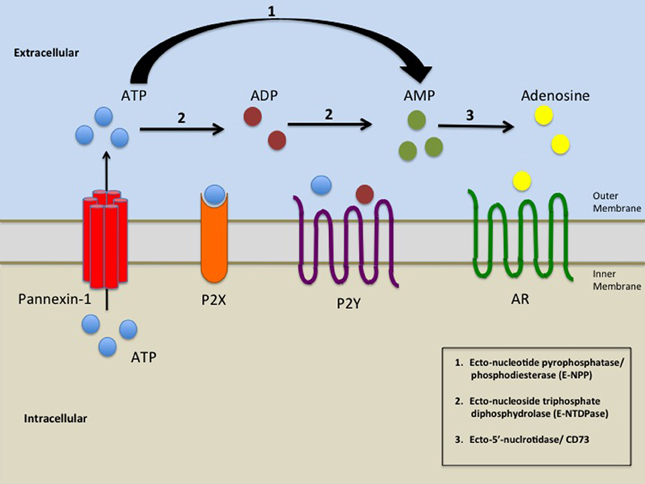
P2Y receptors are activated by extracellular nucleotides, with specific subtypes exhibiting distinct preferences for endogenous ligands. The P2Y1 receptor is primarily activated by adenosine diphosphate (ADP) with an EC50 ≈ 10 μM, while the P2Y12 and P2Y13 receptors are activated by ADP with EC50 values ≈ 60 nM and ≈ 10 nM, respectively.[28] In contrast, the P2Y2 receptor responds equipotently to adenosine triphosphate (ATP) and uridine triphosphate (UTP), with EC50 values around 0.1–1 μM, while the P2Y4 receptor is preferentially activated by UTP.[27] The P2Y6 receptor is selectively activated by uridine diphosphate (UDP), the P2Y11 receptor by ATP, and the P2Y14 receptor by UDP-glucose, a sugar-nucleoside diphosphate.[27] These ligands are released from cells into the extracellular space, often through pannexin-1 or connexin hemichannels during conditions of cellular stress or inflammation, serving as danger signals to modulate immune responses.[29] Ligand selectivity among P2Y subtypes is influenced by structural groupings, with the P2Y1-like receptors (P2Y1, P2Y2, P2Y4, P2Y6, and P2Y11) generally preferring purine or pyrimidine nucleotides such as ATP, ADP, UTP, and UDP, whereas the P2Y12-like receptors (P2Y12, P2Y13, and P2Y14) show higher affinity for diphosphates like ADP or sugar-modified diphosphates like UDP-glucose.[27] For instance, potency at the P2Y2 receptor follows the order ATP ≈ UTP > ADP, highlighting differential responsiveness within the family.[30] Receptor desensitization, which limits prolonged signaling, occurs via agonist-induced phosphorylation by kinases such as protein kinase C or G protein-coupled receptor kinases, leading to reduced responsiveness upon repeated ligand exposure.[31] Exogenous ligands, including synthetic agonists and antagonists, have been developed to probe subtype-specific functions with enhanced selectivity and potency. For the P2Y1 receptor, 2-thio-ADP serves as a selective synthetic agonist, mimicking ADP but with improved stability and potency (EC50 ≈ 10–100 nM).[28] The P2Y6 receptor is targeted by the antagonist MRS2578, which exhibits high selectivity (IC50 ≈ 37 nM at human P2Y6) and no activity at other subtypes like P2Y1, P2Y2, P2Y4, or P2Y11 up to 10 μM.[32] Recent advancements from 2020 to 2025 include the directed evolution of the human P2Y2 receptor for integration into engineered probiotic yeast, enabling these microbes to sense host-derived nucleotides like ATP and UTP for targeted responses in inflammatory bowel disease therapeutics.[33]Mechanism of Receptor Activation
The orthosteric binding pocket of P2Y receptors is located within the transmembrane (TM) bundle, primarily involving TM3, TM6, TM7, and the second extracellular loop (ECL2), which acts as a lid to enclose the ligand. In P2Y1 receptors, for instance, the adenine ring of ADP interacts with hydrophobic residues in TM6 and TM7, while the phosphate groups form ionic bonds with basic residues such as Arg310^{7.39} and nearby ECL2 elements. Similarly, in P2Y12 receptors, the diphosphate moiety of ADP coordinates with Arg256^{6.55} and Lys280^{7.35}, stabilizing the ligand in a deep cavity formed by TM3, TM6, and TM7. These interactions ensure specificity for nucleotide ligands across subtypes, with ECL2 contributing polar contacts that seal the pocket upon binding.[34][35][20] Ligand binding induces a series of conformational changes that transition the receptor from an inactive to an active state, culminating in G protein engagement. The hallmark is the outward movement of TM6 at its intracellular end, typically by 9-11 Å in P2Y subtypes, which opens the intracellular loop 2 (ICL2) region for G protein docking; for example, in P2Y2 receptors, ATP binding triggers a ~10.7 Å TM6 shift relative to inactive structures. Concurrently, the ionic lock—a salt bridge between residues in TM3 and TM6, such as Arg^{3.50} in the DRY motif and Glu^{6.30}—breaks, allowing rearrangement of the toggle switch (Trp^{6.48}) and PIF motif to propagate the signal intracellularly. In P2Y14, agonist binding further involves a ~7.8 Å outward TM6 displacement and inward TM7 tilt, stabilizing the active conformation without the typical TM3 upward shift seen in some GPCRs. These dynamics occur on millisecond to second timescales for conformational dwell times, as inferred from cryo-EM snapshots and simulations.[3][36][37] Allosteric modulation fine-tunes activation in P2Y receptors, with cholesterol and sodium ions influencing the orthosteric pocket's accessibility and stability. Cholesterol binds at the TM interface, stabilizing the active state in P2Y1 and P2Y12 by modulating helix packing, while sodium allosterically binds near Asp^{2.50} in TM2 to inhibit activation in certain conformations. Biased agonism is evident in P2Y12, where some ligands preferentially stabilize Gi coupling over β-arrestin recruitment by altering TM7 positioning, reducing arrestin-binding interfaces. Recent cryo-EM structures from 2022-2025, such as those of ATP-bound P2Y2, reveal subtype-specific features like ECL2 β-hairpin closure upon activation, enhancing pocket enclosure without explicit ECL1 involvement.[34][38][3]G Protein Coupling
Coupling Partners by Subtype
P2Y receptors exhibit subtype-specific coupling to heterotrimeric G proteins, which dictates their downstream signaling profiles. The eight mammalian P2Y subtypes primarily couple to members of the Gq/11, Gi/o, or Gs families, with some displaying promiscuity toward multiple classes. This selectivity arises from interactions at the receptor's intracellular loops and C-terminal domain, enabling tailored physiological responses. The following table summarizes the primary G protein coupling partners for each P2Y subtype, based on pharmacological and structural studies:| Subtype | Primary G Protein Partners |
|---|---|
| P2Y1 | Gq/11 [27] |
| P2Y2 | Gq/11, Gi/o [4] |
| P2Y4 | Gq/11, Gi/o [4] |
| P2Y6 | Gq/11 [27] |
| P2Y11 | Gq/11, Gs [27] |
| P2Y12 | Gi/o [27] |
| P2Y13 | Gi/o [27] |
| P2Y14 | Gi/o [27] |