Hubbry Logo
Diving reflexDiving reflexMain
Open search
Diving reflex
Community hub
Diving reflex
logo
8 pages, 0 posts
0 subscribers
Be the first to start a discussion here.
Diving reflex
Diving reflex
from Wikipedia

Diving reflex in a human baby

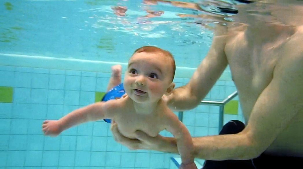
The diving reflex, also known as the diving response and mammalian diving reflex, is a set of physiological responses to immersion that overrides the basic homeostatic reflexes, and is found in all air-breathing vertebrates studied to date.[1][2][3] It optimizes respiration by preferentially distributing oxygen stores to the heart and brain, enabling submersion for an extended time.

The diving reflex is exhibited strongly in aquatic mammals, such as seals,[1][4] otters, dolphins,[5] and muskrats,[6] and exists as a lesser response in other animals, including human babies up to 6 months old (see infant swimming), and diving birds, such as ducks and penguins.[1] Adult humans generally exhibit a mild response, although the dive-hunting Sama-Bajau people[7] and the Haenyeo divers in the South Korean province of Jeju are notable outliers.[8][9]

The diving reflex is triggered specifically by chilling and wetting the nostrils and face while breath-holding,[2][10][11] and is sustained via neural processing originating in the carotid chemoreceptors. The most noticeable effects are on the cardiovascular system, which displays peripheral vasoconstriction, slowed heart rate, redirection of blood to the vital organs to conserve oxygen, release of red blood cells stored in the spleen, and, in humans, heart rhythm irregularities.[2] Although aquatic animals have evolved profound physiological adaptations to conserve oxygen during submersion, the apnea and its duration, bradycardia, vasoconstriction, and redistribution of cardiac output occur also in terrestrial animals as a neural response, but the effects are more profound in natural divers.[1][3]

Physiological response

[edit]

When the face is submerged and water fills the nostrils, sensory receptors sensitive to wetness within the nasal cavity and other areas of the face supplied by the fifth (V) cranial nerve (the trigeminal nerve) relay the information to the brain.[1] The tenth (X) cranial nerve (the vagus nerve) – part of the autonomic nervous system – then produces bradycardia and other neural pathways elicit peripheral vasoconstriction, restricting blood from limbs and all organs to preserve blood and oxygen for the heart, brain, and lungs, concentrating flow in a heart-brain circuit and allowing the animal to conserve oxygen.[3][6]

In humans, the diving reflex is not induced when limbs are introduced to cold water. Mild bradycardia is caused by subjects holding their breath without submerging the face in water.[12][13] When breathing with the face submerged, the diving response increases proportionally to decreasing water temperature.[10] However, the greatest bradycardia effect is induced when the subject is breath-holding with the face wetted.[12] Apnea with nostril and facial cooling are triggers of this reflex.[1][10][12]

Children tend to survive longer than adults when deprived of oxygen underwater. The exact mechanism for this effect has been debated and may be a result of brain cooling similar to the protective effects seen in people treated with deep hypothermia.[13][14]

The diving response in animals, such as the dolphin, varies considerably depending on level of exertion during foraging.[5]

Exceptions in human divers

[edit]

In humans whose historic way of life involves foraging for food underwater by breath-hold diving, there is evidence for more extensive physiological and genetic adaptations of the diving reflex than in typical humans. Having harvested underwater seafood over centuries, the nomadic Sama-Bajau people of Southeast Asia have enlarged spleens and more intense peripheral vasoconstriction during breath-hold diving – giving advantages for prolonged underwater hunting – and display natural selection for the genes controlling these adaptations.[7] Similarly, the Haenyeo women divers of South Korea have pronounced bradycardia and exceptional cold tolerance during breath-hold diving, with evidence of adaptive genetic variation contributing to these advantages.[8][15]

Carotid body chemoreceptors

[edit]

During sustained breath-holding while submerged, blood oxygen levels decline while carbon dioxide and acidity levels rise,[1] stimuli that collectively act upon chemoreceptors located in the bilateral carotid bodies.[16][17] As sensory organs, the carotid bodies convey the chemical status of the circulating blood to brain centers regulating neural outputs to the heart and circulation.[1][17] Preliminary evidence in ducks and humans indicates that the carotid bodies are essential for these integrated cardiovascular responses of the diving response,[16][17] establishing a "chemoreflex" characterized by parasympathetic (slowing) effects on the heart and sympathetic (vasoconstrictor) effects on the vascular system.[1][18]

Circulatory responses

[edit]

Plasma fluid losses due to immersion diuresis occur within a short period of immersion.[19] Head-out immersion causes a blood shift from the limbs and into the thorax. The fluid shift is largely from the extravascular tissues and the increased atrial volume results in a compensatory diuresis. Plasma volume, stroke volume, and cardiac output remain higher than normal during immersion. The increased respiratory and cardiac workload causes increased blood flow to the cardiac and respiratory muscles. Stroke volume is not greatly affected by immersion or variation in ambient pressure, but bradycardia reduces the overall cardiac output, particularly due to the diving reflex in breath-hold diving.[20]

Bradycardia and cardiac output

[edit]

Bradycardia is the response to facial contact with cold water: the human heart rate slows down ten to twenty-five percent.[10] Seals experience changes that are even more dramatic, going from about 125 beats per minute to as low as 10 on an extended dive.[4][21] During breath-holding, humans also display reduced left ventricular contractility and diminished cardiac output,[12][22] effects that may be more severe during submersion due to hydrostatic pressure.[22]

Slowing the heart rate reduces the cardiac oxygen consumption, and compensates for the hypertension due to vasoconstriction. However, breath-hold time is reduced when the whole body is exposed to cold water as the metabolic rate increases to compensate for accelerated heat loss even when the heart rate is significantly slowed.[2]

Splenic contraction

[edit]

The spleen contracts in response to lowered levels of oxygen and increased levels of carbon dioxide, releasing red blood cells and increasing the oxygen capacity of the blood.[23] This may start before the bradycardia.[2]

Blood shift

[edit]

Blood shift is a term used when blood flow to the extremities is redistributed to the head and torso during a breath-hold dive. Peripheral vasoconstriction occurs during submersion by resistance vessels limiting blood flow to muscles, skin, and viscera, regions which are "hypoxia-tolerant", thereby preserving oxygenated blood for the heart, lungs, and brain.[3] The increased resistance to peripheral blood flow raises the blood pressure, which is compensated by bradycardia, conditions which are accentuated by cold water.[2] Aquatic mammals have blood volume that is some three times larger per mass than in humans, a difference augmented by considerably more oxygen bound to hemoglobin and myoglobin of diving mammals, enabling prolongation of submersion after capillary blood flow in peripheral organs is minimized.[2]

Arrhythmias

[edit]

Cardiac arrhythmias are a common characteristic of the human diving response.[2][24] As part of the diving reflex, increased activity of the cardiac parasympathetic nervous system not only regulates the bradycardia, but also is associated with ectopic beats which are characteristic of human heart function during breath-hold dives.[2] Arrhythmias may be accentuated by neural responses to face immersion in cold water, distension of the heart due to central blood shift, and the increasing resistance to left ventricular ejection (afterload) by rising blood pressure.[2] Other changes commonly measured in the electrocardiogram during human breath-hold dives include ST depression, heightened T wave, and a positive U wave following the QRS complex,[2] measurements associated with reduced left ventricular contractility and overall depressed cardiac function during a dive.[12][22]

Renal and water balance responses

[edit]

In hydrated subjects, immersion will cause diuresis and excretion of sodium and potassium. Diuresis is reduced in dehydrated subjects, and in trained athletes in comparison with sedentary subjects.[20]

Respiratory responses

[edit]

Snorkel breathing is limited to shallow depths just below the surface due to the effort required during inhalation to overcome the hydrostatic pressure on the chest.[20] Hydrostatic pressure on the surface of the body due to head-out immersion in water causes negative pressure breathing which shifts blood into the intrathoracic circulation.[19]

Lung volume decreases in the upright position due to cranial displacement of the abdomen due to hydrostatic pressure, and resistance to air flow in the airways increases significantly because of the decrease in lung volume.[19]

Hydrostatic pressure differences between the interior of the lung and the breathing gas delivery, increased breathing gas density due to ambient pressure, and increased flow resistance due to higher breathing rates may all cause increased work of breathing and fatigue of the respiratory muscles.[20]

There appears to be a connection between pulmonary edema and increased pulmonary blood flow and pressure which results in capillary engorgement. This may occur during higher intensity exercise while immersed or submersed.[20]

Facial immersion at the time of initiating breath-hold is a necessary factor for maximising the mammalian diving reflex in humans.[25]

Adaptations of aquatic mammals

[edit]

Diving mammals have an elastic aortic bulb thought to help maintain arterial pressure during the extended intervals between heartbeats during dives, and have high blood volume, combined with large storage capacity in veins and retes of the thorax and head in seals and dolphins.[3] Chronic physiological adaptations of blood include elevated hematocrit, hemoglobin, and myoglobin levels which enable greater oxygen storage and delivery to essential organs during a dive.[3] Oxygen use is minimised during the diving reflex by energy-efficient swimming or gliding behaviour, and regulation of metabolism, heart rate, and peripheral vasoconstriction.[3]

Aerobic diving capacity is limited by available oxygen and the rate at which it is consumed. Diving mammals and birds have a considerably greater blood volume than terrestrial animals of similar size, and in addition have a far greater concentration of haemoglobin and myoglobin, and this haemoglobin and myoglobin is also capable of carrying a higher oxygen load. During diving, the hematocrit and hemoglobin are temporarily increased by reflex splenic contraction, which discharges a large additional amount of red blood cells. The brain tissue of diving mammals also contains higher levels of neuroglobin and cytoglobin than terrestrial animals.[3]

Aquatic mammals seldom dive beyond their aerobic diving limit, which is related to the myoglobin oxygen stored. The muscle mass of aquatic mammals is relatively large, so the high myoglobin content of their skeletal muscles provides a large reserve. Myoglobin-bound oxygen is only released in relatively hypoxic muscle tissue, so the peripheral vasoconstriction due to the diving reflex makes the muscles ischaemic and promotes early use of myoglobin bound oxygen.[3]

History

[edit]

The diving bradycardia was first described by Edmund Goodwyn in 1786 and later by Paul Bert in 1870.[26]

Examples in fiction

[edit]

See also

[edit]

References

[edit]
Revisions and contributorsEdit on WikipediaRead on Wikipedia
from Grokipedia
The diving reflex, also known as the mammalian diving response, is an innate, oxygen-conserving physiological mechanism present in all air-breathing vertebrates that activates during submersion in water to prioritize vital organ perfusion and extend survival time under hypoxic conditions. It integrates multiple autonomic responses to counteract the stress of apnea and immersion, overriding typical homeostatic controls to protect the brain and heart from oxygen deprivation. This reflex comprises three primary components: (a reflex slowing of the , which significantly reduces in humans), apnea (sustained breath-holding that minimizes oxygen expenditure through the lungs), and peripheral (narrowing of blood vessels in non-essential areas like the limbs, redirecting oxygenated blood centrally). These changes are mediated by the via the for bradycardia and sympathetic activation for vasoconstriction, with additional effects like splenic contraction in some species to release stored red blood cells. The diving reflex is primarily triggered by the immersion of the face—especially the forehead, eyes, and nasal area—in cold water (ideally around 10–15°C), which stimulates receptors, combined with voluntary or involuntary apnea. In humans, the response is most pronounced in infants due to their higher sensitivity to facial cooling, diminishing with age but trainable in breath-hold divers to achieve heart rates as low as 10–20 beats per minute. Evolutionarily, the reflex likely originated as an adaptation for amphibious or aquatic lifestyles, being far more robust in marine mammals like seals and whales, where it enables prolonged dives lasting over an hour. In humans, it serves a vestigial role but holds clinical relevance; for instance, cold water facial immersion can enhance tolerance to hypoxia and has been explored in resuscitation protocols for or to mimic oxygen-sparing effects.

Overview

Definition and triggers

The diving reflex, also known as the mammalian diving reflex, is a protective physiological response observed in mammals, including humans, that conserves oxygen during submersion in water by redirecting blood flow to vital organs such as the brain and heart. This reflex involves three primary components: bradycardia (a reduction in heart rate), apnea (voluntary or involuntary cessation of breathing), and peripheral vasoconstriction (narrowing of blood vessels in non-essential tissues to minimize oxygen consumption). These changes collectively lower metabolic demand and prioritize perfusion of critical organs, enabling prolonged survival underwater without air. The primary triggers of the diving reflex are facial immersion in cold water, apnea, and hydrostatic pressure from submersion. Facial immersion, particularly in water below 21°C (70°F), is the most potent stimulus, as it activates sensory receptors in the face and nasal regions, which signal via the to the to initiate the response. In contrast, full-body immersion without facial involvement elicits a weaker response, highlighting the specialized role of the in detecting cold water on the face. Apnea reinforces the reflex by simulating underwater conditions, while hydrostatic pressure contributes to cardiovascular adjustments, though it is less critical than thermal stimulation. In practice, the diving reflex can be elicited in humans by holding the breath and immersing the face in a bowl of ice-cold water, or by pressing a cold pack against the forehead and eyes for as long as tolerable. These methods rapidly activate vagal pathways through cold stimulation of the facial nerves. Secondary triggers, such as hypoxia (low oxygen levels below 60 mm Hg) and hypercapnia (elevated carbon dioxide), amplify the diving reflex but do not initiate it independently; instead, they activate carotid chemoreceptors to enhance vasoconstriction and bradycardia.

Physiological significance

The diving reflex represents an evolutionary adaptation that enhances survival during submersion by conserving limited oxygen supplies and safeguarding critical organs such as the brain and heart. This reflex optimizes resource allocation in hypoxic conditions, prioritizing essential functions over non-vital ones to delay the onset of severe oxygen deprivation. In vertebrates, including humans, it functions as a protective mechanism that evolved to support aquatic excursions, enabling prolonged underwater activity without immediate catastrophic failure. A key benefit lies in oxygen conservation, achieved primarily through peripheral that redirects blood flow—and thus oxygen—away from less essential tissues toward the and cardiovascular structures. This blood shift maintains oxygenation of vital organs, reducing overall oxygen depletion rates during apnea. In humans, the reflex can extend breath-hold times, allowing untrained individuals to sustain submersion for up to 1 minute compared to 30 seconds without it, while trained freedivers may achieve 2-3 minutes by leveraging enhanced reflex activation. By limiting peripheral oxygen use, the reflex effectively slows the progression to hypoxia, preventing cellular damage in oxygen-sensitive tissues. Beyond acute oxygen management, the diving reflex contributes to broader physiological resilience, including heat conservation through that minimizes thermal loss in cold water, thereby mitigating risk during immersion. It also integrates with the body's stress response, promoting a parasympathetic shift that dampens sympathetic overactivation and supports recovery from environmental stressors. These adaptations underscore its role in mammalian across aquatic and terrestrial contexts, linking innate reflexes to enhanced in challenging environments.

Human Physiological Responses

Cardiovascular changes

The diving reflex in humans elicits pronounced , characterized by a 10-25% reduction in upon facial immersion in cold water, typically dropping from a resting rate of around 70 beats per minute to 50-60 beats per minute. This response is primarily mediated by parasympathetic activation through the , triggered by stimulation of afferents in the face and cold receptors. The serves to conserve oxygen by lowering cardiac oxygen demand during the initial phase of submersion. Concomitant with bradycardia, peripheral vasoconstriction occurs, increasing systemic vascular resistance and reducing blood flow to the limbs by approximately 50%. This sympathetic-mediated effect, involving alpha-1 adrenergic receptor activation, redirects blood centrally to protect vital organs such as the brain and heart, enhancing overall oxygen preservation. The vasoconstriction contributes to a rise in mean arterial blood pressure, further supporting perfusion to essential tissues. Despite the , is maintained or only slightly reduced through compensatory increases in , ensuring adequate circulation to critical areas. Additionally, splenic contraction releases stored red blood cells into circulation, boosting oxygen-carrying capacity by 10-20% via elevated levels. This is facilitated by alpha-adrenergic stimulation during apnea. A key aspect of these changes is the redistribution of , where plasma shifts centrally to the and , preventing collapse under hydrostatic pressure and maintaining vital organ oxygenation. This blood shift, combined with the other cardiovascular adjustments, optimizes the body's response to the hypoxic conditions of submersion.

Respiratory and gas exchange responses

The diving reflex in humans triggers an immediate apnea upon submersion of the face in cold water, initiating involuntary breath-holding to conserve oxygen stores. This response typically lasts 30 to 60 seconds in untrained individuals, as observed in experimental facial immersion protocols simulating submersion. The suppression of the urge to breathe is mediated by activation of laryngeal receptors, which elicit the laryngeal chemoreflex—a primitive protective mechanism that promotes central apnea and inhibits respiratory efforts. A key component of this respiratory adjustment is , involving reflexive closure of the and to prevent aspiration of water into the lower airways. This closure, combined with , maintains airway integrity during immersion and contributes to the overall suppression of ventilation post-trigger. These adaptations optimize by halting pulmonary ventilation, allowing stored oxygen to be prioritized for vital organs while accumulates. The resulting hypoxia and are tolerated longer than in non-immersed breath-holding due to a blunted hypercapnic drive, which delays the onset of involuntary breathing movements. Oxygen consumption is thereby reduced, with studies indicating a notable decrease in metabolic demand during the reflex, aiding survival in hypoxic conditions. Selective cooling may further support this by lowering cerebral metabolic rate, though its role in humans remains under investigation primarily through case analyses.

Other systemic adjustments

During breath-hold immersion that triggers the diving reflex, human renal responses include immersion diuresis, mediated by suppressed levels of antidiuretic hormone (ADH) due to increased central blood volume and elevated (ANP). This promotes water excretion in the kidneys, increasing urine output by 50-100% or more to reduce plasma volume and counteract the central fluid shift. The renin-angiotensin-aldosterone system is suppressed, leading to and kaliuresis, which support fluid and balance during immersion. Plasma volume maintenance occurs via hemoconcentration, a consequence of peripheral that redirects blood flow away from non-vital organs and splenic contraction, which releases stored erythrocytes into circulation. This increases and concentration, ensuring adequate oxygen-carrying capacity in central circulation without net loss of plasma volume. The heightened integral to the diving reflex may induce benign ectopic beats or minor arrhythmias, such as premature ventricular contractions, particularly during prolonged apnea; however, these are infrequent and typically in healthy individuals. Endocrine adjustments also encompass catecholamine dynamics, where initial sympathetic activation supports vasoconstriction, but overall metabolic rate is lowered through integrated reflex inhibition, prioritizing oxygen delivery to vital organs like the brain and heart.

Variations and Mechanisms in Humans

Influencing factors and exceptions

The diving reflex exhibits notable variations influenced by age and physical fitness. In infants and young children, the reflex is particularly pronounced, with heart rate reductions reaching up to 51% during facial submersion, aiding in oxygen conservation during potential submersion events. In contrast, the magnitude of the reflex diminishes progressively with age, as autonomic responses weaken, resulting in less bradycardia and vasoconstriction in older adults. Training, particularly in freedivers, can enhance the reflex's intensity through repeated exposure, leading to greater heart rate reductions—often dropping to 20-30 beats per minute in practitioners during deep dives—to optimize oxygen use. Exceptions to the reflex's expression occur in certain individuals, where it may be absent or markedly weak. Genetic factors, such as polymorphisms in genes (e.g., ADRA1A), can alter vascular and cardiac responses, leading to reduced or during immersion. Neurological conditions affecting autonomic function, including , can similarly impair the reflex, resulting in minimal changes. differences also contribute, with the reflex slightly stronger in males, manifesting as more pronounced and better overall performance in breath-hold diving tasks. Environmental modifiers further influence the reflex's potency. Warmer water temperatures attenuate the response by reducing the cold stimulus to endings, leading to weaker compared to immersion in cold water (below 10°C). Additionally, substances like alcohol impair the reflex by blunting reductions during facial immersion.

Neural and sensory control

The diving reflex in humans is primarily initiated by sensory inputs from the face and upper airways, with cold water immersion stimulating cutaneous receptors innervated by the (cranial nerve V). These receptors, particularly cold-sensitive thermoreceptors in the , cheeks, and nasal region, detect the temperature drop and transmit afferent signals to the , triggering the reflex arc. Additional sensory contributions come from laryngeal receptors, whose afferent fibers travel via the (a branch of the , cranial nerve X), enhancing the reflex during apnea and submersion by integrating airway stimulation signals. Peripheral chemoreceptors in the carotid bodies also play a key role by sensing changes in blood gases, particularly hypoxia and that develop during breath-holding. These chemoreceptors activate via afferents, amplifying vagal efferent output to promote and , thereby sustaining the reflex beyond initial sensory triggers. Central integration occurs primarily in the , where the and associated nuclei process these afferent inputs to coordinate autonomic responses. The nucleus tractus solitarius (NTS) in the medulla receives converging signals from trigeminal, vagal, and glossopharyngeal pathways, serving as a primary relay for parasympathetic activation and establishing vagal dominance that overrides sympathetic activity. The contributes to this balance by modulating sympathetic and parasympathetic outflows, ensuring prioritized oxygen conservation through selective and reduced . This neural orchestration results in a potent inhibitory effect on respiration and , exemplified by the reflex observed during immersion.

Adaptations in Aquatic Mammals

Enhanced circulatory and respiratory adaptations

Aquatic mammals exhibit amplified circulatory adaptations during dives, characterized by extreme bradycardia that reduces heart rates to as low as 4-10 beats per minute, far more pronounced than the approximately 50 beats per minute observed in humans. This severe slowing of the heart minimizes cardiac oxygen consumption and work, conserving limited stores for extended submersion. Profound peripheral vasoconstriction accompanies bradycardia, effectively shutting down blood flow to non-vital tissues such as the muscles and digestive organs while prioritizing perfusion to the heart, brain, and lungs. Aquatic mammals possess blood volumes 2-3 times greater than those of comparably sized terrestrial mammals, enabling substantial redirection of this expanded reservoir to central organs without compromising vital function. Respiratory adaptations further enhance dive endurance through voluntary apnea, which can last up to two hours in extreme cases, supported by efficient oxygen management to delay the onset of hypoxia. A compliant thoracic structure allows passive collapse at depth, equalizing internal and external pressures to prevent and limit nitrogen uptake that could lead to decompression issues. Skeletal muscles store substantial oxygen via high concentrations, facilitating aerobic and reducing reliance on anaerobic pathways during prolonged breath-holds. Circulatory efficiency is bolstered by elevated levels, which increase blood oxygen-carrying capacity beyond that of terrestrial counterparts, ensuring sustained delivery to critical tissues. The enlarged serves as a key oxygen reserve, contracting upon submersion to release stored erythrocytes, thereby raising and hemoglobin concentrations by up to 50% to further augment circulating oxygen. These integrated mechanisms collectively enable aquatic mammals to endure dives far exceeding capabilities while maintaining physiological stability.

Species-specific examples

In Weddell seals (Leptonychotes weddellii), the diving reflex enables extreme submergence, with recorded dives reaching depths exceeding 900 meters and durations up to 96 minutes. During these prolonged apneas, profound conserves oxygen by minimizing cardiac workload. Peripheral dramatically redirects blood flow to vital central organs like the heart and brain, thereby protecting against hypoxia. Sperm whales (Physeter macrocephalus) exemplify the reflex's adaptation for ultra-deep foraging, achieving apneas of up to 90 minutes at depths surpassing 2,000 meters. At these pressures, massive lung compression occurs, collapsing alveoli to prevent nitrogen absorption and embolism while maintaining gas exchange via stored oxygen in blood and muscles. Aortic constriction complements this by regulating blood flow and pressure gradients, ensuring perfusion to the brain and heart amid near-total peripheral shutdown. Sea otters (Enhydra lutris) and dolphins (Delphinidae spp.) display intermediate diving reflex traits suited to shallower, more frequent submergences. Sea otters routinely dive to 100 meters for 4–5 minutes, relying on moderate and to manage oxygen debt during bouts, though their smaller body size limits compared to pinnipeds. In dolphins, the reflex integrates a nasal plug mechanism—a sealing the blowhole—that enforces apnea by preventing water ingress and air escape, allowing dives up to 10–15 minutes at depths of 300 meters while drops to 12 beats per minute. Behavioral integration of the diving reflex in these species manifests in surfacing patterns precisely calibrated to oxygen stores, optimizing recovery and minimizing exposure risks. For instance, Weddell seals and sperm whales exhibit U-shaped dive profiles, descending rapidly, gliding at depth to conserve energy, and ascending when muscle and oxygen levels approach critical thresholds, often surfacing for brief 2–5 minute intervals to replenish stores before resuming. Recent studies indicate Weddell seals strategically time their deepest and longest dives earlier in the day to align with levels, enhancing . This rhythmic behavior, driven by feedback, extends overall dive cycles while preventing exhaustive depletion.

Evolutionary and Comparative Perspectives

Evolutionary origins

The diving reflex, characterized by apnea, , and peripheral , has its ancestral basis in hypoxia responses observed in amphibians and other early vertebrates, where submersion triggers oxygen-conserving mechanisms to survive aquatic environments. This primitive reflex is conserved across all mammals, serving as an innate response to facial immersion or that prioritizes vital organ perfusion during potential scenarios. The reflex likely has ancient origins in early vertebrates, conserved and adapted in mammals under selective pressures for survival in variable environments that included occasional submersion risks. It was subsequently enhanced in semi-aquatic mammals, such as early pinnipeds, which transitioned to marine habitats approximately 30 million years ago in the , where prolonged dives demanded more pronounced cardiovascular adjustments to manage oxygen debt. In pinnipeds, the response varies across families, correlating with their aquatic lifestyles and the need for efficient foraging in cold, deep waters. Genetic underpinnings of the reflex involve conserved genes regulating vagal control and autonomic development. This gene's role in visceral reflex circuits, including hypoxia sensing and vagal efferents, demonstrates evolutionary conservation from early vertebrates to modern mammals, with mutations disrupting these pathways in conditions like congenital . Direct fossil evidence for the diving reflex is absent, as physiological responses do not fossilize, but indirect support comes from the shared neural architecture in —analogs within sauropsids—suggesting the reflex predates the mammal-bird divergence around 310 million years ago. For mammals, the focus remains on post-dinosaur Cretaceous-Paleogene boundary adaptations (~66 million years ago), where small, nocturnal synapsid descendants likely retained the primitive reflex amid environmental shifts, with enhancements appearing later in aquatic lineages.

Comparisons across mammals

The diving reflex exhibits considerable variation in intensity and expression across mammalian species, with the strength of the response generally correlating with the degree of to aquatic environments. In strictly terrestrial mammals, the reflex is weak and incomplete, typically manifesting as mild without accompanying apnea or significant peripheral . For instance, in dogs, nasopharyngeal stimulation with water induces under , but the overall response lacks the robust oxygen-conserving features observed in more aquatic species, such as sustained breath-holding or blood flow redistribution. Humans display a moderately developed diving reflex compared to terrestrial counterparts but substantially weaker than that in fully aquatic mammals, based on heart rate reductions. During facial immersion in cold water, human heart rate typically decreases by about 25% (from an average resting rate of 76 beats per minute to 56 beats per minute), whereas Weddell seals exhibit reductions of 40-80% during voluntary dives. Sea otters, as semi-aquatic mammals, show an intermediate profile, with heart rates dropping to 35-50% of resting levels (50-65% reduction) during submergence, bridging the gap between human and pinniped responses. This functional gradient in reflex strength ranges from minimal in large terrestrial species like elephants, where elicitation of bradycardia or other components is negligible due to the absence of aquatic pressures, to maximal in deep-diving cetaceans such as blue whales, which achieve heart rates as low as 2-8 beats per minute during dives (an 80-90% reduction from surface rates of 25-37 beats per minute). Experimental cross-species studies, including comparative analyses of face-immersion apnea, confirm that reflex intensity—measured by metrics like bradycardic magnitude and apnea duration—positively correlates with aquatic lifestyle, with terrestrial species showing the weakest responses and progressively stronger ones in semi-aquatic and obligate aquatic forms.

History and Research

Discovery and early studies

The earliest observations of the diving reflex trace back to 1786, when British physician Edmund Goodwyn described in seals during submersion in water, noting a significant slowing of the compared to air exposure. This initial account highlighted the reflex's potential role in conserving oxygen under hypoxic conditions, though it remained largely overlooked for nearly a century. In the 1870s, French physiologist conducted pioneering experiments demonstrating diving across species, including ducks and humans; he observed heart rates dropping to as low as 10 beats per minute in immersed human subjects, establishing it as a key physiological response to apnea and facial immersion. Building on this, Charles Richet's studies in the 1890s using ducks further confirmed the reflex's involuntary nature, showing that submerged birds survived up to three times longer than those exposed to air alone, due to coordinated cardiovascular adjustments. The mid-20th century saw systematic quantification of the reflex through animal models, particularly in the 1940s work of American physiologist Laurence Irving and Norwegian-American biologist Per F. Scholander on seals; their experiments revealed profound (heart rates reduced by 80-90%), selective peripheral , and splenic contraction, all contributing to oxygen conservation during prolonged dives lasting over 30 minutes. These findings paralleled earlier avian studies and emphasized the reflex's evolutionary significance in aquatic mammals. Initial human data emerged in the 1960s through investigations of breath-hold divers, such as pearl divers, where Scholander and colleagues documented robust reflex activation—including reductions of up to 50% and blood flow redistribution—confirming its presence and functionality in humans during voluntary apnea.

Modern research and applications

Recent studies utilizing (fMRI) have elucidated the neural underpinnings of the diving reflex, particularly its activation during apnea and facial immersion. For instance, breath-holding paradigms, which simulate key components of the reflex, have demonstrated subject-specific activation in respiratory centers, including the , highlighting the reflex's role in central respiratory control under hypoxic conditions. These findings from the 2000s and onward build on earlier work by integrating to map trigeminal--vagal pathways involved in and . Genetic research in the and has explored variability in the diving reflex, identifying polymorphisms that influence vascular responses. A 2016 study revealed that gene variants in genes such as BDKRB2 and determine the magnitude of peripheral during the reflex, with stronger responses in individuals carrying certain alleles. Subsequent work in 2022 confirmed sex-specific differences in reflex intensity linked to ADRA1A polymorphisms, underscoring genetic factors in reflex efficacy across populations. In , training protocols have been shown to enhance the diving reflex, improving oxygen conservation and extending apnea durations for competitive performance. Specialized breath-hold training over two weeks induces earlier onset of and splenic contraction, increasing levels and oxygen-carrying capacity, which supports elite athletes in achieving records exceeding 10 minutes by 2025. For example, men's world records in reached approximately 11 minutes in competitions during this period, attributed in part to reflex optimization through repeated immersion. Medically, the diving reflex is employed as a vagal maneuver to treat heart rhythm disorders, such as paroxysmal supraventricular tachycardia (PSVT). It can be triggered by submerging the face in ice-cold water (0–10°C) while holding the breath, or by applying a cold pack or ice-cold wet towel to the forehead and eyes for as long as tolerable; these methods stimulate the trigeminal nerve, rapidly activating vagal pathways to induce bradycardia and restore sinus rhythm, with success rates of 20–40%. This noninvasive approach mimics direct vagal nerve stimulation and has been applied to treat arrhythmias, including paroxysmal atrial tachycardia, without pharmacological intervention, though it requires medical supervision to mitigate risks such as aspiration.

References

Add your contribution
Related Hubs
User Avatar
No comments yet.