Hubbry Logo
Preterm birthPreterm birthMain
Open search
Preterm birth
Community hub
Preterm birth
logo
8 pages, 0 posts
0 subscribers
Be the first to start a discussion here.
Preterm birth
Preterm birth
from Wikipedia

Preterm birth
Other namesPremature birth, preemies, premmies
Intubated preterm baby in an incubator
SpecialtyNeonatology, Pediatrics, Obstetrics
SymptomsBirth of a baby at younger than 37 weeks' gestational age[1]
ComplicationsCerebral palsy, delays in development, hearing problems, sight problems[1]
CausesOften unknown[2]
Risk factorsDiabetes, high blood pressure, Multiple gestation, obesity or underweight, a number of vaginal infections, celiac disease, tobacco smoking, psychological stress[2][3][4]
PreventionProgesterone[5]
TreatmentCorticosteroids, keeping the baby warm through skin-to-skin contact, supporting breastfeeding, treating infections, supporting breathing[2][6]
Frequency~15 million a year (12% of deliveries)[2]
Deaths805,800[7]

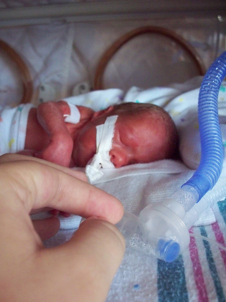
Preterm birth, also known as premature birth, is the birth of a baby at fewer than 37 weeks gestational age, as opposed to full-term delivery at approximately 40 weeks.[1] Extreme preterm[2] is less than 28 weeks, very early preterm birth is between 28 and 32 weeks, early preterm birth occurs between 32 and 34 weeks, late preterm birth is between 34 and 36 weeks' gestation.[8] These babies are also known as premature babies or colloquially preemies (American English)[9] or premmies (Australian English).[10] Symptoms of preterm labor include uterine contractions which occur more often than every ten minutes and/or the leaking of fluid from the vagina before 37 weeks.[11][12] Premature infants are at greater risk for cerebral palsy, delays in development, hearing problems and problems with their vision.[1] The earlier a baby is born, the greater these risks will be.[1]

The cause of spontaneous preterm birth is often not known.[2] Risk factors include diabetes, high blood pressure, multiple gestation (being pregnant with more than one baby), being either obese or underweight, vaginal infections, air pollution exposure, tobacco smoking, and psychological stress.[2][3][13] For a healthy pregnancy, medical induction of labor or cesarean section are not recommended before 39 weeks unless required for other medical reasons.[2] There may be certain medical reasons for early delivery such as preeclampsia.[14]

Preterm birth may be prevented in those at risk if the hormone progesterone is taken during pregnancy.[5] Evidence does not support the usefulness of bed rest to prevent preterm labor.[5][15] Of the approximately 900,000 preterm deaths globally in 2019, it is estimated that at least 75% of these preterm infants would have survived with appropriate cost-effective treatment, and the survival rate is highest among the infants born the latest in gestation.[2] In women who might deliver between 24 and 37 weeks, corticosteroid treatment may improve outcomes.[6][16] A number of medications, including nifedipine, may delay delivery so that a mother can be moved to where more medical care is available and the corticosteroids have a greater chance to work.[17] Once the baby is born, care includes keeping the baby warm through skin-to-skin contact or incubation, supporting breastfeeding and/or formula feeding, treating infections, and supporting breathing.[2] Preterm babies sometimes require intubation.[2]

Preterm birth is the most common cause of death among infants worldwide.[1] About 15 million babies are preterm each year (5% to 18% of all deliveries).[2] Late preterm birth accounts for 75% of all preterm births.[18] This rate is inconsistent across countries. In the United Kingdom 7.9% of babies are born pre-term and in the United States 12.3% of all births are before 37 weeks gestation.[19][20] Approximately 0.5% of births are extremely early periviable births (20–25 weeks of gestation), and these account for most of the deaths.[21] In many countries, rates of premature births have increased between the 1990s and 2010s.[2] Complications from preterm births resulted globally in 0.81 million deaths in 2015, down from 1.57 million in 1990.[7][22] The chance of survival at 22 weeks is about 6%, while at 23 weeks it is 26%, 24 weeks 55% and 25 weeks about 72%.[23] The chances of survival without any long-term difficulties are lower.[24]

Signs and symptoms

[edit]
A new mother holds her premature baby at Kapiolani Medical Center NICU in Honolulu, Hawaii.

Signs and symptoms of preterm labor include four or more uterine contractions in one hour. In contrast to false labour, true labor is accompanied by cervical dilation and effacement. Also, vaginal bleeding in the third trimester, heavy pressure in the pelvis, or abdominal or back pain could be indicators that a preterm birth is about to occur. A watery discharge from the vagina may indicate premature rupture of the membranes that surround the baby. While the rupture of the membranes may not be followed by labor, usually delivery is indicated as infection (chorioamnionitis) is a serious threat to both fetus and mother. In some cases, the cervix dilates prematurely without pain or perceived contractions, so that the mother may not have warning signs until very late in the birthing process.

Causes

[edit]

Preterm labor can be caused by medical conditions including infections, environmental or drug exposures, cervix insufficiency, uterine abnormalities, or amniotic fluid problems.[25] Some women require preterm labor induction for medical conditions.[25] Other preterm births are spontaneous. In some cases, preterm labor occurs spontaneously and there is no identifiable cause.[25] There are maternal and pregnancy-related risk factors that increase the risk of a women experiencing preterm labor.

Risk factors of preterm labor

[edit]

The exact cause of spontaneous preterm birth is difficult to determine and it may be caused by many different factors at the same time as labor is a complex process.[26][27] The research available is limited with regard to the cervix and therefore is limited in discerning what is or is not normal.[12] Four different pathways have been identified that can result in preterm birth and have considerable evidence: precocious fetal endocrine activation, uterine overdistension (placental abruption), decidual bleeding, and intrauterine inflammation or infection.[28]

Identifying women at high risk of giving birth early would enable the health services to provide specialized care for these women and their babies, for example a hospital with a special care baby unit such as a neonatal intensive care unit (NICU). In some instances, it may be possible to delay the birth. Risk scoring systems have been suggested as an approach to identify those at higher risk; however, there is no strong research in this area so it is unclear whether the use of risk scoring systems for identifying mothers would prolong pregnancy and reduce the numbers of preterm births or not.[29]

Maternal factors

[edit]
Risk factor Relative risk[30] 95% confidence
interval
[30]
Fetal fibronectin 4.0 2.9–5.5
Short cervical length 2.9 2.1–3.9
Prenatal Care Absent[31] 2.9 2.8–3.0
Chlamydia 2.2 1.0–4.8
Low socio-economic status 1.9 1.7–2.2
Large or small pregnancy weight gain 1.8 1.5–2.3
Short maternal height 1.8 1.3–2.5
Periodontitis 1.6 1.1–2.3
Celiac disease 1.4[32] 1.2–1.6[32]
Asymptomatic bacteriuria 1.1 0.8–1.5
High or low BMI 0.96 0.66–1.4
odds ratio
History of spontaneous preterm birth 3.6 3.2–4.0
Bacterial vaginosis 2.2 1.5–3.1
Black ethnicity/race 2.0 1.8–2.2
Filipino ancestry[33] 1.7 1.5–2.1
Unwanted pregnancy[34]: 1 1.5 1.41–1.61
Unintended pregnancy[34]: 1 1.31 1.09–1.58
Being single/unmarried[35] 1.2 1.03–1.28
Percentage premature births in England and Wales 2011, by age of mother and whether single or multiple birth

Risk factors in the mother have been identified that are linked to a higher risk of a preterm birth. These include age (either very young or older),[36] high or low body mass index (BMI),[37][38] length of time between pregnancies,[39] endometriosis,[40] previous spontaneous (i.e., miscarriage) or surgical abortions,[41][42] unintended pregnancies,[34] untreated or undiagnosed celiac disease,[32][4] fertility difficulties, heat exposure,[43] and genetic variables.[44]

Studies on type of work and physical activity have given conflicting results, but it is opined that stressful conditions, hard labor, and long hours are probably linked to preterm birth.[36] Obesity does not directly lead to preterm birth;[45] however, it is associated with diabetes and hypertension which are risk factors by themselves.[36] To some degree those individuals may have underlying conditions (i.e., uterine malformation, hypertension, diabetes) that persist. Couples who have tried more than one year versus those who have tried less than one year before achieving a spontaneous conception have an adjusted odds ratio of 1.35 (95% confidence interval 1.22–1.50) of preterm birth.[46] Pregnancies after IVF confers a greater risk of preterm birth than spontaneous conceptions after more than one year of trying, with an adjusted odds ratio of 1.55 (95% CI 1.30–1.85).[46]

Certain ethnicities may have a higher risk as well. For example, in the U.S. and the UK, Black women have preterm birth rates of 15–18%, more than double than that of the white population. Many Black women have higher preterm birth rates due to multiple factors but the most common is high amounts of chronic stress, which can eventually lead to premature birth.[47] Adult chronic disease is not always the case with premature birth in Black women, which makes the main factor of premature birth challenging to identify.[47] Filipinos are also at high risk of premature birth, and it is believed that nearly 11–15% of Filipinos born in the U.S. (compared to other Asians at 7.6% and whites at 7.8%) are premature.[48] Filipinos being a big risk factor is evidenced with the Philippines being the eighth-highest ranking in the world for preterm births, the only non-African country in the top 10.[49] This discrepancy is not seen in comparison to other Asian groups or Hispanic immigrants and remains unexplained.[36] Genetic make-up is a factor in the causality of preterm birth. Genetics has been a big factor into why Filipinos have a high risk of premature birth as the Filipinos have a large prevalence of mutations that help them be predisposed to premature births.[48] An intra- and transgenerational increase in the risk of preterm delivery has been demonstrated.[44] No single gene has been identified.

Marital status has long been associated with risks for preterm birth. A 2005 study of 25,373 pregnancies in Finland revealed that unmarried mothers had more preterm deliveries than married mothers (P=0.001).[35] Pregnancy outside of marriage was associated overall with a 20% increase in total adverse outcomes, even at a time when Finland provided free maternity care. A study in Quebec of 720,586 births from 1990 to 1997 revealed less risk of preterm birth for infants with legally married mothers compared with those with common-law wed or unwed parents.[50][needs update] A study conducted in Malaysia in 2015 showed a similar trend, with marital status being significantly associated with preterm birth.[51] However, the result of a study conducted in the US showed that between 1989 and 2006, marriage became less protective of preterm births which was attributed to the changing social norms and behaviors surrounding marriage.[52]

Factors during pregnancy

[edit]

Medications during pregnancy, living conditions, air pollution, smoking, illicit drugs or alcohol, infection, or physical trauma may also cause a preterm birth.

Air pollution: Living in an area with a high concentration of air pollution is a major risk factor for preterm labor, including living near major roadways or highways where vehicle emissions are high from traffic congestion or are a route for diesel trucks that tend to emit more pollution.[53][54][13]

The use of fertility medication that stimulates the ovary to release multiple eggs and of IVF with embryo transfer of multiple embryos has been implicated as a risk factor for preterm birth. Often labor has to be induced for medical reasons; such conditions include high blood pressure,[55] pre-eclampsia,[56] maternal diabetes,[57] asthma, thyroid disease, and heart disease.

Certain medical conditions in the pregnant mother may also increase the risk of preterm birth. Some women have anatomical problems that prevent the baby from being carried to term. These include a weak or short cervix (the strongest predictor of premature birth).[58][59][60][55] Women with vaginal bleeding during pregnancy are at higher risk for preterm birth. While bleeding in the third trimester may be a sign of placenta previa or placental abruption—conditions that occur frequently preterm—even earlier bleeding that is not caused by these conditions is linked to a higher preterm birth rate.[61] Women with abnormal amounts of amniotic fluid, whether too much (polyhydramnios) or too little (oligohydramnios), are also at risk.[36] Anxiety and depression have been linked as risk factors for preterm birth.[36][62]

The use of tobacco, cocaine, and excessive alcohol during pregnancy increases the chance of preterm delivery. Tobacco is the most commonly used drug during pregnancy and contributes significantly to low birth weight delivery.[63] Babies with birth defects are at higher risk of being born preterm.[64]

Passive smoking and/or smoking before the pregnancy influences the probability of a preterm birth. The World Health Organization published an international study in March 2014.[65]

Presence of anti-thyroid antibodies is associated with an increased risk preterm birth with an odds ratio of 1.9 and 95% confidence interval of 1.1–3.5.[66]

Intimate violence against the mother is another risk factor for preterm birth.[67]

Physical trauma may case a preterm birth. The Nigerian cultural method of abdominal massage has been shown to result in 19% preterm birth among women in Nigeria, plus many other adverse outcomes for the mother and baby.[68] This ought not be confused with massage therapy conducted by a fully trained and certified/licensed massage therapist or by significant others trained to provide massage during pregnancy, which—in a study involving pregnant females with prenatal depression—has been shown to have numerous positive results during pregnancy, including the reduction of preterm birth, less depression, lower cortisol, and reduced anxiety.[69] In healthy women, however, no effects have been demonstrated in a controlled study.

Infection

[edit]

The frequency of infection in preterm birth is inversely related to the gestational age. Mycoplasma genitalium infection is associated with increased risk of preterm birth, and spontaneous abortion.[70]

Infectious microorganisms can be ascending, hematogenous, iatrogenic by a procedure, or retrograde through the fallopian tubes. From the deciduae they may reach the space between the amnion and chorion, the amniotic fluid, and the fetus. A chorioamnionitis also may lead to sepsis of the mother. Fetal infection is linked to preterm birth and to significant long-term disability including cerebral palsy.[71]

It has been reported that asymptomatic colonization of the decidua occurs in up to 70% of women at term using a DNA probe suggesting that the presence of micro-organism alone may be insufficient to initiate the infectious response.

As the condition is more prevalent in black women in the U.S. and the UK, it has been suggested to be an explanation for the higher rate of preterm birth in these populations. It is opined that bacterial vaginosis before or during pregnancy may affect the decidual inflammatory response that leads to preterm birth. The condition known as aerobic vaginitis can be a serious risk factor for preterm labor; several previous studies failed to acknowledge the difference between aerobic vaginitis and bacterial vaginosis, which may explain some of the contradiction in the results.[72]

Untreated yeast infections are associated with preterm birth.[73]

A review into prophylactic antibiotics (given to prevent infection) in the second and third trimester of pregnancy (13–42 weeks of pregnancy) found a reduction in the number of preterm births in women with bacterial vaginosis. These antibiotics also reduced the number of waters breaking before labor in full-term pregnancies, reduced the risk of infection of the lining of the womb after delivery (endometritis), and rates of gonococcal infection. However, the women without bacterial vaginosis did not have any reduction in preterm births or pre-labor preterm waters breaking. Much of the research included in this review lost participants during follow-up so did not report the long-term effects of the antibiotics on mothers or babies. More research in this area is needed to find the full effects of giving antibiotics throughout the second and third trimesters of pregnancy.[74]

A number of maternal bacterial infections are associated with preterm birth including pyelonephritis, asymptomatic bacteriuria, pneumonia, and appendicitis. A review into giving antibiotics in pregnancy for asymptomatic bacteriuria (urine infection with no symptoms) found the research was of very low quality but that it did suggest that taking antibiotics reduced the numbers of preterm births and babies with low birth weight.[75] Another review found that one dose of antibiotics did not seem as effective as a course of antibiotics but fewer women reported side effects from one dose.[76] This review recommended that more research is needed to discover the best way of treating asymptomatic bacteriuria.[75]

A different review found that preterm births happened less for pregnant women who had routine testing for low genital tract infections than for women who only had testing when they showed symptoms of low genital tract infections.[77] The women being routinely tested also gave birth to fewer babies with a low birth weight. Even though these results look promising, the review was only based on one study so more research is needed into routine screening for low genital tract infections.[77]

Also periodontal disease has been shown repeatedly to be linked to preterm birth.[78][79] In contrast, viral infections, unless accompanied by a significant febrile response, are considered not to be a major factor in relation to preterm birth.[36]

Genetics

[edit]

There is believed to be a maternal genetic component in preterm birth.[80] Estimated heritability of timing-of-birth in women was 34%. However, the occurrence of preterm birth in families does not follow a clear inheritance pattern, thus supporting the idea that preterm birth is a non-Mendelian trait with a polygenic nature.[81]

Prenatal care

[edit]

The absence of prenatal care has been associated with higher rates of preterm births. Analysis of 15,627,407 live births in the United States in 1995–1998 concluded that the absence of prenatal care carried a 2.9 (95%CI 2.8, 3.0) times higher risk of preterm births.[31] This same study found statistically significant relative risks of maternal anemia, intrapartum fever, unknown bleeding, renal disease, placental previa, hydramnios, placenta abruption, and pregnancy-induced hypertension with the absence of prenatal care. All these prenatal risks were controlled for other high-risk conditions, maternal age, gravidity, marital status, and maternal education. The absence of prenatal care prior to and during the pregnancy is primarily a function of socioeconomic factors (low family income and education), access to medical consultations (large distance from the place of residence to the healthcare unit and transportation costs), quality of healthcare, and social support.[82] Efforts to decrease rates of preterm birth should aim to increase the deficits posed by the aforementioned barriers and to increase access to prenatal care.

Diagnosis

[edit]

Placental alpha microglobulin-1

[edit]

Placental alpha microglobulin-1 (PAMG-1) has been the subject of several investigations evaluating its ability to predict imminent spontaneous preterm birth in women with signs, symptoms, or complaints suggestive of preterm labor.[83][84][85][86][87][88] In one investigation comparing this test to fetal fibronectin testing and cervical length measurement via transvaginal ultrasound, the test for PAMG-1 (commercially known as the PartoSure test) has been reported to be the single best predictor of imminent spontaneous delivery within 7 days of a patient presenting with signs, symptoms, or complaints of preterm labor. Specifically, the PPV, or positive predictive value, of the tests were 76%, 29%, and 30% for PAMG-1, fFN and CL, respectively (P < 0.01).[89]

Fetal fibronectin

[edit]

Fetal fibronectin (fFN) has become an important biomarker—the presence of this glycoprotein in the cervical or vaginal secretions indicates that the border between the chorion and decidua has been disrupted. A positive test indicates an increased risk of preterm birth, and a negative test has a high predictive value.[36] It has been shown that only 1% of women in questionable cases of preterm labor delivered within the next week when the test was negative.[90]

Ultrasound

[edit]

Obstetric ultrasound has become useful in the assessment of the cervix in women at risk for premature delivery. A short cervix preterm is undesirable: A cervical length of less than 25 mm (0.98 in) at or before 24 weeks of gestational age is the most common definition of cervical incompetence.[91]

Emerging technologies

[edit]

Technologies under research and development to facilitate earlier diagnosis of preterm births include sanitary pads that identify biomarkers such as fFN and PAMG-1 in vaginal secretions. These devices then calculate a risk of preterm birth and send the findings to a smartphone.[92] The notion that risk-scoring systems are accurate in predicting preterm birth has been debated in multiple literature reviews.[93][94]

Classification

[edit]
Stages in prenatal development, with weeks and months numbered from last menstruation

In humans, the usual definition of preterm birth is birth before a gestational age of 37 complete weeks.[95] In the normal human fetus, several organ systems mature between 34 and 37 weeks, and the fetus reaches adequate maturity by the end of this period. One of the main organs greatly affected by premature birth is the lungs. The lungs are one of the last organs to mature in the womb; because of this, many premature babies spend the first days and weeks of their lives on ventilators. Therefore, a significant overlap exists between preterm birth and prematurity. Generally, preterm babies are premature and term babies are mature. Preterm babies born near 37 weeks often have no problems relating to prematurity if their lungs have developed adequate surfactant, which allows the lungs to remain expanded between breaths. Sequelae of prematurity can be reduced to a small extent by using drugs to accelerate maturation of the fetus, and to a greater extent by preventing preterm birth.

Prevention

[edit]

Historically efforts have been primarily aimed to improve survival and health of preterm infants (tertiary intervention). Such efforts, however, have not reduced the incidence of preterm birth. Increasingly primary interventions that are directed at all women, and secondary intervention that reduce existing risks are looked upon as measures that need to be developed and implemented to prevent the health problems of premature infants and children.[96] Smoking bans are effective in decreasing preterm births.[97] Different strategies are used in the administration of prenatal care, and future studies need to determine if the focus can be on screening for high-risk women, or widened support for low-risk women, or to what degree these approaches can be merged.[96]

Before pregnancy

[edit]

Adoption of specific professional policies can immediately reduce risk of preterm birth as the experience in assisted reproduction has shown when the number of embryos during embryo transfer was limited.[96] Many countries have established specific programs to protect pregnant women from hazardous or night-shift work and to provide them with time for prenatal visits and paid pregnancy-leave. The EUROPOP study showed that preterm birth is not related to type of employment, but to prolonged work (over 42 hours per week) or prolonged standing (over 6 hours per day).[98] Also, night work has been linked to preterm birth.[99] Health policies that take these findings into account can be expected to reduce the rate of preterm birth.[96] Preconceptional intake of folic acid is recommended to reduce birth defects. There is also some evidence that folic acid supplement preconceptionally (before becoming pregnant) may reduce premature birth.[100] Reducing smoking is expected to benefit pregnant women and their offspring.[96]

During pregnancy

[edit]

Self-care methods to reduce the risk of preterm birth include proper nutrition, avoiding stress, seeking appropriate medical care, avoiding infections, and the control of preterm birth risk factors (e.g. working long hours while standing on feet, carbon monoxide exposure, domestic abuse, and other factors).[101] Reducing physical activity during pregnancy has not been shown to reduce the risk of a preterm birth.[102] Healthy eating can be instituted at any stage of the pregnancy including nutritional adjustments and consuming suggested vitamin supplements.[96] Calcium supplementation in women who have low dietary calcium may reduce the number of negative outcomes including preterm birth, pre-eclampsia, and maternal death.[103] The World Health Organization (WHO) suggests 1.5–2 g of calcium supplements daily, for pregnant women who have low levels of calcium in their diet.[104] Supplemental intake of C and E vitamins have not been found to reduce preterm birth rates.[105]

While periodontal infection has been linked with preterm birth, randomized trials have not shown that periodontal care during pregnancy reduces preterm birth rates.[96] Smoking cessation has also been shown to reduce the risk.[106] The use of personal at home uterine monitoring devices to detect contractions and possible preterm births in women at higher risk of having a preterm baby have been suggested.[107] These home monitors may not reduce the number of preterm births, however, using these devices may increase the number of unplanned antenatal visits and may reduce the number of babies admitted to special care when compared with women receiving normal antenatal care.[107] Support from medical professionals, friends, and family during pregnancy may be beneficial at reducing caesarean birth and may reduce prenatal hospital admissions; however, these social supports alone may not prevent preterm birth.[108]

Screening during pregnancy

[edit]

Screening for asymptomatic bacteriuria followed by appropriate treatment reduces pyelonephritis and reduces the risk of preterm birth.[109] Extensive studies have been carried out to determine if other forms of screening in low-risk women followed by appropriate intervention are beneficial, including screening for and treatment of Ureaplasma urealyticum, group B streptococcus, Trichomonas vaginalis, and bacterial vaginosis did not reduce the rate of preterm birth.[96] Routine ultrasound examination of the length of the cervix may identify women at risk of preterm labour and tentative evidence suggests ultrasound measurement of the length of the cervix in those with preterm labor can help adjust management and results in the extension of pregnancy by about four days.[110] Screening for the presence of fibronectin in vaginal secretions is not recommended at this time in women at low risk of preterm birth.[medical citation needed]

Reducing existing risks

[edit]

Women are identified to be at increased risk for preterm birth on the basis of their past obstetrical history or the presence of known risk factors. Preconception intervention can be helpful in selected patients in a number of ways. Patients with certain uterine anomalies may have a surgical correction (i.e. removal of a uterine septum), and those with certain medical problems can be helped by optimizing medical therapies prior to conception, be it for asthma, diabetes, hypertension, and others.

Multiple pregnancies

[edit]

In multiple pregnancies, which often result from use of assisted reproductive technology, there is a high risk of preterm birth. Selective reduction is used to reduce the number of fetuses to two or three.[111][112][113]

Reducing indicated preterm birth

[edit]

A number of agents have been studied for the secondary prevention of indicated preterm birth. Trials using low-dose aspirin, fish oil, vitamin C and E, and calcium to reduce preeclampsia demonstrated some reduction in preterm birth only when low-dose aspirin was used.[96] Even if agents such as calcium or antioxidants were able to reduce preeclampsia, a resulting decrease in preterm birth was not observed.[96]

Reducing spontaneous preterm birth

[edit]

Reduction in activity by the mother—pelvic rest, limited work, bed rest—may be recommended although there is no evidence it is useful with some concerns it is harmful.[114] Increasing medical care by more frequent visits and more education has not been shown to reduce preterm birth rates.[108] Use of nutritional supplements such as omega-3 polyunsaturated fatty acids is based on the observation that populations who have a high intake of such agents are at low risk for preterm birth, presumably as these agents inhibit production of proinflammatory cytokines. A randomized trial showed a significant decline in preterm birth rates,[115] and further studies are in the making.

Antibiotics
[edit]

While antibiotics can get rid of bacterial vaginosis in pregnancy, this does not appear to change the risk of preterm birth.[116] It has been suggested that chronic chorioamnionitis is not sufficiently treated by antibiotics alone (and therefore they cannot ameliorate the need for preterm delivery in this condition).[96]

Progestogens
[edit]

Progestogens—often given in the form of vaginal[117] progesterone or hydroxyprogesterone caproate—relax the uterine musculature, maintain cervical length, and possess anti-inflammatory properties; all of which invoke physiological and anatomical changes considered to be beneficial in reducing preterm birth. Two meta-analyses demonstrated a reduction in the risk of preterm birth in women with recurrent preterm birth by 40–55%.[118][119]

Progestogen supplementation also reduces the frequency of preterm birth in pregnancies where there is a short cervix.[120] A short cervix is one that is less than 25mm, as detected during a transvaginal cervical length assessment in the midtrimester.[121] However, progestogens are not effective in all populations, as a study involving twin gestations failed to see any benefit.[122] Despite extensive research related to progestogen effectiveness, uncertainties remain concerning types of progesterone and routes of administration.[123]

Cervical cerclage
[edit]

In preparation for childbirth, the woman's cervix shortens. Preterm cervical shortening is linked to preterm birth and can be detected by ultrasonography. Cervical cerclage is a surgical intervention that places a suture around the cervix to prevent its shortening and widening. Numerous studies have been performed to assess the value of cervical cerclage and the procedure appears helpful primarily for women with a short cervix and a history of preterm birth.[120][124] Instead of a prophylactic cerclage, women at risk can be monitored during pregnancy by sonography, and when shortening of the cervix is observed, the cerclage can be performed.[96]

Treatment

[edit]
Preterm birth at 32 weeks 4 days, with a weight of 2,000 g attached to medical equipment

Tertiary interventions are aimed at women who are about to go into preterm labor, or rupture the membranes or bleed preterm. The use of the fibronectin test and ultrasonography improves the diagnostic accuracy and reduces false-positive diagnosis. While treatments to arrest early labor where there is progressive cervical dilatation and effacement will not be effective to gain sufficient time to allow the fetus to grow and mature further, it may defer delivery sufficiently to allow the mother to be brought to a specialized center that is equipped and staffed to handle preterm deliveries.[125] In a hospital setting women are hydrated via intravenous infusion (as dehydration can lead to premature uterine contractions).[126]

If a baby has cardiac arrest at birth and is less than 22 to 24 weeks gestational age, attempts at resuscitation are not generally indicated.[127]

Steroids

[edit]

Severely premature infants may have underdeveloped lungs because they are not yet producing their own surfactant. This can lead directly to respiratory distress syndrome, also called hyaline membrane disease, in the neonate. To try to reduce the risk of this outcome, pregnant mothers with threatened premature delivery prior to 34 weeks are often administered at least one course of glucocorticoids, an antenatal steroid that crosses the placental barrier and stimulates the production of surfactant in the lungs of the baby.[16] Steroid use up to 37 weeks is also recommended by the American Congress of Obstetricians and Gynecologists.[16] Typical glucocorticoids that would be administered in this context are betamethasone or dexamethasone, often when the pregnancy has reached viability at 23 weeks.[citation needed]

In cases where premature birth is imminent, a second "rescue" course of steroids may be administered 12 to 24 hours before the anticipated birth. There are still some concerns about the efficacy and side effects of a second course of steroids, but the consequences of RDS are so severe that a second course is often viewed as worth the risk. A 2015 Cochrane review (updated in 2022) supports the use of repeat dose(s) of prenatal corticosteroids for women still at risk of preterm birth seven days or more after an initial course.[128]

A Cochrane review from 2020 recommends the use of a single course of antenatal corticosteroids to accelerate fetal lung maturation in women at risk of preterm birth. Treatment with antenatal corticosteroids reduces the risk of perinatal death, neonatal death and respiratory distress syndrome and probably reduces the risk of IVH.[129]

Concerns about adverse effects of prenatal corticosteroids include increased risk for maternal infection, difficulty with diabetic control, and possible long-term effects on neurodevelopmental outcomes for the infants. There is ongoing discussion about when steroids should be given (i.e. only antenatally or postnatally too) and for how long (i.e. single course or repeated administration). Despite these unknowns, there is a consensus that the benefits of a single course of prenatal glucocorticosteroids vastly outweigh the potential risks.[130][131][132]

Antibiotics

[edit]

The routine administration of antibiotics to all women with threatened preterm labor reduces the risk of the baby being infected with group B streptococcus and has been shown to reduce related mortality rates.[133]

When membranes rupture prematurely, obstetrical management looks for development of labor and signs of infection. Prophylactic antibiotic administration has been shown to prolong pregnancy and reduced neonatal morbidity with rupture of membranes at less than 34 weeks.[134] Because of concern about necrotizing enterocolitis, amoxicillin or erythromycin has been recommended but not amoxicillin + clavulanic acid (co-amoxiclav).[134]

Tocolysis

[edit]

A number of medications may be useful to delay delivery including: nonsteroidal anti-inflammatory drugs, calcium channel blockers, beta mimetics, and atosiban.[135] Tocolysis rarely delays delivery beyond 24–48 hours.[136] This delay, however, may be sufficient to allow the pregnant woman to be transferred to a center specialized for management of preterm deliveries and give administered corticosteroids to reduce neonatal organ immaturity. Meta-analyses indicate that calcium-channel blockers and an oxytocin antagonist can delay delivery by 2–7 days, and β2-agonist drugs delay by 48 hours but carry more side effects.[96][137] Magnesium sulfate does not appear to be useful to prevent preterm birth.[138] Its use before delivery, however, does appear to decrease the risk of cerebral palsy.[139]

Mode of delivery

[edit]

The routine use of caesarean section for early delivery of infants expected to have very low birth weight is controversial,[140] and a decision concerning the route and time of delivery probably needs to be made on a case-by-case basis.

Neonatal care

[edit]
Incubator for preterm baby

In developed countries premature infants are usually cared for in a neonatal intensive care unit (NICU). The physicians who specialize in the care of very sick or premature babies are known as neonatologists. In the NICU, premature babies are kept under radiant warmers or in incubators (also called isolettes), which are bassinets enclosed in plastic with climate control equipment designed to keep them warm and limit their exposure to germs. Modern neonatal intensive care involves sophisticated measurement of temperature, respiration, cardiac function, oxygenation, and brain activity. After delivery, plastic wraps or warm mattresses are useful to keep the infant warm on their way to the NICU.[141] Treatments may include fluids and nutrition through intravenous catheters, oxygen supplementation, mechanical ventilation support, and medications.[142] In developing countries where advanced equipment and even electricity may not be available or reliable, simple measures such as kangaroo care (skin-to-skin warming), encouraging breastfeeding, and basic infection control measures can significantly reduce preterm morbidity and mortality. Kangaroo mother care (KMC) can decrease the risk of neonatal sepsis, hypothermia, hypoglycemia and increase exclusive breastfeeding.[143] Bili lights may also be used to treat newborn jaundice (hyperbilirubinemia).

Water can be carefully provided to prevent dehydration but not so much to increase risks of side effects.[144]

Breathing support

[edit]

In terms of respiratory support, there may be little or no difference in the risk of death or chronic lung disease between high flow nasal cannulae (HFNC) and continuous positive airway pressure (CPAP) or nasal intermittent positive pressure ventilation (NPPV).[145] For extremely preterm babies (born before 28 weeks' gestation), targeting a higher versus a lower oxygen saturation range makes little or no difference overall to the risk of death or major disability.[146] Babies born before 32 weeks have been shown to have a lower risk of death from bronchopulmonary dysplasia if they have CPAP immediately after being born, compared to receiving either supportive care or assisted ventilation.[147]

There is insufficient evidence for or against placing preterm stable twins in the same cot or incubator (co-bedding).[148]

Nutrition

[edit]

Meeting the appropriate nutritional needs of preterm infants is important for long-term health. Optimal care may require a balance of meeting nutritional needs and preventing complications related to feeding. The ideal growth rate is not known; however, preterm infants usually require a higher energy intake compared to babies who are born at term.[149] The recommended amount of milk is often prescribed based on approximated nutritional requirements of a similar aged fetus who is not compromised.[150] An immature gastrointestinal tract (GI tract), medical conditions (or co-morbidities), risk of aspirating milk, and necrotizing enterocolitis may lead to difficulties in meeting this high nutritional demand and many preterm infants have nutritional deficits that may result in growth restrictions.[150] In addition, very small preterm infants cannot coordinate sucking, swallowing, and breathing.[151] Tolerating a full enteral feeding (the prescribed volume of milk or formula) is a priority in neonatal care as this reduces the risks associated with venous catheters including infection, and may reduce the length of time the infant requires specialized care in the hospital.[150] Different strategies can be used to optimize feeding for preterm infants. The type of milk/formula and fortifiers, route of administration (by mouth, tube feeding, venous catheter), timing of feeding, quantity of milk, continuous or intermittent feeding, and managing gastric residuals are all considered by the neonatal care team when optimizing care. The evidence in the form of high quality randomized trials is generally fairly weak in this area, and for this reason different neonatal intensive care units may have different practices and this results in a fairly large variation in practice. The care of preterm infants also varies in different countries and depends on resources that are available.[150]

Human breast milk and formula

[edit]

The American Academy of Pediatrics recommended feeding preterm infants human milk, finding "significant short- and long-term beneficial effects," including lower rates of necrotizing enterocolitis (NEC).[152] In the absence of evidence from randomised controlled trials about the effects of feeding preterm infants with formula compared with mother's own breast milk, data collected from other types of studies suggest that mother's own breast milk is likely to have advantages over formula in terms of the baby's growth and development.[153][149] Milk from human donors also reduces the risk of NEC by half in very low birth rate infants and very preterm infants.[23]

Breast milk or formula alone may not be sufficient to meet the nutritional needs of some preterm infants. Fortification of breast milk or formula by adding extra nutrients is an approach often taken for feeding preterm infants, with the goal of meeting the high nutritional demand.[149] High quality randomized controlled trials are needed in this field to determine the effectiveness of fortification.[154] It is unclear if fortification of breast milk improves outcomes in preterm babies, though it may speed growth.[154] Supplementing human milk with extra protein may increase short-term growth but the longer-term effects on body composition, growth and brain development are uncertain.[155][156] Higher protein formula (between 3 and 4 grams of protein per kilo of body weight) may be more effective than low protein formula (less than 3 grams per kilo per day) for weight gain in formula-fed low-birth-weight infants.[157] There is insufficient evidence about the effect on preterm babies' growth of supplementing human milk with carbohydrate,[158] fat,[159][160] and branched-chain amino acids.[161] Conversely, there is some indication that preterm babies who cannot breastfeed may do better if they are fed only with diluted formula compared to full strength formula but the clinical trial evidence remains uncertain.[162]

Individualizing the nutrients and quantities used to fortify enteral milk feeds in infants born with very low birth weight may lead to better short-term weight gain and growth but the evidence is uncertain for longer term outcomes and for the risk of serious illness and death.[163] This includes targeted fortification (adjusting the level of nutrients in response to the results of a test on the breast milk) and adjustable fortification (adding nutrients based on testing the infant).[163]

Multi-nutrient fortifier used to fortify human milk and formula has traditionally been derived from bovine milk.[164] Fortifier derived from humans is available; however, the evidence from clinical trials is uncertain and it is not clear if there are any differences between human-derived fortifier and bovine-derived fortifier in terms of neonatal weight gain, feeding intolerance, infections, or the risk of death.[164]

Timing of feeds

[edit]

For very preterm infants, most neonatal care centres start milk feeds gradually, rather than starting with a full enteral feeding right away; however, it is not clear if starting full enteral feeding early affects the risk of necrotising enterocolitis.[150] In these cases, the preterm infant would be receiving the majority of their nutrition and fluids intravenously. The milk volume is usually gradually increased over the following weeks.[150] Research into the ideal timing of enteral feeding and whether delaying enteral feeding or gradually introducing enteral feeds is beneficial at improving growth for preterm infants or low birth weight infants is needed.[150] In addition, the ideal timing of enteral feeds to prevent side effects such as necrotising enterocolitis or mortality in preterm infants who require a packed red blood cell transfusion is not clear.[165] Potential disadvantages of a more gradual approach to feeding preterm infants associated with less milk in the gut and include slower GI tract secretion of hormones and gut motility and slower microbial colonization of the gut.[150]

Regarding the timing of starting fortified milk, preterm infants are often started on fortified milk/formula once they are fed 100 mL/kg of their body weight. Other some neonatal specialists feel that starting to feed a preterm infant fortified milk earlier is beneficial to improve intake of nutrients.[166] The risks of feeding intolerance and necrotising enterocolitis related to early versus later fortification of human milk are not clear.[166] Once the infant is able to go home from the hospital there is limited evidence to support prescribing a preterm (fortified) formula.[167]

Intermittent feeding versus continuous feeding

[edit]

For infants who weigh less than 1500 grams, tube feeding is usually necessary.[151] Most often, neonatal specialists feed preterm babies intermittently with a prescribed amount of milk over a short period of time. For example, a feed could last 10–20 minutes and be given every 3 hours. This intermittent approach is meant to mimic conditions of normal bodily functions involved with feeding and allow for a cyclic pattern in the release of gastrointestinal tract hormones to promote development of the gastrointestinal system.[151] In certain cases, continuous nasogastric feeding is sometimes preferred. There is low to very low certainty evidence to suggest that low birth weight babies who receive continuous nasogastic feeding may reach the benchmark of tolerating full enteral feeding later than babies fed intermittently and it is not clear if continuous feeding has any effect on weight gain or the number of interruptions in feedings.[151] Continuous feeding may have little to no effect on length of body growth or head circumference and the effects of continuous feeding on the risk of developing necrotising enterocolitis is not clear.[151]

Since preterm infants with gastro-oesophageal reflux disease do not have a fully developed antireflux mechanism, deciding on the most effective approach for nutrition is important. It is not clear if continuous bolus intragastric tube feeding is more effective compared to intermittent bolus intragastric tube feeding for feeding preterm infants with gastroesophageal reflux disease.[168]

For infants who would benefit from intermittent bolus feeding, some infants may be fed using the "push feed" method using a syringe to gently push the milk or formula into the stomach of the infant. Others may be fed using a gravity feeding system where the syringe is attached directly to a tube and the milk or formula drips into the infant's stomach. It is not clear from medical studies which approach to intermittent bolus feeding is more effective or reduces adverse effects such as apnea, bradycardia, or oxygen desaturation episodes.[169][170]

High volume feeds

[edit]

High-volume (more than 180 mL per kilogram per day) enteral feeds of fortified or non-fortified human breast milk or formula may improve weight gain while the pre-term infant is hospitalized, however, there is insufficient evidence to determine if this approach improves growth of the neonate and other clinical outcomes including length of hospital stay.[149] The risks or adverse effects associated with high-volume enteral feeding of preterm infants including aspiration pneumonia, reflux, apnea, and sudden oxygen desaturation episodes have not been reported in the trials considered in a 2021 systematic review.[149]

Parenteral (intraveneous) nutrition

[edit]

For preterm infants who are born after 34 weeks of gestation ("late preterm infants") who are critically ill and cannot tolerate milk, there is some weak evidence that the infant may benefit from including amino acids and fats in the intravenous nutrition at a later time point (72 hours or longer from hospital admission) versus early (less than 72 hours from admission to hospital), however further research is required to understand the ideal timing of starting intravenous nutrition.[171]

Gastric residuals

[edit]

For preterm infants in neonatal intensive care on gavage feeds, monitoring the volume and colour of gastric residuals, the milk and gastrointestinal secretions that remain in the stomach after a set amount of time, is common standard of care practice.[172] Gastric residual often contains gastric acid, hormones, enzymes, and other substances that may help improve digestion and mobility of the gastrointestinal tract.[172] Analysis of gastric residuals may help guide timing of feeds.[172] Increased gastric residual may indicate feeding intolerance or it may be an early sign of necrotizing enterocolitis.[172] Increased gastric residual may be caused by an underdeveloped gastrointestinal system that leads to slower gastric emptying or movement of the milk in the intestinal tract, reduced hormone or enzyme secretions from the gastrointestinal tract, duodenogastric reflux, formula, medications, and/or illness.[172] The clinical decision to discard the gastric residuals (versus re-feeding) is often individualized based on the quantity and quality of the residual.[172] Some experts also suggest replacing the fresh milk or curded milk and bile-stained aspirates, but not replacing haemorrhagic residual.[172] Evidence to support or refute the practice of re-feeding preterm infants with gastric residuals is lacking.[172]

Hyponatraemia and hypernatraemia

[edit]

Imbalances of sodium (hyponatraemia and hypernatraemia) are common in babies born preterm.[173] Hypernatraemia (sodium levels in the serum of more than 145-150 mmol/L) is common early on in preterm babies and the risk of hyponatraemia (sodium levels of less than 135 nmol/L) increases after about a week of birth if left untreated and prevention approaches are not used.[173] Preventing complications associated with sodium imbalances is part of standard of care for preterm infants and includes careful monitoring of water and sodium given to the infant.[173] The optimal sodium dose given immediately after birth (first day) is not clear and further research is needed to understand the idea management approach.[173]

Hearing assessment

[edit]

The Joint Committee on Infant Hearing (JCIH) state that for preterm infants who are in the neonatal intensive care unit (NICU) for a prolonged time should have a diagnostic audiologic evaluation before they are discharged from the hospital.[174] Well babies follow a 1-2-3-month benchmark timeline where they are screened, diagnosed, and receiving intervention for a hearing loss. However, for very premature babies, it might not be possible to complete a hearing screen at one month of age due to several factors. Once the baby is stable, an audiologic evaluation should be performed. For premature babies in the NICU, auditory brainstem response (ABR) testing is recommended. If the infant does not pass the screen, they should be referred for an audiologic evaluation by an audiologist.[174] If the infant is on aminoglycosides such as gentamicin for less than five days they should be monitored and have a follow-up 6–7 months of being discharged from the hospital to ensure there is no late onset hearing loss due to the medication.[174]

Outcomes and prognosis

[edit]
Preterm infants survival rates[175][176][177][178][179][180]

Preterm births can result in a range of problems including mortality and physical and mental delays.[181][182]

Mortality and morbidity

[edit]

In the U.S. where many neonatal infections and other causes of neonatal death have been markedly reduced, prematurity is the leading cause of neonatal mortality at 25%.[183] Prematurely born infants are also at greater risk for having subsequent serious chronic health problems as discussed below.

The earliest gestational age at which the infant has at least a 50% chance of survival is referred to as the limit of viability. As NICU care has improved over the last 40 years, the limit of viability has reduced to approximately 24 weeks.[184][185] Most newborns who die, and 40% of older infants who die, were born between 20 and 25.9 weeks (gestational age), during the second trimester.[21]

As risk of brain damage and developmental delay is significant at that threshold even if the infant survives, there are ethical controversies over the aggressiveness of the care rendered to such infants. The limit of viability has also become a factor in the abortion debate.[186]

Specific risks for the preterm neonate

[edit]

Preterm infants usually show physical signs of prematurity in reverse proportion to the gestational age. As a result, they are at risk for numerous medical problems affecting different organ systems.

Survival

[edit]

The chance of survival at 22 weeks is about 6%, while at 23 weeks it is 26%, 24 weeks 55% and 25 weeks about 72% as of 2016.[190] With extensive treatment up to 30% of those who survive birth at 22 weeks survive longer term as of 2019.[191] The chances of survival without long-term difficulties is less.[24] Of those who survive following birth at 22 weeks 33% have severe disabilities.[191] In the developed world, overall survival is about 90% while in low-income countries survival rates are about 10%.[192]

Some children will adjust well during childhood and adolescence,[181] although disability is more likely nearer the limits of viability. A large study followed children born between 22 and 25 weeks until the age of 6 years old. Of these children, 46% had moderate to severe disabilities such as cerebral palsy, vision or hearing loss and learning disabilities, 34% had mild disabilities, and 20% had no disabilities; 12% had disabling cerebral palsy.[193] Up to 15% of premature infants have significant hearing loss.[194]

As survival has improved, the focus of interventions directed at the newborn has shifted to reduce long-term disabilities, particularly those related to brain injury.[181] Some of the complications related to prematurity may not be apparent until years after the birth. A long-term study demonstrated that the risks of medical and social disabilities extend into adulthood and are higher with decreasing gestational age at birth and include cerebral palsy, intellectual disability, disorders of psychological development, behavior, and emotion, disabilities of vision and hearing, and epilepsy.[195] Standard intelligence tests showed that 41% of children born between 22 and 25 weeks had moderate or severe learning disabilities when compared to the test scores of a group of similar classmates who were born at full term.[193] It is also shown that higher levels of education were less likely to be obtained with decreasing gestational age at birth.[195] People born prematurely may be more susceptible to developing depression as teenagers.[196] Some of these problems can be described as being within the executive domain and have been speculated to arise due to decreased myelinization of the frontal lobes.[197] Studies of people born premature and investigated later with MRI brain imaging, demonstrate qualitative anomalies of brain structure and grey matter deficits within temporal lobe structures and the cerebellum that persist into adolescence.[198] Throughout life they are more likely to require services provided by physical therapists, occupational therapists, or speech therapists.[181] They are more likely to develop type 1 diabetes (roughly 1.2 times the rate) and type 2 diabetes (1.5 times).[199]

Despite the neurosensory, mental and educational problems studied in school age and adolescent children born extremely preterm, the majority of preterm survivors born during the early years of neonatal intensive care are found to do well and to live fairly normal lives in young adulthood.[200] Young adults born preterm seem to acknowledge that they have more health problems than their peers, yet feel the same degree of satisfaction with their quality of life.[201]

Beyond the neurodevelopmental consequences of prematurity, infants born preterm have a greater risk for many other health problems. For instance, children born prematurely have an increased risk for developing chronic kidney disease.[202]

Epidemiology

[edit]
Disability-adjusted life year for prematurity and low birth weight per 100,000 inhabitants in 2004:[203]
  No data
  Less than 120
  120–240
  240–360
  360–480
  480–600
  600–720
  720–840
  840–960
  960–1080
  1080–1200
  1200–1500
  More than 1500

Preterm birth complicates 5–18% of births worldwide.[73] In Europe and many developed countries the preterm birth rate is generally 5–9%,[204] while in the U.S. from 2007 to 2022 the rate fluctuated from 9.6 to 10.5 per cent.[205]

As weight is easier to determine than gestational age, the World Health Organization tracks rates of low birth weight (< 2,500 grams), which occurred in 16.5% of births in less developed regions in 2000.[206] It is estimated that one third of these low birth weight deliveries are due to preterm delivery. Weight generally correlates to gestational age; however, infants may be underweight for other reasons than a preterm delivery. Neonates of low birth weight (LBW) have a birth weight of less than 2,500 g (5 lb 8 oz) and are mostly but not exclusively preterm babies as they also include small for gestational age (SGA) babies. Weight-based classification further recognizes Very Low Birth Weight (VLBW) which is less than 1,500 g, and Extremely Low Birth Weight (ELBW) which is less than 1,000 g.[207] Almost all neonates in these latter two groups are born preterm.

About 75% of nearly a million deaths due to preterm delivery would survive if provided warmth, breastfeeding, treatments for infection, and breathing support.[192] Complications from preterm births resulted in 740,000 deaths in 2013, down from 1.57 million in 1990.[22]

Society and culture

[edit]

Economics

[edit]

Preterm birth is a significant cost factor in healthcare, not even considering the expenses of long-term care for individuals with disabilities due to preterm birth. A 2003 study in the U.S. determined neonatal costs to be $224,400 for a newborn at 500–700 g versus $1,000 at over 3,000 g. The costs increase exponentially with decreasing gestational age and weight.[208] The 2007 Institute of Medicine report Preterm Birth[209] found that the 550,000 premature babies born each year in the U.S. run up about $26 billion in annual costs, mostly related to care in neonatal intensive care units, but the real tab may top $50 billion.[210]

Notable cases

[edit]

James Elgin Gill (born on 20 May 1987 in Ottawa, Ontario, Canada) was the earliest premature baby in the world, until that record was broken in 2004. He was 128 days premature, 21 weeks 5 days gestation, and weighed 624 g (1 lb 6 oz). He survived.[211][212]

In 2014, Lyla Stensrud, born in San Antonio, Texas, U.S., became the youngest premature baby in the world. She was born at 21 weeks 4 days and weighed 410 grams (less than a pound). Kaashif Ahmad resuscitated the baby after she was born. As of November 2018, Lyla was attending preschool. She had a slight delay in speech, but no other known medical issues or disabilities.[213]

Amillia Taylor is also often cited as the most premature baby.[214] She was born on 24 October 2006 in Miami, Florida, U.S., at 21 weeks and 6 days' gestation.[215] This report has created some confusion as her gestation was measured from the date of conception (through in vitro fertilization) rather than the date of her mother's last menstrual period, making her appear 2 weeks younger than if gestation was calculated by the more common method. At birth, she was 23 cm (9 in) long and weighed 280 g (10 oz).[214] She had digestive and respiratory problems, together with a brain hemorrhage. She was discharged from the Baptist Children's Hospital on 20 February 2007.[214]

The record for the smallest premature baby to survive was held for a considerable amount of time by Madeline Mann, who was born in 1989 at 26 weeks, weighing 280.0 g (9.875 oz) and measuring 24 cm (9.5 in) long.[216] This record was broken in September 2004 by Rumaisa Rahman, who was born in the same hospital, Loyola University Medical Center in Maywood, Illinois.[217] at 25 weeks' gestation. At birth, she was 20 cm (8 in) long and weighed 261 g (9.2 oz).[218] Her twin sister was also a small baby, weighing 563 g (1 lb 3.9 oz) at birth. During pregnancy their mother had pre-eclampsia, requiring birth by caesarean section. The larger twin left the hospital at the end of December, while the smaller remained there until 10 February 2005 by which time her weight had increased to 1.18 kg (2 lb 10 oz).[219] Generally healthy, the twins had to undergo laser eye surgery to correct vision problems, a common occurrence among premature babies.

In May 2019, Sharp Mary Birch Hospital for Women & Newborns in San Diego announced that a baby nicknamed "Saybie" had been discharged almost five months after being born at 23 weeks' gestation and weighing 244 g (8.6 oz). Saybie was confirmed by Dr. Edward Bell of the University of Iowa, which keeps the Tiniest Babies Registry, to be the new smallest surviving premature baby in that registry.[220]

Born in February 2009, at Children's Hospitals and Clinics of Minnesota, Jonathon Whitehill was just 25 weeks' gestation with a weight of 310 g (11 oz). He was hospitalized in a neonatal intensive care unit for five months, and then discharged.[221]

Richard Hutchinson was born at Children's Hospitals and Clinics of Minnesota in Minneapolis, Minnesota, on June 5, 2020, at 21 weeks 2 days gestation. At birth he weighed 340 g (12 oz). He remained hospitalized until November 2020, when he was then discharged.[222][223]

On 5 July 2020 Curtis Means was born at the University of Alabama at Birmingham hospital at 21 weeks 1 day, and weighed 420 g (15 oz). He was discharged in April 2021.[224]

Born on 5 July 2024 at 21st week of gestation with a weight of 10 ounces, Nash Keen currently holds a title of the world's most premature child according to Guinness World Records. He was discharged from the hospital at the age of six months.[225]

Historical figures who were born prematurely include Johannes Kepler (born in 1571 at seven months' gestation), Isaac Newton (born in 1642, small enough to fit into a quart mug, according to his mother), Winston Churchill (born in 1874 at seven months' gestation), and Anna Pavlova (born in 1885 at seven months' gestation).[226]

Effect of the coronavirus pandemic

[edit]

During the COVID-19 pandemic, a drastic drop in the rate of premature births was reported in many countries, ranging from a 20% reduction to a 90% drop in the starkest cases. Studies in Ireland and Denmark first noticed the phenomenon, and it has been confirmed elsewhere. There is no universally accepted explanation for this drop as of August 2020. Hypotheses include additional rest and support for expectant mothers staying at home, less air pollution due to shutdowns and reduced car fumes, and reduced likelihood of catching other diseases and viruses in general due to the lockdowns.[227][needs update]

Research

[edit]

Brain injury is common among preterms, ranging from white matter injury to intraventricular and cerebellar haemorrhages.[228] The characteristic neuropathology of preterms has been described as the "encephalopathy of prematurity".[229] The number of preterms that receive special education is doubled compared to the general population. School marks are lower and so are verbal learning, executive function, language skills, and memory performance scores,[230][231][232][233] as well as IQ scores.[231][233][234][235][236][237][238] Behaviourally, adolescents who were born very preterm and/or very low birth weight have similar self-reports of quality of life, health status and self-esteem as term controls.[239][240][241][242]

Various structural magnetic resonance studies found consistent reductions in whole brain volume.[233][234][236][237][243] The extensive list of particular regions with smaller volumes compared to controls includes many cortical areas (temporal, frontal, parietal, occipital and cingulate), the hippocampal regions, thalamus, basal ganglia, amygdala, brain stem, internal capsule, corpus callosum, and cerebellum. Brain volume reduction seems to be present throughout the whole brain. In contrast, larger volumes were found in some of the same areas including medial/anterior frontal, parietal and temporal cortex, cerebellum, middle temporal gyrus, parahippocampal gyrus, and fusiform gyrus, as well as larger lateral ventricles on average.[244] The cause of these inconsistencies are unknown. Additionally, reductions in cortical surface area/cortical thickness were found in the temporal lobes bilaterally and in left frontal and parietal areas.[235][245] Thicker cortex was found bilaterally in the medial inferior and anterior parts of the frontal lobes and in the occipital lobes. Gestational age was positively correlated with volumes of the temporal and fusiform gyri and sensorimotor cortex bilaterally, left inferior parietal lobule, brain stem, and various white matter tracts, as well as specific positive associations with the cerebellum and thalamus. Several structural brain alterations have been linked back to cognitive and behavioural outcome measures. For example, total brain tissue volume explained between 20 and 40% of the IQ and educational outcome differences between extremely preterm born adolescents and control adolescents.[236][237] In another study, a 25% quartile decrease in white matter values in middle temporal gyrus was associated with a 60% increase in the risk of cognitive impairment.[230] Nosarti and colleagues previously hypothesised that maturational patterns in preterm brains were consistent with the age-related stages typically observed in younger subjects. Their most recent study suggests, however, that their trajectory may not only be delayed but also fundamentally distinctive. Since both smaller and larger regional volumes were found in very preterm individuals compared to controls.[231]

The evidence to support the use of osteopathic manipulations to provide benefit in neonatal care is weak.[246][247]

See also

[edit]

References

[edit]
[edit]
Revisions and contributorsEdit on WikipediaRead on Wikipedia
from Grokipedia
Preterm birth is the delivery of a live infant before 37 completed weeks of gestational age, encompassing subcategories such as extremely preterm (less than 28 weeks), very preterm (28 to less than 32 weeks), and moderate to late preterm (32 to less than 37 weeks). Globally, preterm birth affects approximately one in ten newborns, with an estimated 13.4 million preterm deliveries in 2020 representing a 9.9% rate that has shown little decline since 2010 despite medical advances, meaning that approximately 90% of pregnancies reach at least 37 weeks without preterm delivery. Rates vary widely by region and country, ranging from 4% to 16%, with higher burdens in low- and middle-income nations where access to neonatal care is limited. The condition arises from spontaneous preterm labor, preterm premature rupture of membranes, or medically indicated delivery due to maternal or fetal risks, with robust risk factors including prior preterm birth, multifetal gestation, assisted reproductive technologies, maternal infections, smoking, low pre-pregnancy BMI, and certain genetic or anatomical anomalies like isolated single umbilical artery. Complications from preterm birth remain the leading cause of death among children under five years old, accounting for nearly one million fatalities annually, while survivors often face lifelong disabilities such as cerebral palsy, developmental delays, respiratory issues, and sensory impairments. Neonatal survival rates improve with increasing gestational age and access to specialized care like surfactant therapy and mechanical ventilation, yet extreme prematurity carries mortality risks exceeding 50% in resource-poor settings.

Definition and Classification

Gestational Age Categories and Subtypes

Preterm birth is defined as delivery before 37 completed weeks of , with calculated from the first day of the mother's last menstrual period or more precisely via early . This threshold aligns with the point at which fetal organ systems, particularly lungs and , are generally sufficiently mature for extrauterine survival without excessive morbidity, though risks persist even near term. Subcategories of preterm birth are delineated by gestational age to reflect escalating risks of neonatal complications, such as respiratory distress, intraventricular hemorrhage, and long-term neurodevelopmental deficits, which intensify with decreasing maturity. The World Health Organization (WHO) standardizes these as follows:
CategoryGestational Age RangeKey Characteristics and Risks
Extremely pretermLess than 28 weeksHighest mortality and morbidity; survival rates below 50% before 24 weeks, rising to ~80% at 27 weeks; profound immaturity of multiple organs.
Very preterm28 to less than 32 weeksImproved survival (~90%+ at 30-31 weeks) but elevated risks of chronic lung disease and sepsis.
Moderate preterm32 to less than 34 weeksGenerally better outcomes, though jaundice, feeding issues, and readmissions common.
Late preterm34 to less than 37 weeksAccounts for ~75% of preterm births; subtle risks like hypoglycemia and apnea, often underestimated.
These classifications guide clinical management, resource allocation, and research, as earlier gestations correlate with greater healthcare burdens; for instance, extremely preterm infants often require prolonged NICU stays exceeding 60 days. Variations in subcategory definitions exist across guidelines—e.g., some U.S. sources merge moderate and late preterm under "early preterm" before 34 weeks—but WHO criteria provide a globally consistent framework for comparability. Subtypes within these categories may further specify onset mechanisms (e.g., spontaneous labor or preterm premature ), but remains the primary stratifier for and intervention thresholds, such as antenatal corticosteroids optimal between 24-34 weeks. Accurate dating via reduces misclassification, which can affect up to 10-15% of cases if reliant on last menstrual period alone.

Spontaneous versus Indicated Preterm Birth

Spontaneous preterm birth refers to delivery before 37 weeks of resulting from the onset of preterm labor with intact membranes or preterm premature (PPROM), accounting for approximately 70 to 80 percent of all preterm births. In contrast, indicated preterm birth involves medical intervention, such as or cesarean section, prompted by maternal or fetal conditions necessitating early delivery to mitigate risks, comprising the remaining 20 to 30 percent. This distinction is critical for understanding underlying etiologies, as spontaneous cases often stem from intrinsic uterine or placental dysfunction, whereas indicated cases arise from extrinsic complications identifiable through antenatal monitoring. Globally, preterm birth affects about 10 percent of deliveries, with spontaneous subtypes predominating; for instance, preterm labor contributes 40 to 50 percent and PPROM another 30 percent of spontaneous events. , spontaneous preterm births represented roughly two-thirds of cases as of recent analyses, though exact proportions vary by region and population due to differences in healthcare access and profiles. Trends indicate stability or slight declines in overall preterm rates in high-income settings from 2009 to 2020, but subtype shifts may occur with rising indicated deliveries linked to improved detection of conditions like . Risk factors diverge markedly between subtypes. Spontaneous preterm birth is associated with prior preterm delivery, infections, cervical insufficiency, uterine overdistension (e.g., multiples), and inflammatory pathways, often unpredictable without targeted screening. Indicated preterm birth correlates with maternal comorbidities such as , , heart disease, advanced age, and fetal issues like growth restriction or distress, enabling earlier intervention but reflecting chronic health burdens. Pre-conception factors like tobacco use and influence both but more strongly predict indicated cases. Neonatal outcomes differ, with indicated preterm births sometimes showing lower rates of respiratory distress due to planned timing and antenatal corticosteroids, yet higher overall morbidity from underlying conditions; spontaneous cases carry elevated risks of infection-related complications. Recurrence risks persist across subtypes, as a history of spontaneous preterm birth elevates odds for both future spontaneous and indicated events, underscoring shared vascular or inflammatory pathways. Indicated preterm birth is linked to poorer maternal cardiovascular long-term compared to spontaneous. Preventive strategies thus require subtype-specific approaches, such as progesterone for spontaneous risk reduction versus aggressive management of hypertensive disorders.

Historical Context

Early Observations and Medical Recognition

In , preterm births were recognized through mythological accounts, historical texts, and archeological findings, with infants termed elitomina to denote those born prematurely, often after missing months of . Viability was acknowledged for births after approximately seven months, though a classical held that eight-month fetuses had poorer prognoses than seven-month ones due to incomplete development. Examples include mythological figures like Dionysos, depicted as a preterm nurtured in a with incubator-like warmth by nymphs, and archeological evidence from sites in and revealing burials of preterm infants (24-37 weeks ) in wells or pots, indicating awareness of their distinct fragility. During the 17th and 18th centuries in , particularly , early-born or small were frequently classified as , miscarriages, or malformed "aborted monsters" rather than viable premature beings, limiting targeted medical intervention. A shift began with François Mauriceau's 1691 description of a seven-month, eight-day surviving to age four or five, highlighting potential for viability with care. By 1757, Antoine Petit explicitly differentiated prematurity from , asserting that post-seven-month could survive under proper nurturing, while Nicolas Puzos in 1759 categorized prematurity into three groups based on and weight, advancing descriptive precision. Medical recognition solidified in the late as preterm infants were distinguished from other low-viability neonates around , with the term "premature infant" entering English medical lexicon to denote births before full term gestation. Stéphane Tarnier's 1880 introduction of closed incubators in marked a pivotal technological acknowledgment, reducing mortality from 66% in 1879 to 38% by 1882 through controlled warming. Pierre Budin's 1901 publication of the first major textbook on premature care further formalized the field, emphasizing gavage feeding and prevention, though survival remained dismal—often below two pounds was deemed the viability limit—and infants were labeled "weaklings" implying inherent debility.

Milestones in Survival and Care Advances

The development of infant incubators in the late marked an initial advance in preterm care, with devices introduced in European hospitals around 1880 by figures such as Alexandre Lion, who demonstrated reduced mortality for under 2000 grams by maintaining warmth and humidity. These incubators gained public and medical attention through exhibitions, including those by Dr. Martin Couney starting in 1896, where over 6,500 premature were reportedly saved by 1943 through controlled environments that prevented and supported basic oxygenation. By 1922, the first permanent hospital-based premature infant unit opened in the United States, emphasizing warmth, , and infection prevention as core interventions. The mid-20th century saw the establishment of specialized neonatal units, with expansions in the 1930s incorporating and improved feeding techniques, though risks were not yet fully understood. The modern (NICU) emerged in the 1960s, exemplified by Louis Gluck's unit at Yale in 1960, which integrated monitoring, , and to address respiratory and metabolic failures in preterm infants. Miniaturized blood gas analysis and infusion pumps in the 1960s and 1970s enabled precise management of acid-base balance and fluid delivery, reducing complications from immaturity. A pivotal pharmacological milestone occurred in 1972 when Sir Graham Liggins and Ross Howie demonstrated that antenatal administration of betamethasone to pregnant women at risk of preterm delivery accelerated fetal lung maturation, reducing neonatal respiratory distress syndrome (RDS) incidence by up to 50% and mortality in trials. This intervention, targeting surfactant production deficiency, became standard for gestations between 24 and 34 weeks. Exogenous replacement , introduced clinically in the 1980s following animal studies, revolutionized RDS treatment by replenishing deficient in preterm lungs, decreasing the need for and rates; by the early 1990s, it was established as safe and effective, with prophylaxis in very preterm infants improving short-term survival. Advances in less invasive techniques, such as (CPAP) from the 1970s onward, further minimized risks. Since the mid-1990s, integrated care protocols—including antenatal steroids, , and optimized ventilation—have substantially boosted survival rates, with infants born before 28 weeks now achieving over 80% survival in high-resource settings, compared to under 50% in earlier decades, driven by reduced RDS and incidences. Ongoing refinements in , control, and have lowered overall preterm mortality, though long-term neurodevelopmental risks persist.

Epidemiology

An estimated 13.4 million infants (95% 12.3–15.2 million) were born preterm worldwide in 2020, representing 9.9% of the approximately 135.8 million live births that year. Preterm birth rates vary substantially by country, ranging from 4% in some high-income nations with advanced to 16% in regions with higher burdens of infectious diseases and limited healthcare access, such as parts of and . These estimates derive from models integrating vital registration data, national surveys, and hospital records, though underreporting persists in low-resource settings where many births occur outside formal facilities, potentially understating true incidence in high-burden areas. Global preterm birth rates have shown minimal change over the past decade, remaining stable at around 1 in 10 live births from 2010 to 2020, with the absolute number of preterm births decreasing slightly from 13.8 million to 13.4 million amid rising global birth volumes. rate of reduction was just 0.14% during this period, insufficient to offset underlying risk factors like increasing maternal age and in some populations. Longitudinal analyses from 1990 to 2021 indicate an overall declining trend in crude incidence and associated disability-adjusted life years (DALYs), attributed partly to improved survival through neonatal interventions, but age-standardized incidence rates began rising after 2016, possibly reflecting better detection or shifts in obstetric practices like elective early deliveries. Data limitations, including inconsistent assessment methods (e.g., last menstrual period vs. ), contribute to uncertainty in trend attribution, with calls for enhanced in low- and middle-income countries where over 60% of preterm births occur.

Demographic and Geographic Disparities

Preterm birth rates vary substantially by geography, with a global average of 9.9% in 2020, equating to 13.4 million preterm live births. 00878-4/fulltext) Rates range from 4% in select high-income countries to 16% or higher in low-resource settings, reflecting differences in healthcare , , and infectious . Sub-Saharan Africa and southern bear the heaviest burden, with preterm births comprising about 13% of deliveries and accounting for over 65% of global cases in 2020; these regions reported 38.8 million and 36.1 million live births respectively, amid high rates of maternal infections, , and poverty-related stressors. 00878-4/fulltext) In contrast, European nations exhibit lower incidences, such as 5.94% in and 5.88% in , attributable to advanced and lower exposure to environmental risks. Demographic disparities are evident across racial, ethnic, and socioeconomic lines. , non-Hispanic faced a preterm birth rate of 14.6% in 2022—approximately 55% higher than non-Hispanic White women at 9.4% and Hispanic women at 10.1%—a pattern persisting into 2023 with rates of 14.65%, 9.44%, and 10.14% respectively. These racial differences endure after controlling for and utilization, pointing to multifactorial contributors including potential genetic vulnerabilities and chronic physiological stress, though definitive causal pathways require further empirical scrutiny beyond access-related explanations. Socioeconomic gradients amplify risks, with preterm birth incidence rising in lower-income groups and deprived neighborhoods; for instance, non-Hispanic in high-deprivation U.S. areas experience rates up to 16%, compared to under 10% for non-Hispanic White women in affluent locales. Maternal nativity influences outcomes, as U.S. immigrants exhibit lower preterm rates (9%) than U.S.-born women (9.7%), possibly due to healthier baseline profiles or cultural prior to .
Maternal Race/Ethnicity (U.S., 2022)Preterm Birth Rate
Non-Hispanic Black14.6%
Non-Hispanic White9.4%
10.1%

and

Core Mechanisms and Pathways

Preterm birth arises from the premature activation of the physiologic labor cascade, which normally occurs at term through coordinated hormonal, , and mechanical signals at the maternal- interface. This cascade involves cervical remodeling (softening and dilation via collagen degradation and accumulation), rupture of (through activity), and myometrial contractions (driven by increased gap junctions, oxytocin receptors, and synthesis). In spontaneous preterm cases, these processes are triggered pathologically, often converging on a final common pathway of unchecked that overrides progesterone-mediated quiescence, leading to functional progesterone withdrawal and labor initiation. Infection and sterile inflammation represent a primary pathway, accounting for up to 40% of spontaneous preterm births, particularly those with preterm premature rupture of membranes (PPROM). Microbial invasion of the amniotic cavity or lower genital tract elicits release (e.g., IL-1β, IL-6, TNF-α) from decidual cells, trophoblasts, and immune cells, activating the inflammasome and Toll-like receptors. This inflammatory cascade upregulates prostaglandins (PGDH inhibition fails), , and proteases, promoting influx, membrane weakening, and ; even non-infectious damage-associated molecular patterns (DAMPs) like or heat shock proteins can mimic this via alarmin signaling. Systemic infections or amplify this through hematogenous spread or oral microbiome translocation. Vascular and decidual hemorrhage pathways contribute in 15-20% of cases, often linked to or ischemia from uterine-placental vascular malperfusion. Hypoxia-reoxygenation injury releases fetal stress signals (e.g., S100B protein), triggering decidual hemorrhage and generation, which activates protease-activated receptors (PARs) to induce myometrial and production independently of . This pathway intersects with or fetal growth restriction, where and exacerbate inflammatory mediator release from the . Uterine overdistension and cervical insufficiency pathways mechanically initiate labor in multifetal gestations or , stretching and to release stretch-sensitive cytokines (e.g., IL-8) and activate stretch-activated channels, mimicking term signals prematurely. Fetal stress from anomalies or hypoxia can signal via CRH surges or proteins, amplifying maternal pathways. These heterogeneous triggers underscore multifactorial , with genetic-epigenetic modifiers influencing susceptibility across pathways.

Genetic and Heritable Components

Heritability estimates for preterm birth derived from twin and family studies range from 17% to 40%, indicating a moderate genetic contribution amid multifactorial etiology. A Swedish registry-based study of over 244,000 individuals using an extended twin-sibling design attributed 13.1% of genetic variation to fetal factors, with maternal effects also prominent. These figures underscore that while environmental and obstetric factors predominate, inherited susceptibility plays a substantive role, particularly in spontaneous preterm birth subtypes. Familial recurrence patterns further evidence heritable components, with women whose mothers or sisters experienced preterm delivery facing elevated risks independent of personal obstetric history. For instance, maternal history of preterm birth correlates with increased across daughters' pregnancies, even among those born at term, suggesting transgenerational genetic transmission rather than solely intrauterine programming. studies confirm that preterm-born individuals or those with preterm siblings exhibit heightened recurrence risks, with adjusted ratios approximating 1.5 to 2.0 in cohorts. This pattern holds across diverse ancestries, though absolute risks vary with baseline incidence. Genome-wide association studies (GWAS) have identified candidate loci influencing gestational duration and spontaneous preterm birth risk, often implicating pathways in immune regulation, prostaglandin synthesis, and uterine contractility. A meta-analysis of European-ancestry cohorts pinpointed variants near genes such as EBF1 and AGO3, explaining a fraction of variance in birth timing akin to twin-derived estimates of 30-40%. Subsequent analyses in multi-ancestry samples, including East Asians, highlight alleles in WNT4 and ADCY5, with effect sizes modest but consistent for extreme preterm events. Rare variants in protein-coding regions, detected via , contribute marginally but may amplify risk in compound heterozygotes, particularly for inflammatory-mediated preterm birth. Polygenic risk scores (PRS) aggregating common variants show promise for stratification but remain weakly predictive for preterm birth, capturing less than 5% of liability in validation sets due to polygenicity and gene-environment interplay. Efforts to refine PRS by integrating maternal, fetal, and placental genotypes aim to enhance utility, though clinical translation lags pending larger, diverse genomic datasets. Overall, genetic influences manifest heterogeneously, with stronger signals in familial clusters than sporadic cases, emphasizing the need for causal variant prioritization over candidate gene approaches historically prone to false positives.

Inflammatory, Infectious, and Vascular Factors

Intra-amniotic inflammation represents a central pathway in the of spontaneous preterm birth, often triggered by microbial invasion or sterile insults leading to release and uterine contractility. Dysregulated inflammatory responses, including elevated levels of interleukin-6 (IL-6) and tumor necrosis factor-alpha (TNF-α), activate proteases that degrade in , promoting rupture and labor initiation. This process can occur independently of infection, as in sterile inflammation driven by innate immune activation via damage-associated molecular patterns (DAMPs) from placental or decidual injury. Infectious factors predominantly involve ascending polymicrobial colonization from the lower genital tract, culminating in chorioamnionitis, an acute inflammation of the choriodecidual space affecting up to 40% of preterm deliveries before 32 weeks gestation. Common pathogens include Ureaplasma species, group B Streptococcus, and , which breach intact or ruptured membranes to elicit Toll-like receptor-mediated responses, releasing endotoxins that amplify synthesis and fetal inflammatory syndrome. Chorioamnionitis correlates with histological evidence in 10-20% of term births but rises sharply in preterm cases, contributing to and risks through fetal systemic inflammation. Vascular factors center on uteroplacental ischemia, where impaired spiral artery remodeling or maternal vascular maladaptation reduces placental , releasing anti-angiogenic factors like (sFlt-1) and triggering decidual . This ischemia-hypoxia cascade promotes generation and hemorrhage, linking to 15-20% of spontaneous preterm births via pathways overlapping with etiology. Maternal conditions such as chronic hypertension or thrombophilias exacerbate vascular insufficiency, evidenced by Doppler showing elevated uterine artery pulsatility indices in early predicting preterm delivery. These factors interconnect causally: infections often induce vascular compromise through endothelial damage, while ischemia fosters a pro-inflammatory milieu amplifying susceptibility, underscoring preterm birth as a of multifactorial disruption rather than isolated triggers. Empirical data from placental studies confirm in 50% of cases, in 25%, and vascular lesions in 20%, with overlaps exceeding additive risks.

Risk Factors

Maternal Health and Lifestyle Contributors

Prior preterm birth represents the strongest predictor of recurrence, with risks increasing 2- to 7-fold depending on prior gestational age and whether the delivery was spontaneous or indicated; meta-analyses confirm adjusted odds ratios of 2.5 for any prior preterm and up to 6 for very early prior events. Extremes of maternal age, particularly under 18 or over 35 years, elevate preterm birth risk through mechanisms including cervical immaturity or comorbidities, with cohort data showing 20-50% higher incidence in these groups after adjustment. A short cervical length, typically <25 mm measured by transvaginal ultrasound in the second trimester, independently predicts spontaneous preterm birth with sensitivity around 30-50% and positive predictive value up to 50% for delivery before 35 weeks, often warranting interventions like cerclage. Maternal chronic is a well-established for preterm birth, as it can impair placental blood flow and lead to or , necessitating early delivery. A study of traditional cardiovascular risk factors found that pre-pregnancy doubled the odds of preterm birth compared to normotensive women. Similarly, pre-existing or mellitus elevates risk through mechanisms like macrosomia, , or vascular complications, with meta-analyses indicating a 20-30% increased for spontaneous preterm delivery in affected pregnancies. Obesity, defined as pre-pregnancy BMI ≥30 kg/m², independently heightens preterm birth risk via inflammatory pathways and endothelial dysfunction, with systematic reviews reporting a J-shaped association where both obesity and underweight (BMI <18.5 kg/m²) confer elevated odds, though obesity shows stronger links to indicated preterm deliveries. Maternal infections, particularly genitourinary or periodontal, contribute through ascending inflammation or systemic cytokine release, accounting for up to 40% of cases in some cohorts; robust evidence from umbrella reviews confirms associations with bacterial vaginosis and chorioamnionitis. Among lifestyle factors, cigarette smoking during pregnancy exhibits a dose-dependent relationship with preterm birth, increasing risk by 20-50% via nicotine-induced vasoconstriction and carbon monoxide hypoxia; meta-analyses of cohort studies affirm this for both active and passive exposure, with quitting before 15 weeks mitigating much of the hazard. Illicit drug use, including amphetamines and cocaine, robustly elevates odds (up to threefold for amphetamines per umbrella reviews), through uteroplacental insufficiency and abruption. Alcohol consumption, even moderate, correlates with higher preterm rates in dose-response patterns, though confounding by socioeconomic factors tempers causal inference. Black race is associated with elevated preterm birth rates, approximately 50% higher than White women in U.S. data, persisting after adjustment for socioeconomic and clinical factors in multivariate analyses. Inadequate prenatal care exacerbates these risks by delaying detection of complications, with women receiving late or no care facing 1.5-2 times higher preterm incidence per national surveillance data. Psychological stressors, including depression or intimate partner violence, show associative links (odds ratios 1.2-1.5) potentially via cortisol-mediated pathways, though prospective studies emphasize multifactorial interplay over direct causation.

Fetal and Placental Anomalies

Multiple gestation, such as twins or higher-order multiples, substantially increases preterm birth risk to 50-60% due to uterine overdistension, placental insufficiency, and heightened inflammatory responses, independent of maternal factors. Fetal congenital anomalies, particularly major structural malformations, confer a substantially elevated risk of preterm birth through mechanisms including polyhydramnios-induced uterine distension, fetal distress prompting iatrogenic delivery, and shared pathophysiological pathways such as vascular insufficiency or genetic disruptions. In a large U.S. cohort analysis of singleton live births from 1995–2000, the prevalence of major birth defects among preterm infants (24–36 weeks gestation) was approximately 8%, yielding a prevalence ratio (PR) of 2.65 (95% CI: 2.62–2.68) compared to term births. For very preterm births (24–31 weeks), the defect prevalence rose to 16%, with a PR of 5.25 (95% CI: 5.15–5.35). Risk varies by anomaly type and multiplicity; central nervous system defects exhibited the strongest association, with a PR of 16.23 (95% CI: 15.49–17.00) for very preterm birth, while cardiovascular defects followed at PR 9.29 (95% CI: 9.03–9.56). Pregnancies involving multiple major anomalies demonstrated the highest vulnerability, with an adjusted odds ratio (aOR) of 8.0 (95% CI: 4.6–14.1) for preterm delivery. Overall, neonates with any major congenital anomaly face roughly twofold higher odds of preterm birth (aOR: 2.0, 95% CI: 1.3–2.9). Placental anomalies disrupt maternal-fetal nutrient and oxygen exchange or provoke hemorrhage, often necessitating emergent or planned preterm intervention to avert fetal hypoxia or maternal instability. Placenta previa, characterized by low-lying placental implantation over the cervical os, correlates with preterm delivery rates of 43.5% in affected singleton gestations, driven by antepartum bleeding and cesarean requirements. Placental abruption, involving premature separation, accounts for approximately 10% of all preterm births and yields an adjusted relative risk of 3.9 (95% CI: 3.5–4.4) for delivery before 37 weeks, reflecting acute vascular rupture and coagulopathy. Placenta accreta spectrum disorders, where trophoblast invades beyond the decidua, are linked to preterm birth in up to 74.7% of suspected cases, primarily via scheduled cesarean hysterectomies around 34–36 weeks to mitigate hemorrhage risks. Low-lying or marginal placentas confer intermediate risks, with preterm birth before 37 weeks occurring in about 27–30% of cases. These associations underscore placental pathology's causal role in spontaneous and indicated preterm events, independent of confounding maternal factors in multivariate analyses.

Iatrogenic Factors from Medical Interventions

Iatrogenic preterm birth refers to the intentional initiation of delivery before 37 weeks' gestation through medical interventions such as labor induction or cesarean section, typically to address maternal or fetal compromise including preeclampsia, eclampsia, severe fetal growth restriction, or placental insufficiency. These interventions account for 30% to 40% of all preterm births. Common underlying conditions prompting such decisions include hypertensive disorders (present in 72.8% of iatrogenic cases) and small-for-gestational-age fetuses (21.7% prevalence). Assisted reproductive technologies contribute indirectly via multiple embryo transfers, which elevate the risk of twin or higher-order gestations; twin pregnancies exhibit a 60% preterm birth rate, often requiring iatrogenic delivery due to associated complications like growth discordance or preterm labor threats. Globally, iatrogenic preterm birth represents up to 50% of cases in certain regions, with modifiable factors including promotion of single embryo transfer to reduce multiples. Invasive prenatal diagnostic procedures, such as amniocentesis or chorionic villus sampling, carry a low risk of iatrogenic preterm premature rupture of membranes (PPROM), reported in less than 1% to 2% of amniocentesis cases, which may precipitate preterm labor or necessitate early delivery; however, large studies find no overall increase in preterm delivery rates from these tests. Fetoscopic interventions pose higher risks, with PPROM rates of 3% to 5%. Non-medically indicated interventions, including elective cesarean sections or inductions at 34 to 36 weeks, constitute avoidable iatrogenic factors, linked to sevenfold higher neonatal morbidity risks such as respiratory distress compared to term deliveries; rising global cesarean rates (from 6.7% in 1990 to 19.1% in 2014) amplify this when performed preterm without clear necessity. Guidelines recommend accurate gestational dating via first-trimester ultrasound and evidence-based timing to minimize such occurrences.

Diagnosis and Risk Stratification

Clinical Signs and Symptomatic Evaluation

Preterm labor, defined as regular uterine contractions leading to cervical changes prior to 37 weeks of gestation, presents with symptoms including regular contractions occurring every 10 minutes or more frequently (such as ≥6 contractions per 30 minutes), often accompanied by low back pain, pelvic pressure, or menstrual-like cramps. Additional indicators include vaginal bleeding, increased vaginal discharge, or leakage of amniotic fluid, which may signal membrane rupture. Patients reporting more than six contractions per hour, particularly with persistent pain, warrant immediate assessment to distinguish true labor from false alarms, as up to 30% of symptomatic women between 24 and 34 weeks do not deliver preterm. Initial evaluation begins with a detailed history to confirm gestational age, typically verified against early ultrasound or last menstrual period, and to identify risk factors such as prior or multiple gestation. Contractions are assessed via external tocodynamometry or manual palpation, aiming for documentation of at least four contractions in 20 minutes or eight in 60 minutes over two hours, or ≥6 in 30 minutes. A sterile speculum examination follows to evaluate for cervical discharge, bleeding, or pooling of amniotic fluid suggestive of preterm premature rupture of membranes (PPROM), tested via nitrazine or ferning if indicated; digital cervical examination is deferred in suspected PPROM to minimize infection risk. Cervical status is then appraised digitally if safe, diagnosing preterm labor by dilation of at least 2 cm with 80% effacement or progressive change, per American College of Obstetricians and Gynecologists criteria for gestations from 20 weeks to 36 weeks 6 days. Transvaginal ultrasound measures cervical length, with lengths under 25-30 mm indicating heightened risk, though not diagnostic alone; it also assesses fetal presentation and placental position. Adjunctive tests like fetal fibronectin sampling from cervicovaginal secretions may aid in ruling out imminent delivery if negative (negative predictive value >95% for delivery within 7-14 days), but positive results (e.g., ≥50 ng/mL) require correlation with clinical findings due to lower specificity. Laboratory evaluation includes urinalysis and screens for urinary tract infection, sexually transmitted infections, group B Streptococcus, or inflammatory markers if infection is suspected, guiding targeted . This multifaceted approach balances sensitivity for intervention with avoidance of unnecessary tocolysis, as overtreatment risks maternal side effects without altering outcomes in low-risk cases.

Predictive Biomarkers and Imaging Modalities

(fFN), detected via cervicovaginal swab between 22 and 35 weeks gestation, serves as a for disrupted maternal-fetal interface, with a negative predictive value exceeding 95% for spontaneous preterm birth (sPTB) before 34 weeks in symptomatic women. In asymptomatic high-risk women, fFN negativity predicts low risk of delivery within 7-14 days, though positive predictive value remains modest at 20-30%, limiting its utility for confirming imminent birth. Quantitative fFN thresholds (e.g., ≥50 ng/mL) improve positive predictive value to 32-61% for sPTB <34 weeks, but recent commercial withdrawal of certain fFN assays has prompted exploration of alternatives. Phosphorylated insulin-like growth factor-binding protein-1 (phIGFBP-1), measured in cervicovaginal fluid during mid-trimester, identifies membrane rupture risk and predicts sPTB with sensitivity around 70-80% in asymptomatic women, particularly when combined with clinical history. Emerging maternal serum biomarkers, such as biglycan and decorin, show elevated levels in sPTB cases, with area under the curve (AUC) values of 0.75-0.85 for prediction before 37 weeks, though validation in diverse cohorts is ongoing. Metabolomic profiles from maternal plasma or urine, analyzed via mass spectrometry, reveal altered lipid and amino acid patterns predictive of sPTB as early as first trimester, with some panels achieving AUC >0.80, but across populations remains a challenge due to confounding factors like and diet. Transvaginal ultrasound (TVUS) measurement of cervical length (CL) between 16-24 weeks is the primary imaging modality for sPTB risk stratification, with CL <25 mm indicating 20-50% risk of delivery <34 weeks in singleton pregnancies, outperforming digital exams. Serial TVUS in high-risk women (e.g., prior preterm birth) detects progressive shortening, guiding interventions like progesterone; guidelines recommend screening at 16-20 weeks for those with history, with interobserver variability minimized via standardized protocols. Transabdominal or transperineal ultrasound offers less invasive alternatives but yields higher variability and lower accuracy compared to TVUS. Magnetic resonance imaging (MRI) of the cervix provides superior visualization of internal os funneling and tissue integrity, predicting sPTB with sensitivity up to 90% in selected cohorts, though its higher cost and limited availability restrict routine use over TVUS. Advanced ultrasound techniques, including automated texture analysis of cervical images, enhance predictive AUC to 0.85-0.90 for mid-trimester sPTB by quantifying echogenicity patterns linked to remodeling. Integrating biomarkers with imaging via machine learning models, as in recent cohorts from 2019-2022, achieves prediction accuracies of 80-90% for sPTB in low-risk women under 35, but prospective validation is needed to address overfitting and generalizability. Despite advances, no single modality or biomarker universally predicts all preterm birth subtypes, with ongoing research emphasizing multi-omic panels to overcome limitations in positive predictive power.

Prevention Approaches

Preconception and Lifestyle Optimization

Preconception optimization involves addressing modifiable risk factors prior to conception to mitigate the likelihood of preterm birth, defined as delivery before 37 weeks of gestation. Evidence from systematic reviews indicates that preconception interventions, including lifestyle modifications, can reduce preterm birth rates by improving maternal health status and minimizing inflammatory or metabolic stressors that contribute to early labor. For instance, achieving optimal preconception health through targeted counseling has been linked to lower incidences of adverse perinatal outcomes, including preterm delivery, particularly in high-risk groups such as women with diabetes. Folic acid supplementation before conception plays a key role in risk reduction. Periconceptional folic acid use is associated with a 14% overall decrease in preterm birth risk, with longer-term supplementation (one year or more) yielding 50-70% reductions in early spontaneous preterm births. Higher dietary folate intake during the preconception period further supports this protective effect against preterm delivery. Maintaining a healthy preconception body mass index (BMI) of 18.5-24.9 kg/m² is crucial, as both underweight (BMI <18.5 kg/m²) and obesity (BMI ≥30 kg/m²) elevate preterm birth risks through mechanisms like impaired placentation or chronic inflammation. Preconception weight management, including diet and exercise, can normalize these risks, with adherence to health-conscious dietary patterns—rich in vegetables, fruits, protein sources, and whole grains—correlating with lower preterm birth incidence independent of BMI. Cessation of tobacco, alcohol, and illicit drug use prior to conception substantially lowers preterm birth probability. Women who quit smoking before pregnancy exhibit preterm birth risks comparable to never-smokers, with cessation yielding up to a 26% risk reduction in subsequent pregnancies. Similarly, abstaining from alcohol and drugs mitigates associated vascular and neurotoxic effects that precipitate preterm labor, as substance use during the preconception window heightens overall adverse outcome risks. Physical activity and management of chronic conditions, such as optimizing glycemic control in diabetes, further enhance preconception resilience against preterm birth triggers. Comprehensive preconception counseling integrating these elements—nutrition, weight control, substance avoidance, and exercise—offers a multifaceted approach grounded in causal pathways like reduced oxidative stress and improved endothelial function.

Antenatal Screening and Prophylactic Measures

Transvaginal ultrasound measurement of cervical length between 16 and 24 weeks gestation serves as a key antenatal screening tool for identifying women at risk of spontaneous preterm birth, particularly those with a prior history of preterm delivery. A cervical length below 25 mm is associated with a significantly elevated risk of preterm birth before 34 weeks, with evidence from randomized trials showing that targeted interventions in this group can mitigate outcomes. Routine universal cervical length screening is not recommended for low-risk asymptomatic women due to lack of proven benefit in reducing overall preterm birth rates, though selective screening in high-risk populations is supported by professional guidelines. Fetal fibronectin testing, performed via cervicovaginal swab between 22 and 35 weeks in women with symptoms of preterm labor such as contractions or cervical changes, aids in risk stratification by detecting the protein's presence, which indicates potential placental detachment and labor onset. A negative test result rules out delivery within 7-14 days with high negative predictive value (approximately 95-99%), allowing avoidance of unnecessary hospitalizations or tocolysis, though it has lower positive predictive value and is not endorsed for routine asymptomatic screening. Prophylactic vaginal progesterone supplementation, initiated from 16 weeks gestation in women with a singleton pregnancy and either a prior spontaneous preterm birth or a short cervix (<25 mm) on ultrasound, reduces the relative risk of preterm birth before 35 weeks by 30-40% based on meta-analyses of randomized controlled trials. Guidelines from the American College of Obstetricians and Gynecologists recommend 200 mg daily vaginal progesterone for these indications, with intramuscular 17-alpha-hydroxyprogesterone caproate as an alternative for history-indicated cases, though vaginal administration shows superior efficacy in short cervix subgroups without increasing adverse neonatal outcomes. Cervical cerclage, a surgical stitch placed around the cervix, is indicated prophylactically in select high-risk cases: history-indicated for women with prior second-trimester losses due to cervical insufficiency, ultrasound-indicated for shortening cervix (<25 mm before 24 weeks) with preterm birth history, or emergency for advanced dilation. Meta-analyses indicate cerclage reduces preterm birth before 34 weeks by about 26% in ultrasound-indicated cases, with stronger benefits when combined with vaginal progesterone, though prophylactic use in low-risk or multifetal pregnancies lacks robust evidence and may increase intervention risks without net gain. Other measures, such as serial monitoring in specialist preterm birth clinics for high-risk women, incorporate these screenings and interventions, with observational data suggesting reduced preterm birth rates through multidisciplinary care, though randomized evidence remains limited. No broad population-based prophylactic strategies beyond targeted use have demonstrated consistent efficacy in preventing preterm birth across diverse risk groups.

Interventions for High-Risk Pregnancies

For women with a history of spontaneous , vaginal progesterone supplementation, administered daily from 16-20 weeks gestation until 36 weeks or delivery, reduces the risk of recurrent preterm birth before 34 weeks by approximately 30-40% in singleton pregnancies, based on meta-analyses of randomized trials. This approach is recommended by guidelines for those without contraindications, as intramuscular 17-alpha hydroxyprogesterone caproate showed no benefit in large trials like the 2020 PROLONG study and was discontinued by the FDA in 2023. Vaginal progesterone is particularly effective when combined with cervical length screening, showing greater absolute risk reduction in women with a short cervix (<25 mm) detected via transvaginal ultrasound before 24 weeks. Cervical cerclage, a surgical procedure to reinforce the cervix, is indicated for high-risk cases such as prior preterm birth before 34 weeks or cervical insufficiency, with history-indicated placement typically at 12-14 weeks. Ultrasound-indicated cerclage, performed when cervical length shortens to <25 mm in women with prior spontaneous preterm birth, lowers preterm birth rates by 30-50% compared to expectant management, per randomized controlled trials and FIGO guidelines. Shirodkar or McDonald techniques are used, with removal planned at 36-37 weeks or earlier if labor ensues; however, cerclage does not benefit twin gestations or those without prior preterm history, as evidenced by trials like OPPTIMUM and CERNET. Complications include infection or membrane rupture, occurring in <5% of cases. In multiple gestations, a high-risk category comprising 3-5% of pregnancies but 20-30% of preterm births, interventions are limited; vaginal progesterone may modestly delay delivery in select singletons but lacks consistent efficacy in twins, and routine cerclage or pessaries are not recommended due to neutral or adverse outcomes in meta-analyses. For women with asymptomatic bacteriuria or smoking, targeted treatments like antibiotics or cessation programs reduce preterm risk by 20-50%, though these are adjunctive rather than primary interventions. Bed rest remains unsupported by evidence and may increase thrombosis risk without preventing preterm birth. Ongoing trials explore combined therapies, such as progesterone plus cerclage, which preliminary data suggest further lowers rates of birth before 32 weeks in ultra-high-risk cases. Overall, personalized risk stratification via history and ultrasound guides intervention selection, prioritizing those with proven reductions in neonatal morbidity.

Clinical Management

Labor Suppression and Delay Tactics

Tocolysis involves the administration of pharmacological agents to inhibit uterine contractions and temporarily delay preterm labor, primarily to allow time for antenatal corticosteroids to enhance fetal lung maturity or for maternal transfer to a tertiary care facility. According to (ACOG) guidelines, tocolysis is recommended for gestations between 24 and 33 weeks 6 days when preterm labor is diagnosed and there are no contraindications, aiming for a delay of at least 48 hours rather than indefinite prolongation, as evidence does not support reduced rates of or improved neonatal outcomes beyond this window. Common tocolytic classes include beta-2 adrenergic agonists (e.g., ritodrine or terbutaline), which relax uterine smooth muscle via cyclic AMP elevation but are associated with maternal side effects such as tachycardia, pulmonary edema, and hyperglycemia; calcium channel blockers like nifedipine, which inhibit calcium influx to reduce contractility and demonstrate superior efficacy over beta-agonists and magnesium sulfate in delaying delivery by 48 hours or more with fewer adverse effects; cyclooxygenase inhibitors such as indomethacin, effective short-term (up to 48 hours) by blocking prostaglandin synthesis but limited by fetal risks including ductal-dependent closure after 32 weeks; and magnesium sulfate, used intravenously for its neuromuscular blocking effects, though it provides minimal prolongation compared to alternatives and carries risks of maternal respiratory depression and hypotension. Systematic reviews indicate that while individual agents like nifedipine and prostaglandin inhibitors offer the highest probability of short-term delay and improved maternal tolerance, no tocolytic class consistently reduces perinatal mortality, respiratory distress syndrome, or long-term neurodevelopmental impairment, with benefits largely confined to facilitating corticosteroid administration rather than altering overall preterm birth incidence. Combination therapies, such as ritodrine with nifedipine, show promise in select trials for extended delay beyond seven days, but broader adoption lacks robust endorsement due to insufficient large-scale data on safety and synergy. , an oxytocin receptor antagonist, has not demonstrated improvements in neonatal outcomes when initiated between 30 and 33 weeks. Non-pharmacological tactics include activity restriction or bed rest, which lack empirical support for suppressing labor or preventing preterm birth and may increase risks of venous thromboembolism, muscle atrophy, and gestational weight loss without prolonging gestation. , a surgical stitch to reinforce the cervix, is not a general tactic for active preterm labor but serves as a delay strategy in cases of cervical insufficiency or short cervix (<25 mm before 24 weeks), reducing preterm birth risk before 35 weeks by approximately 30-40% in high-risk singleton pregnancies when placed prophylactically or emergently, though it carries complications like infection or membrane rupture in 1-2% of procedures. Contraindications for all tactics encompass advanced labor (cervical dilation >4 cm), infection, abruption, or fetal demise, prioritizing maternal-fetal safety over prolongation.

Antenatal Corticosteroids and Maternal Therapies

Antenatal corticosteroids, typically betamethasone or dexamethasone, are administered intramuscularly to pregnant women at risk of preterm delivery between 24 and 34 weeks of to accelerate fetal maturation and organ development. A standard single course consists of two 12 mg doses of betamethasone given 24 hours apart or four 6 mg doses of dexamethasone every 12 hours. This intervention reduces the incidence of respiratory distress syndrome by approximately 34%, by 46%, and neonatal mortality by 31%, based on meta-analyses of randomized controlled trials involving over 3,900 participants. The foundational evidence derives from the 1972 Liggins and Howie trial, with subsequent Cochrane reviews confirming these benefits for gestations under 34 weeks. For late preterm births (34 to 36+6 weeks), the 2016 ALPS trial demonstrated that betamethasone reduces neonatal respiratory complications from 11.6% to 8.1% in women with planned delivery in this window, prompting updated guidelines to extend use selectively when delivery is anticipated within 7 days and no contraindications exist. However, benefits diminish if delivery occurs more than 7 days after administration, and evidence indicates potential harms in non-delivering cases, including transient and possible long-term neurodevelopmental risks, though large cohort studies show no overall increase in impairment up to age 6 years. Repeat courses are reserved for persistent threat after 7 days from initial treatment, as the MACS trial found marginal additional respiratory benefits outweighed by reduced fetal growth. Dexamethasone and betamethasone exhibit comparable , with some observational suggesting dexamethasone may confer a slight edge in reducing perinatal death ( 0.88). Maternal therapy, administered intravenously prior to preterm delivery before 32 weeks, provides fetal by mitigating excitotoxic brain injury, reducing the risk of by 32% and gross motor dysfunction by 30%, per the 2009 Magpie and 2010 PREMAG trials meta-analyzed in Cochrane reviews. A of 4-6 g followed by 1-2 g/hour maintenance for 24 hours or until delivery is standard, with monitoring for maternal side effects like and respiratory depression. Unlike corticosteroids, magnesium does not promote maturity but complements it in imminent preterm scenarios. For preterm premature (PPROM), maternal broad-spectrum antibiotics (e.g., erythromycin or plus ) for 48 hours or 7 days extend latency by 7 days and reduce chorioamnionitis, though they do not alter overall . These therapies prioritize fetal benefit over maternal comfort, with decisions guided by , fetal , and maternal status to avoid overuse amid variable prediction accuracy of delivery timing.

Delivery and Immediate Neonatal Support

The mode of delivery for preterm births is determined by , fetal presentation, maternal condition, and fetal well-being, with preferred for cephalic presentations absent contraindications to optimize outcomes. For breech presentations at or below 32 weeks' , cesarean delivery is associated with lower compared to , based on meta-analyses of observational data showing reduced risks of neonatal death and severe morbidity.00683-5/fulltext) Cesarean section may also decrease the incidence of in uncomplicated deliveries before 32 weeks, though overall evidence for routine cesarean in preterm cephalic births remains insufficient to establish superiority over . Following delivery, delayed clamping for at least 30 seconds is recommended for preterm infants to enhance placental transfusion, increasing neonatal levels and reducing risks of and without increasing or requiring therapy. Immediate neonatal support begins with a multidisciplinary team prepared for according to (NRP) guidelines, initiating with warming, drying, and stimulation while assessing heart rate, respirations, and color. For preterm infants, particularly those below 32 weeks' gestation, is critical; placement in occlusive bags or wraps prevents heat loss, as correlates with increased mortality. Respiratory support escalates as needed: positive pressure ventilation with 21-30% oxygen for preterm infants not breathing adequately, titrated to target saturations, followed by (CPAP) or for persistent apnea or . Chest compressions and epinephrine are employed if remains below 60 beats per minute post-ventilation. Stable preterm newborns benefit from immediate skin-to-skin contact (kangaroo mother care) to stabilize temperature, cardiorespiratory function, and promote bonding, as endorsed by WHO guidelines reducing mortality in low-birth-weight infants. Transfer to a follows for ongoing monitoring and specialized interventions such as administration or .

Outcomes and Long-Term Prognosis

Short-Term Morbidity and Mortality Rates

Short-term mortality for preterm infants, defined as death within the first 28 days of life, varies inversely with gestational age (GA), with extremely preterm infants (born before 28 weeks) exhibiting the highest rates. Globally, complications from preterm birth accounted for approximately 900,000 neonatal deaths in 2019, representing the leading cause of under-5 mortality. In high-resource settings like the United States, the preterm-specific infant mortality rate rose slightly from 33.59 to 34.78 per 1,000 live births between 2021 and 2022, reflecting persistent vulnerabilities despite advances in neonatal care. Survival to discharge from neonatal intensive care units (NICUs) for infants born at 22-25 weeks' GA reached 24.9% overall in recent U.S. cohorts, with cumulative mortality peaking at 41.7% in the first three months for extremely preterm cases.
Gestational AgeApproximate Survival Rate to NICU Discharge
22 weeks7-25%
24 weeks30%
25 weeks68%
31 weeks94%
Survival rates derived from population-based studies in developed countries, with lower figures in resource-limited settings due to disparities in access to resuscitation and intensive care. These improvements stem from NICU advancements, including antenatal corticosteroids and surfactant therapy, which have incrementally boosted outcomes since the 1990s, though gains have plateaued for the most immature infants. Short-term morbidity encompasses acute conditions requiring NICU intervention, with incidence decreasing as GA approaches term but remaining elevated compared to full-term peers. Respiratory distress syndrome (RDS) affects up to 80% of infants below 28 weeks, often necessitating , while occurs in 20-30% of very preterm neonates, correlating with neurological insult. complicates 20-36% of admissions, and impacts 5-10% of very low birth weight infants, both contributing to prolonged hospitalization and heightened mortality risk; for infants born at 27 weeks' gestation, the mean length of NICU or hospital stay is approximately 80–90 days, often until near the original due date at a corrected gestational age of 36–38 weeks, with the median similar to the mean but slightly lower if skewed by longer stays in complicated cases. Late preterm infants (34-36 weeks) experience lower but notable rates of respiratory issues and longer stays than those at 37 weeks. Overall, major morbidity (e.g., , severe IVH) declines with increasing GA; for infants born at 27 weeks, survival without major neonatal morbidity is approximately 50–70% in high-resource settings, with improvements over time, where major morbidities include severe lung disease, brain injury, eye problems, or infection. but minor issues like hyperbilirubinemia peak around 31 weeks, affecting over 80% in some cohorts. NICU admission rates for preterm infants hovered at 51.6% in recent U.S. data, underscoring the resource intensity of managing these conditions.

Neurodevelopmental and Health Sequelae

Preterm infants face substantially elevated risks of neurodevelopmental impairments, with prevalence inversely proportional to at birth. In very preterm infants (born before 32 weeks), rates of range from 7% to 19%, depending on the subgroup; for example, among extremely preterm infants (22-27 weeks), affects up to 18.8% of survivors. Cognitive deficits are also common, including lower IQ scores and executive function impairments; meta-analyses indicate that preterm birth, particularly below 32 weeks, is associated with standardized mean differences in cognitive scores of -0.5 to -1.0 standard deviations compared to term-born peers, persisting into and adulthood. Behavioral issues, such as attention-deficit/hyperactivity disorder and autism spectrum traits, occur at 1.5- to 2-fold higher rates in preterm cohorts, often linked to early brain injuries like or damage. Beyond neurodevelopment, preterm birth confers lifelong health risks across multiple systems. Respiratory sequelae predominate, with evolving into chronic obstructive patterns; adults born preterm exhibit reduced lung function, including lower forced expiratory volume, predisposing to early chronic lung disease. Cardiovascular abnormalities persist, including smaller cardiac structures and , with preterm survivors showing 1.5- to 3-fold increased odds of ischemic heart disease in adulthood. Metabolic and renal issues are elevated, encompassing higher incidences of , , and , attributed to disrupted organ maturation and programming effects from neonatal stressors. Overall mortality remains higher into adulthood, with preterm birth linked to 1.5- to 2-fold excess risk of death from cardiorespiratory and other causes. These outcomes vary by and neonatal interventions, with extreme preterm infants bearing the highest burden despite advances in survival.

Prognostic Modifiers and Survival Data

Survival rates for preterm infants are strongly correlated with at birth, with rates increasing markedly from below 50% for deliveries before 24 weeks to over 90% at 28 weeks or later. In a global pooled analysis of studies up to , to discharge for infants born at 22 weeks averaged 27.6% (95% CI: 19.77–35.43). For those at 24 weeks, approximates 60–70%, rising to 70–80% at 25 weeks, 80% at 26 weeks, and 85–90% at 27–28 weeks. Infants born between 32 and 36 weeks exhibit exceeding 95%, approaching term levels. , overall preterm declined across all gestational age strata from 1995 to 2020, reflecting advancements in neonatal intensive care.
Gestational AgeApproximate Survival to Discharge
20–30%
50–70%
70–80%
85–90%
>95%
Key prognostic modifiers beyond include fetal sex, , and . Male preterm infants consistently demonstrate higher mortality rates than females, attributable to physiological vulnerabilities such as delayed lung maturation and greater susceptibility to respiratory distress. Small-for-gestational-age () preterm infants face elevated neonatal mortality risks, with hazard ratios up to 5.43 compared to appropriate-for-gestational-age counterparts, due to compounded effects. Multiple gestation pregnancies often yield slightly lower survival per infant owing to resource competition and higher rates of complications like twin-to-twin transfusion. Antenatal interventions substantially modify outcomes. Administration of antenatal corticosteroids to mothers reduces preterm neonatal mortality by approximately 30%, enhancing lung maturation and decreasing respiratory distress incidence, with greatest benefits observed before 34 weeks gestation. Active at periviable gestations (22–25 weeks) increases survival likelihood, though it correlates with higher initial morbidity. Systemic postnatally may further influence survival in ventilated very preterm infants, modulated by factors like chorioamnionitis absence. Healthcare system variations, including access to level III/IV neonatal units, explain global survival disparities, with high-income settings achieving 10–20% higher rates at extreme preterm gestations than low-resource areas.

Societal and Economic Dimensions

Healthcare Costs and

Preterm births impose a disproportionate economic burden on healthcare systems due to extended (NICU) stays, specialized interventions, and lifelong follow-up care for complications such as respiratory distress syndrome and neurodevelopmental impairments. In the United States, the average medical costs for preterm infants born before 37 weeks exceed $76,000 per case, compared to far lower figures for term births, with these expenses driven primarily by initial ization and readmissions. A California-based analysis of indicated a cost of $269,974 per preterm with complications, reflecting the intensity of resource use for lower gestational ages. Nationally, the annual societal economic impact, encompassing medical, educational, and lost productivity costs, reaches approximately $25.2 billion. These costs escalate with decreasing gestational age; for extremely preterm infants (born at or before 28 weeks), healthcare resource utilization—including , , and surgical interventions—intensifies, often extending NICU stays beyond 60 days and incurring lifetime societal costs estimated at over $50,000 per survivor in adjusted historical terms, though and advancing therapies likely amplify contemporary figures. Employer-sponsored data further highlight that preterm cohorts generate billions in incremental expenditures, with $16.9 billion attributed to healthcare alone for a single year's births in 2005 dollars, underscoring the fiscal strain on public and private payers. Resource allocation challenges are acute in NICUs, where preterm infants, particularly those at 22 weeks , now consume a growing proportion of beds, staff, and equipment amid rising resuscitation efforts and rates; from 2008 to 2021, U.S. NICUs shifted such that these extreme preterm cases represented an increasing share of total utilization despite their high mortality risk. In low- and middle-income countries, where preterm births account for over 75% of neonatal deaths, the absence of scalable NICU results in mortality rates exceeding 50% for infants born at or below 32 weeks, as basic interventions like warmth, feeding support, and infection control remain under-resourced despite their cost-effectiveness. This disparity highlights systemic inefficiencies: high-income settings allocate vast sums to marginal gains for borderline viable infants, while resource-poor environments prioritize term births implicitly through , perpetuating global inequities in outcomes. Preventive strategies, such as targeted antenatal care, could mitigate these burdens by reducing incidence, with some evaluations showing net savings of over $5,000 per averted preterm case through reduced NICU admissions.

Public Policy and Family Structure Influences

Unmarried mothers face elevated risks of preterm birth compared to married mothers, with studies indicating adjusted odds ratios ranging from 1.3 to 1.9 after controlling for socioeconomic and demographic factors. This disparity persists across populations, attributed to chronic stress, reduced paternal involvement, and lower prenatal care adherence among single mothers, which exacerbate physiological pathways like inflammation and cortisol dysregulation leading to earlier labor. Longitudinal data from over 2.4 million U.S. births (1989–2006) show that while preterm birth rates among unmarried mothers declined slightly, the absolute risk remained higher than for married counterparts, highlighting family stability as a modifiable protective factor independent of income alone. Family instability, including or separation, correlates with increased preterm birth incidence through bidirectional : preconceptional relational stress elevates risk, while preterm delivery itself heightens subsequent parental breakup by 10–20% within two years. Children born preterm experience greater household transitions, with very preterm infants showing 1.5–2 times higher parental instability rates by age 12, perpetuating cycles of socioeconomic disadvantage and health vulnerabilities. These patterns underscore that intact two-parent households provide buffering effects via shared resources and emotional support, reducing preterm risks by up to 30% in stable marital unions versus cohabiting or single arrangements. Public policies influencing family formation, such as welfare expansions, have been critiqued for inadvertently subsidizing non-marital childbearing, correlating with rises in single motherhood from 8% of U.S. births in 1960 to 40% by 2020, alongside stagnant or increasing preterm rates in affected demographics. Evaluations of eligibility expansions for pregnant women found no reductions in preterm birth rates, suggesting limited causal impact from expanded access alone without addressing underlying family dynamics. Paid maternity leave policies show inconsistent effects; extensions in duration (e.g., from 6 to 12 months in some European contexts) yielded no significant improvements in birth outcomes or long-term child health, though shorter leaves (under 10 weeks) in the U.S. associate with higher maternal stress and potential preterm risks via inadequate recovery. Policies promoting paternal involvement, like paternity leave mandates, demonstrate modest benefits in retention—fathers taking leave are 25% less likely to separate post-birth—but evidence linking these directly to lower preterm rates remains indirect, mediated through improved household stability rather than biological mechanisms. In contrast, incentives for (e.g., tax credits for joint filers) lack robust preterm-specific trials but align with observational data favoring stable unions; systemic expansions in family-supportive policies without marriage disincentives could mitigate risks, as preterm births impose $26 billion annual U.S. public costs, disproportionately borne by fragmented families. Overall, causal evidence prioritizes policies reinforcing two-parent structures over isolated leave or income supports, given the former's stronger ties to reduced stress-induced preterm pathways.

Controversies and Critical Perspectives

Debates on Iatrogenic Contributions from ART

(ART), including fertilization (IVF) and (ICSI), has been linked to elevated rates of preterm birth, prompting debates over the extent to which procedural interventions directly induce adverse outcomes independent of underlying or multiple gestations. While multiple embryo transfers historically amplified risks through twinning or higher-order multiples—which account for a substantial portion of ART-related preterms—studies adjusting for plurality demonstrate persistent elevations in singleton pregnancies, suggesting iatrogenic contributions from ovarian hyperstimulation, , or endometrial preparation. Critics argue that these risks reflect patient selection biases, such as or subfertility, yet meta-analyses controlling for confounders affirm an independent association, with odds ratios for preterm delivery ranging from 1.5 to 2.0 in ART singletons versus spontaneous conceptions. In singleton pregnancies, the risk of very preterm birth (before 32 weeks) is approximately 68% higher than in naturally conceived counterparts, based on large cohort analyses of over 6 million U.S. births from 2016–2018, even after adjustments for maternal demographics, parity, and comorbidities. Similarly, a 2024 of IVF/ICSI singletons reported a twofold increased of preterm birth overall (OR 2.0, 95% CI 1.5–2.7), attributing much of this to iatrogenic indications like indicated cesarean sections for or hypertensive disorders, which occur at rates 1.3–1.7 times higher in gestations. Frozen transfers, increasingly common, show a 29% elevated of preterm birth (OR 1.29, 95% CI 1.21–1.37) compared to natural cycles, potentially due to supraphysiologic hormonal exposures altering implantation dynamics. These findings challenge assertions that risks dissipate with elective single embryo transfer policies, as implemented in many clinics since the early , since singleton preterm rates remain 40–50% above baseline. Mechanistic hypotheses center on ART-induced disruptions, including abnormal from manipulated embryos or endometrial asynchrony, leading to higher incidences of (OR 1.5–2.0) and , which often necessitate early delivery. A 2022 systematic review of cohort studies found IVF/ICSI associated with increased iatrogenic preterm birth in singletons (pooled OR 1.49, 95% CI 1.28–1.74), distinct from spontaneous preterm labor. Debates persist regarding : proponents of minimal intervention advocate for natural cycle IVF to mitigate risks, citing reduced hypertensive complications, while defenders of ART emphasize that itself confers baseline elevations (e.g., 10–20% higher preterm in subfertile non-ART pregnancies), though this does not fully explain the gradient observed across ART subtypes. Empirical from national registries, such as U.S. ART surveillance indicating ART singletons comprise 5–6% of late preterm births despite representing under 2% of total deliveries, underscore the procedure's disproportionate iatrogenic footprint. Policy-oriented critiques highlight how 's expansion—now accounting for 2–3% of U.S. births annually—may inadvertently inflate population-level preterm rates without proportional gains, particularly as success rates plateau beyond age 40. Some researchers call for enhanced preconception stratification and mandatory singleton protocols, noting that while multiples have declined 30–50% since 2000, singleton preterm contributions from have not proportionally decreased. Conversely, industry responses emphasize improving techniques like selection to lower implantation failures, though long-term data on epigenetic or genomic perturbations from remain inconclusive and warrant caution in attributing all excesses to procedure alone. These tensions reflect broader causal realism in weighing 's benefits against empirically documented obstetric trade-offs.

Explanations for Persistent Racial Disparities

Non-Hispanic have preterm birth rates approximately 1.5 to 2 times higher than non-Hispanic women, with cohort data showing 11.9% versus 7.8% for births before 37 weeks . This gap endures after adjusting for (SES), education, insurance, and utilization, with the widest disparities observed among high-SES groups: 9.9% for versus 5.5% for women at <37 weeks, and odds ratios of 1.72 (95% CI 1.54-1.92) even after covariate controls. Persistence across SES levels indicates that traditional risk factors like income or access do not fully account for the difference, prompting investigation into unmeasured social, environmental, and biological mechanisms. Psychosocial stress, often linked to experiences of and segregation, is a leading proposed explanation, with studies estimating it elevates preterm birth risk 1.5- to 3-fold through neuroendocrine pathways like hypothalamic-pituitary-adrenal axis activation. The "weathering" hypothesis posits that cumulative lifetime stress accelerates physiological aging in , increasing vulnerability to preterm birth via chronic inflammation and telomere shortening, supported by higher measures in affected populations. Environmental exposures tied to residential segregation, such as or neighborhood disadvantage, contribute plausibly, with pregnancy-specific factors explaining up to three times more variance in among compared to Whites. However, causal attribution to remains debated, as immigrant (e.g., from ) exhibit rates closer to those of White women, suggesting U.S.-specific contextual influences over inherent traits. Biological and genetic factors also play roles, though their contribution to racial gaps is smaller and contested. of preterm birth is estimated at 14-40% from twin and studies, with maternal genetic effects higher in (accounting for ~1.04% variance versus 0.50% in Whites) and specific variants like COL24A1 interacting with to elevate risk. Epigenetic modifications, such as differences at loci influencing (e.g., ADAMTS genes), show racial patterns potentially amplified by stress or exposures, but explain less than 1% of variance in genome-wide analyses. Conditions like hypertensive disorders (pre-pregnancy 18.7% in Black versus 8.2% in White women) and (51.4% versus 23.2%) are more prevalent and trigger preterm labor, yet do not fully bridge the gap after controls. Overall, no single mechanism dominates; empirical models indicate environmental heterogeneity drives most racial variance in , with modulating susceptibility rather than determining outcomes.

Ongoing Research Directions

Novel Preventive and Therapeutic Trials

A randomized controlled trial initiated in recent years is assessing the preventive potential of oral probiotics containing Lactobacillus crispatus to enhance its vaginal microbiome abundance, thereby reducing spontaneous preterm birth risk in at-risk populations. Phase III multicenter trials are evaluating aspirin dose escalation regimens for secondary prevention of recurrent preterm birth among women with a history of delivery before 35 weeks' gestation, involving approximately 1,800 participants in a double-blind, randomized design to determine efficacy in prolonging gestation. Investigations into maternal immune responsiveness are testing novel screening methods to identify women at elevated for inflammatory-driven preterm birth, with subsequent of tailored preventive interventions to mitigate this pathway. In therapeutic contexts, oxytocin receptor antagonists such as retosiban have demonstrated preliminary efficacy in small placebo-controlled trials of women in preterm labor, extending pregnancy latency by an average of 8.2 days without significant adverse effects, though larger confirmatory studies are warranted. Emerging anti-inflammatory agents targeting (TLR-4) and related pathways are under preclinical and early clinical exploration as to interrupt inflammatory cascades leading to preterm labor, with expert consensus highlighting their alignment with unmet needs in precision medicine approaches. Cervical pessaries as mechanical reinforcement have shown mixed results in recent randomized studies for women with short , reducing preterm birth rates in select subgroups but failing to achieve consistent broad efficacy, prompting refined trial designs focused on biomarkers.

Emerging Technologies Including Artificial Wombs

Artificial womb technology, also known as partial or extra-uterine life support systems, seeks to provide a womb-like environment for extremely preterm infants born between 22 and 28 weeks , potentially reducing complications associated with conventional (NICU) interventions such as . These systems typically involve fluid-filled biobags or amniotic-like chambers that maintain via an interface, delivering oxygenated blood and nutrients while allowing natural to support organ maturation without invasive respiratory support. Proponents argue this approach mimics intrauterine conditions more closely than current incubators, which often lead to , , and long-term neurodevelopmental deficits due to and volutrauma from ventilators. Pioneering work includes the "biobag" developed by researchers at the , demonstrated in preterm lamb models equivalent to human fetuses at 23-24 weeks . In 2017 trials, lambs were supported for up to four weeks, achieving growth comparable to utero development, with preserved fluid secretion, normal cardiovascular function, and no evidence of infection or neurologic injury upon delivery. Subsequent refinements, such as volume-adjustable prototypes, have addressed growth accommodation over extended periods, simulating physiologic expansion for infants up to a four-week bridge to viability. Similar systems, like the EXTEND (EXTrauterine Environment for Neonatal Development) model, integrate artificial technology to prioritize organ maturation over immediate pulmonary independence, potentially lowering mortality and morbidity rates for infants under 1,000 grams . As of 2025, no human clinical trials have commenced, with regulatory bodies like the U.S. (FDA) outlining AWT as a investigational device for extremely preterm infants (EPIs) to bridge the "22- to 25-week viability gap," but emphasizing needs for , sterility, and long-term safety data from scaled models. Ethical concerns persist, including risks of unforeseen physiologic disruptions, equitable access, and societal implications for reproductive norms, though data suggest feasibility without or developmental arrest. Critics highlight prematurity in transitioning to trials, citing gaps in understanding human-specific responses like immune modulation and brain wiring in non-primate models. Complementary emerging technologies include advanced artificial placenta systems using pumpless (ECMO) to decouple from , tested in ovine models to mitigate ventilator-induced injury. Innovations in AI-driven neonatal monitoring and therapies for maturation represent broader supportive advancements, though AWT remains the most transformative for direct preterm extension. Real-world implementation hinges on interdisciplinary trials balancing innovation with rigorous safety validation.

References

Add your contribution
Related Hubs
User Avatar
No comments yet.