Hubbry Logo
HomeoboxHomeoboxMain
Open search
Homeobox
Community hub
Homeobox
logo
8 pages, 0 posts
0 subscribers
Be the first to start a discussion here.
Homeobox
Homeobox
from Wikipedia

Homeodomain
The Antennapedia homeodomain protein from Drosophila melanogaster bound to a fragment of DNA.[1] The recognition helix and unstructured N-terminus are bound in the major and minor grooves respectively.
Identifiers
SymbolHomeodomain
PfamPF00046
Pfam clanCL0123
InterProIPR001356
SMARTSM00389
PROSITEPDOC00027
SCOP21ahd / SCOPe / SUPFAM
Available protein structures:
Pfam  structures / ECOD  
PDBRCSB PDB; PDBe; PDBj
PDBsumstructure summary
PDB1ahd​, 1akh​, 1apl​, 1au7​, 1b72​, 1b8i​, 1bw5​, 1cqt​, 1du0​, 1du6​, 1e3o​, 1enh​, 1f43​, 1fjl​, 1ftt​, 1ftz​, 1gt0​, 1hdd​, 1hdp​, 1hf0​, 1hom​, 1ic8​, 1ig7​, 1jgg​, 1k61​, 1kz2​, 1le8​, 1lfb​, 1lfu​, 1mh3​, 1mh4​, 1mnm​, 1nk2​, 1nk3​, 1o4x​, 1ocp​, 1oct​, 1p7i​, 1p7j​, 1pog​, 1puf​, 1qry​, 1s7e​, 1san​, 1uhs​, 1vnd​, 1wi3​, 1x2m​, 1x2n​, 1yrn​, 1yz8​, 1zq3​, 1ztr​, 2cqx​, 2cra​, 2cue​, 2cuf​, 2dmq​, 2e1o​, 2ecb​, 2ecc​, 2h8r​, 2hdd​, 2hi3​, 2hoa​, 2jwt​, 2lfb​, 2p81​, 2r5y​, 2r5z​, 3hdd​, 9ant

A homeobox is a DNA sequence, around 180 base pairs long, that regulates large-scale anatomical features in the early stages of embryonic development. Mutations in a homeobox may change large-scale anatomical features of the full-grown organism.

Homeoboxes are found within genes that are involved in the regulation of patterns of anatomical development (morphogenesis) in animals, fungi, plants, and numerous single cell eukaryotes.[2] Homeobox genes encode homeodomain protein products that are transcription factors sharing a characteristic protein fold structure that binds DNA to regulate expression of target genes.[3][4][2] Homeodomain proteins regulate gene expression and cell differentiation during early embryonic development, thus mutations in homeobox genes can cause developmental disorders.[5]

Homeosis is a term coined by William Bateson to describe the outright replacement of a discrete body part with another body part, e.g. antennapedia—replacement of the antenna on the head of a fruit fly with legs.[6] The "homeo-" prefix in the words "homeobox" and "homeodomain" stems from this mutational phenotype, which is observed when some of these genes are mutated in animals. The homeobox domain was first identified in a number of Drosophila homeotic and segmentation proteins, but is now known to be well-conserved in many other animals, including vertebrates.[3][7][8]

Discovery

[edit]
Drosophila with the antennapedia mutant phenotype exhibit homeotic transformation of the antennae into leg-like structures on the head.

The existence of homeobox genes was first discovered in Drosophila by isolating the gene responsible for a homeotic transformation where legs grow from the head instead of the expected antennae. Walter Gehring identified a gene called antennapedia that caused this homeotic phenotype.[9] Analysis of antennapedia revealed that this gene contained a 180 base pair sequence that encoded a DNA binding domain, which William McGinnis termed the "homeobox".[10] The existence of additional Drosophila genes containing the antennapedia homeobox sequence was independently reported by Ernst Hafen, Michael Levine, William McGinnis, and Walter Jakob Gehring of the University of Basel in Switzerland and Matthew P. Scott and Amy Weiner of Indiana University in Bloomington in 1984.[11][12] Isolation of homologous genes by Edward de Robertis and William McGinnis revealed that numerous genes from a variety of species contained the homeobox.[13][14] Subsequent phylogenetic studies detailing the evolutionary relationship between homeobox-containing genes showed that these genes are present in all bilaterian animals.

Homeodomain structure
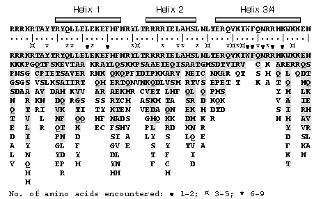
[edit]

The characteristic homeodomain protein fold consists of a 60-amino acid long domain composed of three alpha helices. The following shows the consensus homeodomain (~60 amino acid chain):[15]

            Helix 1          Helix 2         Helix 3/4
         ______________    __________    _________________
RRRKRTAYTRYQLLELEKEFHFNRYLTRRRRIELAHSLNLTERHIKIWFQNRRMKWKKEN
....|....|....|....|....|....|....|....|....|....|....|....|
         10        20        30        40        50        60
The vnd/NK-2 homeodomain-DNA complex. Helix 3 of the homeodomain binds in the major groove of the DNA and the N-terminal arm binds in the minor groove, in analogy with other homeodomain-DNA complexes.

Helix 2 and helix 3 form a so-called helix-turn-helix (HTH) structure, where the two alpha helices are connected by a short loop region. The N-terminal two helices of the homeodomain are antiparallel and the longer C-terminal helix is roughly perpendicular to the axes of the first two. It is this third helix that interacts directly with DNA via a number of hydrogen bonds and hydrophobic interactions, as well as indirect interactions via water molecules, which occur between specific side chains and the exposed bases within the major groove of the DNA.[7]

Homeodomain proteins are found in eukaryotes.[2] Through the HTH motif, they share limited sequence similarity and structural similarity to prokaryotic transcription factors,[16] such as lambda phage proteins that alter the expression of genes in prokaryotes. The HTH motif shows some sequence similarity but a similar structure in a wide range of DNA-binding proteins (e.g., cro and repressor proteins, homeodomain proteins, etc.). One of the principal differences between HTH motifs in these different proteins arises from the stereochemical requirement for glycine in the turn which is needed to avoid steric interference of the beta-carbon with the main chain: for cro and repressor proteins the glycine appears to be mandatory, whereas for many of the homeotic and other DNA-binding proteins the requirement is relaxed.

Sequence specificity

[edit]

Homeodomains can bind both specifically and nonspecifically to B-DNA with the C-terminal recognition helix aligning in the DNA's major groove and the unstructured peptide "tail" at the N-terminus aligning in the minor groove. The recognition helix and the inter-helix loops are rich in arginine and lysine residues, which form hydrogen bonds to the DNA backbone. Conserved hydrophobic residues in the center of the recognition helix aid in stabilizing the helix packing. Homeodomain proteins show a preference for the DNA sequence 5'-TAAT-3'; sequence-independent binding occurs with significantly lower affinity. The specificity of a single homeodomain protein is usually not enough to recognize specific target gene promoters, making cofactor binding an important mechanism for controlling binding sequence specificity and target gene expression. To achieve higher target specificity, homeodomain proteins form complexes with other transcription factors to recognize the promoter region of a specific target gene.

Biological function

[edit]

Homeodomain proteins function as transcription factors due to the DNA binding properties of the conserved HTH motif. Homeodomain proteins are considered to be master control genes, meaning that a single protein can regulate expression of many target genes. Homeodomain proteins direct the formation of the body axes and body structures during early embryonic development.[17] Many homeodomain proteins induce cellular differentiation by initiating the cascades of coregulated genes required to produce individual tissues and organs. Other proteins in the family, such as NANOG are involved in maintaining pluripotency and preventing cell differentiation.

Regulation

[edit]

Hox genes and their associated microRNAs are highly conserved developmental master regulators with tight tissue-specific, spatiotemporal control. These genes are known to be dysregulated in several cancers and are often controlled by DNA methylation.[18][19] The regulation of Hox genes is highly complex and involves reciprocal interactions, mostly inhibitory. Drosophila is known to use the polycomb and trithorax complexes to maintain the expression of Hox genes after the down-regulation of the pair-rule and gap genes that occurs during larval development. Polycomb-group proteins can silence the Hox genes by modulation of chromatin structure.[20]

Mutations

[edit]

Mutations to homeobox genes can produce easily visible phenotypic changes in body segment identity, such as the Antennapedia and Bithorax mutant phenotypes in Drosophila. Duplication of homeobox genes can produce new body segments, and such duplications are likely to have been important in the evolution of segmented animals.

Evolution

[edit]

Phylogenetic analysis of homeobox gene sequences and homeodomain protein structures suggests that the last common ancestor of plants, fungi, and animals had at least two homeobox genes.[21] Molecular evidence shows that some limited number of Hox genes have existed in the Cnidaria since before the earliest true Bilatera, making these genes pre-Paleozoic.[22] It is accepted that the three major animal ANTP-class clusters, Hox, ParaHox, and NK (MetaHox), are the result of segmental duplications. A first duplication created MetaHox and ProtoHox, the latter of which later duplicated into Hox and ParaHox. The clusters themselves were created by tandem duplications of a single ANTP-class homeobox gene.[23] Gene duplication followed by neofunctionalization is responsible for the many homeobox genes found in eukaryotes.[24][25] Comparison of homeobox genes and gene clusters has been used to understand the evolution of genome structure and body morphology throughout metazoans.[26]

Types of homeobox genes

[edit]

Hox genes

[edit]
Hox gene expression in Drosophila melanogaster.

Hox genes are the most commonly known subset of homeobox genes. They are essential metazoan genes that determine the identity of embryonic regions along the anterior-posterior axis.[27] The first vertebrate Hox gene was isolated in Xenopus by Edward De Robertis and colleagues in 1984.[28] The main interest in this set of genes stems from their unique behavior and arrangement in the genome. Hox genes are typically found in an organized cluster. The linear order of Hox genes within a cluster is directly correlated to the order in which they are expressed in both time and space during development. This phenomenon is called colinearity.

Mutations in these homeotic genes cause displacement of body segments during embryonic development. This is called ectopia. For example, when one gene is lost the segment develops into a more anterior one, while a mutation that leads to a gain of function causes a segment to develop into a more posterior one. Famous examples are Antennapedia and bithorax in Drosophila, which can cause the development of legs instead of antennae and the development of a duplicated thorax, respectively.[29]

In vertebrates, the four paralog clusters are partially redundant in function, but have also acquired several derived functions. For example, HoxA and HoxD specify segment identity along the limb axis.[30][31] Specific members of the Hox family have been implicated in vascular remodeling, angiogenesis, and disease by orchestrating changes in matrix degradation, integrins, and components of the ECM.[32] HoxA5 is implicated in atherosclerosis.[33][34] HoxD3 and HoxB3 are proinvasive, angiogenic genes that upregulate b3 and a5 integrins and Efna1 in ECs, respectively.[35][36][37][38] HoxA3 induces endothelial cell (EC) migration by upregulating MMP14 and uPAR. Conversely, HoxD10 and HoxA5 have the opposite effect of suppressing EC migration and angiogenesis, and stabilizing adherens junctions by upregulating TIMP1/downregulating uPAR and MMP14, and by upregulating Tsp2/downregulating VEGFR2, Efna1, Hif1alpha and COX-2, respectively.[39][40] HoxA5 also upregulates the tumor suppressor p53 and Akt1 by downregulation of PTEN.[41] Suppression of HoxA5 has been shown to attenuate hemangioma growth.[42] HoxA5 has far-reaching effects on gene expression, causing ~300 genes to become upregulated upon its induction in breast cancer cell lines.[42] HoxA5 protein transduction domain overexpression prevents inflammation shown by inhibition of TNFalpha-inducible monocyte binding to HUVECs.[43][44]

LIM genes

[edit]

LIM genes (named after the initial letters of the names of three proteins where the characteristic domain was first identified) encode two 60 amino acid cysteine and histidine-rich LIM domains and a homeodomain. The LIM domains function in protein-protein interactions and can bind zinc molecules. LIM domain proteins are found in both the cytosol and the nucleus. They function in cytoskeletal remodeling, at focal adhesion sites, as scaffolds for protein complexes, and as transcription factors.[45]

Pax genes

[edit]

Most Pax genes contain a homeobox and a paired domain that also binds DNA to increase binding specificity, though some Pax genes have lost all or part of the homeobox sequence.[46] Pax genes function in embryo segmentation, nervous system development, generation of the frontal eye fields, skeletal development, and formation of face structures. Pax 6 is a master regulator of eye development, such that the gene is necessary for development of the optic vesicle and subsequent eye structures.[47]

POU genes

[edit]

Proteins containing a POU region consist of a homeodomain and a separate, structurally homologous POU domain that contains two helix-turn-helix motifs and also binds DNA. The two domains are linked by a flexible loop that is long enough to stretch around the DNA helix, allowing the two domains to bind on opposite sides of the target DNA, collectively covering an eight-base segment with consensus sequence 5'-ATGCAAAT-3'. The individual domains of POU proteins bind DNA only weakly, but have strong sequence-specific affinity when linked. The POU domain itself has significant structural similarity with repressors expressed in bacteriophages, particularly lambda phage.

Plant homeobox genes

[edit]

As in animals, the plant homeobox genes code for the typical 60 amino acid long DNA-binding homeodomain or in case of the TALE (three amino acid loop extension) homeobox genes for an atypical homeodomain consisting of 63 amino acids. According to their conserved intron–exon structure and to unique codomain architectures they have been grouped into 14 distinct classes: HD-ZIP I to IV, BEL, KNOX, PLINC, WOX, PHD, DDT, NDX, LD, SAWADEE and PINTOX.[24] Conservation of codomains suggests a common eukaryotic ancestry for TALE[48] and non-TALE homeodomain proteins.[49]

Human homeobox genes

[edit]

The Hox genes in humans are organized in four chromosomal clusters:

name chromosome gene
HOXA (or sometimes HOX1) - HOXA@ chromosome 7 HOXA1, HOXA2, HOXA3, HOXA4, HOXA5, HOXA6, HOXA7, HOXA9, HOXA10, HOXA11, HOXA13
HOXB - HOXB@ chromosome 17 HOXB1, HOXB2, HOXB3, HOXB4, HOXB5, HOXB6, HOXB7, HOXB8, HOXB9, HOXB13
HOXC - HOXC@ chromosome 12 HOXC4, HOXC5, HOXC6, HOXC8, HOXC9, HOXC10, HOXC11, HOXC12, HOXC13
HOXD - HOXD@ chromosome 2 HOXD1, HOXD3, HOXD4, HOXD8, HOXD9, HOXD10, HOXD11, HOXD12, HOXD13

ParaHox genes are analogously found in four areas. They include CDX1, CDX2, CDX4; GSX1, GSX2; and PDX1. Other genes considered Hox-like include EVX1, EVX2; GBX1, GBX2; MEOX1, MEOX2; and MNX1. The NK-like (NKL) genes, some of which are considered "MetaHox", are grouped with Hox-like genes into a large ANTP-like group.[50][51]

Humans have a "distal-less homeobox" family: DLX1, DLX2, DLX3, DLX4, DLX5, and DLX6. Dlx genes are involved in the development of the nervous system and of limbs.[52] They are considered a subset of the NK-like genes.[50]

Human TALE (Three Amino acid Loop Extension) homeobox genes for an "atypical" homeodomain consist of 63 rather than 60 amino acids: IRX1, IRX2, IRX3, IRX4, IRX5, IRX6; MEIS1, MEIS2, MEIS3; MKX; PBX1, PBX2, PBX3, PBX4; PKNOX1, PKNOX2; TGIF1, TGIF2, TGIF2LX, TGIF2LY.[50]

In addition, humans have the following homeobox genes and proteins:[50]

  1. ^ Grouped as Lmx 1/5, 2/9, 3/4, and 6/8.
  2. ^ Grouped as Six 1/2, 3/6, and 4/5.
  3. ^ Questionable, per [50]
  4. ^ The Pax genes. Grouped as Pax2/5/8, Pax3/7, and Pax4/6.
  5. ^ Nk4.
  6. ^ Nk5.

See also

[edit]

References

[edit]

Further reading

[edit]
[edit]
Revisions and contributorsEdit on WikipediaRead on Wikipedia
from Grokipedia
A homeobox is a highly conserved DNA sequence approximately 180 base pairs in length that encodes a 60-amino-acid protein domain known as the homeodomain, which functions as a sequence-specific DNA-binding motif in transcription factors. These sequences are integral to homeobox genes, a diverse family of regulatory genes that control key aspects of development, including morphogenesis, cell differentiation, and pattern formation, across eukaryotes such as animals, plants, and fungi. The homeobox was first discovered in 1983 through studies on homeotic mutations in the fruit fly Drosophila melanogaster, with the sequence identified via low-stringency hybridization in genes from the and bithorax complexes by researchers including Matthew Scott, Amy Weiner, William McGinnis, Michael Levine, and Walter Gehring; the findings were published in 1984. This breakthrough demonstrated the homeobox's presence in developmental control genes across distant species—from insects to vertebrates—via "zoo blot" hybridizations, underscoring its evolutionary conservation over more than 600 million years and revealing a shared genetic toolkit for animal body patterning. In animals, homeobox genes have diversified into at least 11 classes and over 100 families, with prominent examples like the clusters that specify segmental identities along the anterior-posterior body axis during embryogenesis. Homeodomain proteins act primarily as transcription factors, binding to specific DNA motifs to activate or repress downstream genes involved in , tissue specification, and cellular proliferation. In , homeobox genes such as those in the KNOX and HD-ZIP classes regulate maintenance and development, while in fungi, they govern differentiation, , and pathogenicity. Mutations or dysregulation of homeobox genes can lead to dramatic homeotic transformations, congenital defects (e.g., limb malformations), and diseases including cancers and cardiovascular disorders, highlighting their ongoing relevance in both and .

Overview and Discovery

Definition and General Role

The homeobox is a conserved DNA sequence of approximately 180 base pairs that encodes a 60-amino-acid DNA-binding domain known as the homeodomain. This motif, characterized by a helix-turn-helix structure, enables the encoded proteins to recognize and bind specific DNA sequences, thereby functioning as transcription factors that modulate gene expression. Homeobox-containing genes are integral to the precise spatiotemporal control of developmental programs, distinguishing them from other regulatory elements through their high degree of sequence conservation. In their general role, homeobox genes serve as master regulators of embryogenesis, , and cell differentiation across eukaryotic organisms. These genes orchestrate the activation or repression of downstream targets to ensure coordinated cellular responses, thereby establishing foundational patterns in tissue formation and organ specification. For instance, they play a pivotal part in body patterning, such as directing the anterior-posterior axis in animals by specifying segmental identities along the embryonic . The evolutionary conservation of the homeobox underscores its ancient origin, with sequences identifiable in diverse eukaryotic lineages including animals, , and fungi. This widespread presence, dating back to early eukaryotic , reflects the domain's fundamental importance in adapting developmental strategies to varying environmental and morphological demands across taxa. Such conservation highlights how homeobox genes have been co-opted for analogous regulatory functions despite the divergence of major eukaryotic groups.

Historical Discovery

The discovery of the homeobox sequence emerged from studies on homeotic mutations in the fruit fly Drosophila melanogaster, which cause dramatic transformations in body segment identity, such as legs developing in place of antennae. In early 1983, researchers in Walter Gehring's laboratory at the isolated (cDNA) clones of the (Antp) gene, responsible for one such , and sequenced a conserved 180-base-pair (bp) DNA segment within it. This sequence, encoding a 60-amino-acid motif predicted to bind DNA, was identified through low-stringency hybridization experiments that revealed similar repeats in other homeotic genes. Independently, in Matthew Scott and Amy Weiner's work under Thomas Kaufman's supervision at , the same conserved sequence was found in the (Ubx) gene from the bithorax complex, confirming its presence across homeotic loci. These findings were reported in parallel publications in 1984, marking the initial identification of what would be termed the homeobox. The term "homeobox" was coined in 1984 by Andrew Laughon and Matthew Scott, reflecting the sequence's location in homeotic genes and its box-like conservation. Key experiments involved cloning and sequencing homeotic genes from mutants, using Southern blots and zoo blots to detect homologous sequences across species, which suggested a fundamental role in developmental regulation. This conserved motif was quickly extended beyond insects; by mid-1984, Bill McGinnis and colleagues used homeobox probes to isolate similar sequences in genomic DNA, revealing homeobox-containing genes clustered on chromosomes analogous to the fly's and bithorax complexes. Similar discoveries in s, including the cloning of homeobox genes in by Andres Carrasco and Eddy De Robertis, underscored the motif's evolutionary conservation. These vertebrate findings, published in 1984, laid the groundwork for identifying mammalian clusters in the late 1980s. The homeobox discovery built on earlier genetic analyses of homeotic genes, notably Edward B. Lewis's pioneering work in the 1940s and 1950s on the bithorax complex, which demonstrated how mutations alter segment identity along the anterior-posterior axis. In the 1970s, Christiane Nüsslein-Volhard and Eric Wieschaus conducted saturation mutagenesis screens in Drosophila embryos, identifying genes that control segmentation and segment identity, including homeotic selectors. Their collective contributions to understanding homeotic gene function earned Lewis, Nüsslein-Volhard, and Wieschaus the 1995 Nobel Prize in Physiology or Medicine "for their discoveries concerning genetic control of early embryonic development." This recognition highlighted how molecular cloning in the 1980s, inspired by their genetic frameworks, unveiled the homeobox as a shared regulatory element across homeotic genes.

Molecular Structure

Homeodomain Architecture

The homeodomain is a compact globular protein domain typically comprising approximately 60 amino acids that folds into a characteristic three-dimensional structure consisting of three alpha-helices connected by two short loops. The first helix (helix 1, residues ~8-22) and second helix (helix 2, residues ~26-35) are linked by a flexible loop, while helix 2 connects to the third helix (helix 3, residues ~42-58) via a short turn, forming a conserved helix-turn-helix (HTH) motif where helices 2 and 3 pack against each other at an angle of about 120 degrees. This HTH motif positions helix 3, known as the recognition helix, to interact directly with the major groove of DNA, enabling sequence-specific binding. The overall fold is stabilized by hydrophobic interactions between the helices. Key residues within the homeodomain contribute to its DNA-binding capability, particularly in helix 3, which contains highly conserved amino acids such as at position 51 (Asn51) and at position 53 (Arg53). Asn51 forms hydrogen bonds with bases in the DNA major groove, while Arg53 contacts residues, contributing to the preference for AT-rich sequences. These residues, along with at position 48 (Trp48) and at position 49 (Phe49), form the primary DNA recognition interface and are invariant across most homeodomains, ensuring structural integrity and binding affinity. Additionally, an at position 5 (Arg5) in the N-terminal extension often inserts into the minor groove, enhancing specificity through electrostatic interactions with the DNA backbone. The homeodomain forms a 1:1 stoichiometric complex with DNA, where helix 3 docks into the major groove of a TAAT core motif within AT-rich sequences, while the N-terminal arm wraps around the DNA helix to contact the minor groove. This architecture was first elucidated through nuclear magnetic resonance (NMR) spectroscopy for the Antennapedia homeodomain from Drosophila melanogaster in 1989, revealing the three-helix bundle in solution, and confirmed by X-ray crystallography of the engrailed homeodomain-DNA complex at 2.8 Å resolution in 1990, which highlighted the precise contacts between protein side chains and DNA bases. Water-mediated hydrogen bonds further stabilize the interface, allowing subtle adjustments for sequence discrimination. Structural variations in homeodomains arise primarily from flexible N- and C-terminal extensions, which can extend up to 15-20 residues and influence binding specificity without altering the core fold. The N-terminal arm, rich in basic residues, varies in length and composition across homeodomain subclasses, enabling additional minor groove interactions that fine-tune target site selection, as seen in Hox proteins. In contrast, the C-terminal arm is generally shorter and more variable, often involved in protein-protein interactions rather than direct DNA contact, contributing to the modular nature of homeodomain function. These extensions maintain flexibility in the unbound state, allowing conformational adaptation upon DNA binding.

DNA Sequence Specificity

Homeodomains primarily recognize DNA through interactions in the major groove, with a strong preference for the core motif TAAT, where the recognition (helix 3) inserts directly into the groove to form base-specific contacts. This motif is conserved across many homeodomain proteins, but flanking introduce variations that refine binding preferences; for instance, the (C/G)TAATTG is common. These sequence rules enable selective by distinguishing subtle differences in target sites. Key determinants of specificity lie in the amino acid residues of helix 3, particularly position 50, which acts as a discriminator by contacting bases 3' to the TAAT core via hydrogen bonds and van der Waals interactions. In (Antp), (Gln) at position 50 promotes binding to sites with or immediately following TAAT, whereas (Lys) at this position in Engrailed favors , altering target site selection and functional outcomes. studies confirm that substituting residues at position 50 can redirect binding to non-native sites, underscoring its role in diversification. Experimental approaches, such as bacterial one-hybrid assays akin to SELEX, have mapped specificities for numerous homeodomains, revealing at least 17 distinct profiles; for example, Antp and Engrailed share T(A/T)AT(T/G)(A/G), while Abd-B prefers TTATGG. combined with binding assays further demonstrates that single changes in 3, like those at position 50, shift target recognition, as seen in altered affinities for variant motifs . These methods highlight how sequence variations correlate with functional specificity without exhaustive enumeration. Cofactors enhance DNA sequence specificity by stabilizing complexes and exposing latent preferences; notably, PBX proteins interact with Hox homeodomains to form heterodimers that bind bipartite sites like 5'-ATGATTNATNN-3', where PBX contacts the 5' half and Hox the 3' half. This partnership modulates the Hox amino-terminal arm, enabling progressive core preferences from TTAT (anterior Hox) to TGAT (posterior Hox), which alone exhibit lower specificity. Such interactions are essential for precise target discrimination in development.

Biological Roles

Developmental Functions

Homeobox genes play pivotal roles in during embryonic development, primarily by establishing the anterior-posterior body axis and specifying segment identities through their collinear expression patterns. In bilaterian animals, —a major subclass of homeobox genes—are transcribed in a spatially ordered manner that mirrors their , a phenomenon known as spatial colinearity, which ensures precise regional specification along the body axis. This colinearity is evident in , where homeotic mutations cause transformations, such as the conversion of into wings by bithorax complex mutations or legs into antennae by complex mutations, demonstrating how these genes dictate segmental identity. In vertebrates, similar Hox colinearity patterns the , with anterior Hox genes expressed in cervical regions and posterior ones in thoracic and areas, thereby conferring distinct vertebral identities. Beyond axial patterning, homeobox genes drive by regulating spatiotemporal expression that guides tissue morphogenesis in structures like limbs, eyes, and the . In limb development, Hoxd cluster genes exhibit biphasic expression: an early phase patterns proximal elements (e.g., ), while a later phase specifies distal ones (e.g., digits), with collinear activation ensuring proper anterior-posterior asymmetry. For eye formation, the Rx homeobox gene initiates retinal progenitor specification in the anterior , cooperating with to induce optic vesicle outgrowth and maintain identity. In neural development, subdivide the into rhombomeres, with specific combinations (e.g., Hoxa1 in r4) directing neuronal subtype differentiation and cranial nerve positioning.01611-2) Homeobox genes also influence cell fate determination, balancing pluripotency and differentiation in various lineages, including hematopoiesis. In hematopoietic stem cells (HSCs), medial HOXA genes (e.g., HOXA5–HOXA7) mark the transition from primitive to definitive hematopoiesis, promoting self-renewal and multilineage potential during human embryonic development. For instance, retinoic acid-induced HOXA expression restricts HSC fate, preventing reversion to earlier mesodermal states and enabling long-term engraftment. In somitogenesis, a process integral to vertebral patterning, Hox genes pre-specify paraxial mesoderm identity before segmentation clock activation, ensuring coordinated formation of somites that give rise to the axial skeleton. These functions underscore the homeobox genes' role as master regulators, integrating positional cues to orchestrate developmental outcomes across species.

Regulatory Mechanisms

Homeobox genes are primarily regulated at the transcriptional level through intricate networks involving auto-regulation, cross-regulation, and responses to extracellular signaling pathways. , a prominent subset of homeobox genes, form regulatory networks where individual members auto-activate their own expression or cross-activate/repress others to establish and maintain spatial expression domains along the anterior-posterior axis. For instance, Hoxb3 expression in the posterior and is controlled by IIIa, which contains binding sites for Hoxb3 and Hoxb4 proteins, enabling auto-regulation and cross-regulation that restrict expression up to the rhombomere 5/6 boundary. Enhancers associated with homeobox genes often integrate inputs from signaling pathways such as Wnt and FGF, which modulate accessibility and promoter interactions to fine-tune expression during development. In limb development, FGF8 and Shh signaling synergistically activate via a regulatory switch from proximal (T-DOM) to distal (C-DOM) enhancers, involving increased H3K27 acetylation in enhancer regions. Wnt signaling similarly patterns early Hox expression in the by activating Cdx and through β-catenin-dependent mechanisms, ensuring collinear activation. Epigenetic modifications play a crucial role in silencing homeobox loci, particularly through modifications and that maintain heritable repression states. H3K27 trimethylation by Polycomb group proteins, specifically the Polycomb repressive complex 2 (PRC2) through the SET domain of its subunit, enforces silencing of Hox clusters. While defects in H3K4 monomethylation lead to reduced expression at promoters of genes like Hoxd4 and Hoxc8. at CpG islands within Hox loci, often in concert with hypoacetylation, contributes to stable inactivation; for example, hyper- or hypomethylation at the Hoxd4 locus disrupts normal expression patterns without global methylation changes. These mechanisms ensure that inappropriate of homeobox genes is prevented outside their specified domains, with long-range interactions facilitating enhancer-promoter looping for precise control. Post-transcriptional regulation further refines homeobox gene output via microRNAs (miRNAs) and , generating functional diversity and temporal precision. miR-196, embedded within Hox clusters, directly targets Hoxb8 mRNA by binding its 3' UTR, leading to cleavage and repression that restricts Hoxb8 expression in the posterior limb and ; disruption causes homeotic transformations in chick and mouse models. Alternative splicing produces multiple isoforms with distinct activities, as seen in mouse Hoxa1, Hoxa9, and Hoxa10, where isoform variations alter protein domains and regulatory potential without affecting overall cluster organization. Feedback loops, encompassing positive and negative autoregulation, are integral to sustaining homeobox expression domains over time. Hoxb1 maintains its segmental expression in rhombomere 4 through a conserved autoregulatory loop involving direct binding to three motifs in its regulatory regions, requiring cofactors like Pbx/Exd for cooperative activation. Such loops often integrate cross-regulatory inputs, where posterior Hox proteins repress anterior ones (posterior prevalence), forming networks that stabilize patterns during embryogenesis. , such as miRNA-mediated repression, counterbalances positive autoregulation to prevent and ensure domain boundaries.

Genetic Types and Classification

Hox Gene Family

The Hox gene family represents the prototypical subset of homeobox genes, encoding transcription factors that play essential roles in establishing body plans during animal development. These genes are characterized by their conserved homeodomain and are organized into genomic clusters that exhibit spatial and temporal collinearity, a hallmark feature linking gene order to expression patterns along the anterior-posterior axis. In vertebrates, including humans, the family comprises 39 genes, which are highly conserved across bilaterian species, underscoring their fundamental importance in metazoan evolution. In vertebrates, Hox genes are clustered into four paralogous genomic loci designated HoxA, HoxB, HoxC, and HoxD, located on separate chromosomes. Each cluster typically contains 8 to 11 genes, arranged in a linear fashion with a consistent 3' to 5' transcriptional orientation, totaling the 39 functional members in humans. This clustered organization arose through two rounds of whole-genome duplication early in vertebrate evolution, resulting in paralogous genes (e.g., HoxA1, HoxB1, HoxC1, HoxD1) that share sequence similarity and often overlapping functions. A key organizational principle is colinearity, where the physical order of genes within each cluster corresponds to their expression domains: genes at the 3' end are expressed in more anterior regions of the embryo, while 5' genes are restricted to posterior domains. This spatial collinearity is complemented by temporal collinearity, with 3' genes activating earlier in development than their 5' counterparts. Hox genes orchestrate axial patterning and through precise regulation of downstream targets. For instance, HoxA1 is crucial for segmentation, where its expression in rhombomere 4 helps specify neuronal identities and cranial development; mutations disrupt this patterning, leading to severe defects in the model. Similarly, , a 5' posterior gene, is vital for limb , particularly in forming distal digits and skeletal elements, as evidenced by its role in regulating chondrogenesis and joint specification in developing autopods. These functions highlight the family's role in translating positional information into morphological outcomes. A distinctive feature of the Hox family is the functional redundancy among paralogs, which provides robustness to developmental processes. For example, the Hox4 paralogs (HoxA4, HoxB4, HoxD4) collectively maintain cervical vertebral identity, with triple mutants exhibiting homeotic transformations to atlas-like structures. Hox proteins often require cofactors such as PBX and MEIS family members to achieve full transcriptional activity; these TALE-homeodomain proteins form heterodimeric complexes that enhance DNA-binding specificity and recruit chromatin-modifying machinery to Hox target enhancers. This cofactor interaction is essential for the precise spatiotemporal control of Hox-regulated genes during embryogenesis.

Non-Hox Homeobox Genes

Non-Hox homeobox genes comprise a broad collection of transcription factors that extend beyond the clustered Hox family, enabling specialized regulation of development in diverse tissues and organs. These genes are dispersed throughout the and classified into major classes such as LIM, PRD, POU, and specific subclasses like NKL within the ANTP class, distinguished by unique domain structures that facilitate interactions, and transcriptional control. In humans, over 200 such genes exist across 11 classes and more than 100 families, underscoring their evolutionary diversification for precise developmental roles. The LIM-homeodomain class includes genes encoding proteins with two N-terminal LIM domains—cysteine- and histidine-rich motifs that bind and mediate protein-protein interactions—adjacent to a homeodomain. This architecture allows LIM proteins to integrate signaling pathways and assemble transcriptional complexes. The class encompasses six families, including LHX and ISL, with 12 members in humans. A prominent example is LHX1, which drives specification of the renal progenitor field from during kidney . In Lhx1-null mice, ureteric bud formation and nephron induction fail, resulting in , as LIM domains facilitate interactions with cofactors like LDB1 to activate downstream targets such as Wt1. Genes of the PRD class feature a PRD domain, often a paired-like motif involved in DNA recognition, combined with a homeodomain, and include the PAX subfamily distinguished by an additional paired box for enhanced specificity. This class contains 50 human genes across 31 families, such as PAX, OTX, and CRX. The PAX genes, in particular, orchestrate through combinatorial domain usage. For instance, PAX6 acts as a master regulator in eye and development; in Pax6 mutant mice (Small eye model), lens placode induction and neural formation are absent, while in the pancreas, Pax6 loss disrupts glucagon-producing alpha-cell differentiation by failing to activate endocrine progenitors. The POU class is defined by a bipartite POU domain—a POU-specific region and a POU-homeodomain—that cooperatively binds an octamer DNA motif, enabling high-affinity transcriptional regulation. Human POU genes number 16 across seven families, including POU1 and POU5. POU5F1 (Oct4) maintains pluripotency in embryonic stem cells by repressing differentiation genes and sustaining self-renewal circuits; Oct4-null mouse embryos arrest at the blastocyst stage with inner cell mass failure. Conversely, POU1F1 (Pit1) specifies anterior pituitary lineages, driving somatotropes, lactotropes, and thyrotropes; mutations in Pit1 cause combined pituitary hormone deficiency, impairing growth, lactation, and thyroid function due to defective cell commitment.00348-7)90072-1) Other notable non-Hox families include the NKL subclass of the ANTP class, which features a standard homeodomain with NK-specific extensions for target selectivity, comprising genes like NKX2-5 essential for cardiogenesis. In Nkx2-5-knockout mice, cardiac progenitors form but fail to undergo looping and chamber septation, leading to embryonic lethality from severe heart defects, as NKX2-5 activates myocardial genes like Nppa. The PRD class further includes non-PAX members such as those regulating segmentation in invertebrates; for example, Drosophila paired (prd), a PRD gene, defines odd-numbered parasegment boundaries by coordinating pair-rule gene expression. Collectively, non-Hox homeobox genes exhibit functional diversity through tissue-restricted expression and context-dependent interactions, governing organ-specific processes like nephrogenesis, ocular and endocrine differentiation, stem cell maintenance, cardiogenesis, and segmental patterning, in contrast to the ' emphasis on axial body organization.

Evolutionary Aspects

Origin and Conservation

Homeobox genes trace their origins to the last eukaryotic common ancestor (LECA), which existed approximately 1.5–2 billion years ago, as evidenced by the presence of proto-homeobox sequences in diverse eukaryotic lineages including fungi and . Genomic analyses of fungal , such as those in microsporidians and nucleariids, and algal genomes from unicellular chlorophytes within , reveal conserved homeobox-like motifs that predate multicellularity and support an ancient eukaryotic origin for these regulatory elements. These findings indicate that a rudimentary homeobox system was already functional in the LECA, likely involved in basic before the diversification of complex body plans. The homeodomain, the defining DNA-binding motif of homeobox proteins, exhibits remarkable conservation across metazoans, with key residues—particularly those in the three alpha-helices responsible for DNA recognition—showing over 90% sequence identity between distantly related like and vertebrates. This high fidelity in critical positions underscores the structural and functional stability of the homeodomain, enabling precise spatiotemporal control of despite evolutionary divergence spanning hundreds of millions of years. In s, the homeobox gene repertoire expanded dramatically through whole-genome duplications, with two successive events (known as 1R and 2R) occurring near the base of the vertebrate lineage around 500 million years ago, resulting in the characteristic four . These duplications provided raw genetic material for subfunctionalization and neofunctionalization, enhancing developmental complexity. Expression patterns of in modern animals mirror the diverse body plans that emerged during the approximately 540 million years ago, suggesting that homeobox diversification contributed to the rapid evolution of bilaterian morphologies preserved in the fossil record.

Diversification Across Species

Homeobox genes have undergone significant diversification across metazoan lineages through mechanisms such as gene duplication, loss, and functional divergence, adapting to the specific developmental needs of diverse species. In insects like Drosophila melanogaster, the ancestral Hox cluster has undergone rearrangements and partial dispersal, resulting in a single, fragmented cluster of eight genes that maintain collinear expression along the anterior-posterior axis but lack the tight linkage seen in other groups. In contrast, vertebrate genomes exhibit extensive duplication events, with mammals possessing four paralogous Hox clusters (HoxA, HoxB, HoxC, and HoxD) arising from two rounds of whole-genome duplication in the vertebrate ancestor, each containing 7–13 genes that collectively specify regional identities during embryogenesis. These duplications have allowed for subcluster-specific innovations, while losses, such as the absence of certain non-Hox homeobox genes like Msxlx in Olfactores (tunicates and vertebrates), reflect lineage-specific streamlining. In non-bilaterian metazoans, such as cnidarians, Hox-like homeobox genes exhibit early diversification adapted to radial body plans. For instance, in the Nematostella vectensis, two Hox-like genes (NvAx6 and NvAx1) pattern the oral-aboral axis, with NvAx6 promoting oral development and NvAx1 specifying aboral structures through mutual inhibition during early embryogenesis. These genes, orthologous to anterior and posterior bilaterian Hox classes, suggest that homeobox diversification predates bilaterian axial complexity, enabling directive patterning in radially symmetric ancestors over 600 million years ago. Functional shifts following duplication have driven neofunctionalization, particularly in vertebrates, where certain Hox paralogs acquired novel roles in morphological transitions. During the fin-to-limb , duplicated Hoxd genes in the HoxD cluster neofunctionalized to regulate distal limb elements, with promoting digit-like structures absent in fish fins through enhanced expression in mesenchymal condensations. Similarly, Hoxa11 and Hoxd11 paralogs diverged to coordinately pattern nervous system and skeletal elements in limbs, illustrating how post-duplication changes in protein interactions and regulatory landscapes facilitated the emergence of weight-bearing appendages. Comparative genomics has elucidated this diversification through ortholog identification, primarily via sequence alignment of the conserved homeodomain motif using tools like BLAST and phylogenetic reconstruction. Such analyses reveal extensive losses in parasitic lineages; for example, tapeworms (Cestoda) have lost over 20 homeobox gene families, including key Hox and ParaHox members, correlating with simplified body plans and obligate parasitism in flatworms. These reductions, inferred from alignments with free-living relatives, highlight how gene loss contributes to evolutionary adaptation in degenerate morphologies.

Pathological Implications

Mutations and Disorders

Mutations in homeobox genes, which transcription factors critical for developmental patterning, frequently result in congenital disorders characterized by limb malformations, craniofacial abnormalities, and organ defects. These mutations disrupt the precise spatiotemporal during embryogenesis, leading to phenotypes that reflect the genes' roles in specifying body segment identity and tissue differentiation. Common types of mutations include point mutations that alter the DNA-binding homeodomain and polyalanine tract expansions within the protein's N-terminal region. For instance, polyalanine expansions in the gene, such as duplications of 7 to 24 residues, cause synpolydactyly type 1, a condition featuring webbed fingers and toes with extra digits. Similarly, missense mutations or polyalanine expansions in HOXA13 lead to hand-foot-genital syndrome, involving short thumbs, small feet, and urogenital anomalies. Point mutations in the homeodomain, including nonsense and frameshift variants, underlie congenital , marked by iris and increased risk. The pathological mechanisms often involve , where a single functional fails to provide sufficient protein for normal development, or dominant-negative effects, in which mutant proteins interfere with wild-type counterparts. In synpolydactyly, polyalanine expansions promote protein aggregation and sequestration of normal , exerting a dominant-negative influence that perturbs limb patterning. Haploinsufficiency of HOXA13 similarly disrupts anterior-posterior limb axis formation, contributing to the skeletal and genital defects in hand-foot-genital syndrome. mutations typically cause , reducing transcriptional activation of genes and leading to ocular . Animal models, particularly knockout mice, have recapitulated these human phenotypes and provided insights into mutation effects. Targeted disruption of , such as Hoxa-2, results in homeotic transformations where second structures develop rhombomere 2-like identities, including cleft palate and skeletal defects. knockout mice exhibit limb reductions and delayed chondrogenesis, mirroring aspects of synpolydactyly and underscoring the genes' dosage-sensitive roles in formation. These studies confirm that loss-of-function mutations disrupt axial patterning, often through altered Hox expression gradients.

Therapeutic and Research Applications

Homeobox genes have emerged as promising targets in for cancers driven by their misexpression, particularly in solid tumors like where HOXB7 overexpression promotes cell proliferation and invasion. For instance, a 2024 study demonstrated an innovative extracellular vesicle-based approach to engineer CD8+ T cells specifically against HOXB7-expressing tumor cells, enhancing anti-tumor immunity in preclinical models of . Similarly, in general have been identified as potential therapeutic targets in , where their promotion of proliferation suggests opportunities for silencing strategies to inhibit tumor growth. In stem cell research, homeobox transcription factors such as NANOG play a critical role in induced pluripotent stem (iPS) cell reprogramming by overcoming epigenetic barriers and stabilizing pluripotency. Overexpression of NANOG in minimal factor conditions has been shown to induce full pluripotency in somatic cells, facilitating the generation of naive-like iPS cells for regenerative applications. Recent reviews highlight NANOG's integration into reprogramming cocktails to enhance efficiency and epigenetic resetting, underscoring its utility in deriving patient-specific stem cells for disease modeling and therapy. Drug development efforts have focused on small molecules that modulate homeodomain-DNA interactions to disrupt oncogenic homeobox activity, particularly in hematological malignancies like . Screening strategies using technology have identified compounds that inhibit homeodomain binding, offering a foundation for targeting aberrant transcription in cancer cells. Notably, small-molecule inhibitors of MEIS1, a TALE-class homeobox overexpressed in , have been developed to reduce self-renewal and leukemic propagation in preclinical models. Post-2020 advances include CRISPR-based editing of homeobox loci to dissect their functions in disease models, such as genome-wide screens targeting HOXA9-bound regions in mixed-lineage leukemia-rearranged cells, revealing noncoding regulatory elements as potential therapeutic vulnerabilities. In neurodevelopment research, 2023 studies have linked loss-of-function variants in homeobox genes like LHX2 to variable neurodevelopmental disorders, informing targeted interventions through improved genetic models. These efforts highlight the growing integration of gene editing in systems to study homeobox-driven processes, paving the way for precision therapies in developmental and oncogenic contexts.

Homeobox in Plants

Key Plant Homeobox Families

In plants, homeobox genes have evolved independently from those in animals, arising through the fusion of a homeodomain with diverse protein domains that confer specificity to developmental processes. This evolutionary divergence resulted in several unique families, including the KNOX, HD-ZIP, and WOX families, which are absent in animal lineages and play pivotal roles in regulating architecture and growth. The KNOX (KNOTTED-like homeobox) family belongs to the three-amino-loop-extension (TALE) superclass of homeodomain proteins and is characterized by a conserved KNOX domain adjacent to the homeodomain, which is involved in protein-protein interactions. KNOX genes are divided into two classes: Class I, which includes genes like SHOOTMERISTEMLESS (STM) expressed in the shoot apical meristem to maintain indeterminate cell fates, and Class II, exemplified by ARABIDOPSIS THALIANA HOMEOBOX 3 (KNAT3), which contributes to leaf development and differentiation. These classes differ in their expression patterns and regulatory targets, with Class I genes predominantly active in meristematic tissues and Class II more broadly distributed in differentiated organs. The HD-ZIP (homeodomain-leucine zipper) family is exclusive to land plants and features a homeodomain fused to a leucine zipper motif that facilitates DNA binding as dimers, enabling responses to environmental cues like light. This family is subdivided into four classes (I–IV), but Classes I–III are particularly prominent in vascular and organ development; for instance, Class III members such as ATHB8 regulate vascular tissue differentiation, while REVOLUTA establishes adaxial-abaxial polarity in leaves. Class I genes, like ATHB1, often respond to abiotic stresses, whereas Class II genes integrate hormonal signals for growth modulation. The WOX (WUSCHEL-related homeobox) family comprises plant-specific transcription factors with a homeodomain and a WUS-box motif that mediates short-range signaling in maintenance. WOX genes are grouped into ancient, intermediate, and modern , with key members like WUSCHEL (WUS) sustaining niches in shoot and floral . Other WOX proteins, such as those in the modern , influence flowering transitions by modulating identity and phase changes.

Roles in Plant Development

Plant homeobox genes play crucial roles in regulating developmental processes, particularly through families like KNOX and HD-ZIP III, which maintain atic identity and establish organ polarity. In the shoot apical (SAM), class I KNOX genes, such as SHOOTMERISTEMLESS (STM) in , are essential for sustaining undifferentiated populations by promoting biosynthesis and repressing signaling, thereby preventing premature differentiation of meristem cells. These genes orchestrate a balance of growth regulators to ensure continuous organ initiation from the SAM. For organ polarity, class III HD-ZIP genes, including PHABULOSA (PHB), PHAVOLUTA (PHV), and REVOLUTA (REV), specify adaxial-abaxial axes in lateral organs like leaves and by promoting adaxial (upper) identity while antagonizing abaxial (lower) fate through interactions with KANADI genes. In leaves, this patterning ensures proper lamina outgrowth and vascular organization, whereas in , it contributes to radial patterning and tissue specification. Misregulation disrupts polarity, leading to radialized or inversely oriented organs. Homeobox genes also mediate environmental responses, integrating developmental cues with abiotic stresses. In drought conditions, the HD-ZIP I ATHB6 in acts downstream of signaling via protein phosphatases ABI1 and ABI2, enhancing stress tolerance by modulating responses and stomatal closure. Overexpression of ATHB6 reduces malondialdehyde levels and activates and ABA pathways, promoting root growth under water deficit. For signaling, the TALE homeobox ARABIDOPSIS THALIANA HOMEOBOX 1 (ATH1) converges with energy and pathways to control rosette architecture, restricting elongation and promoting compact growth in response to blue and far-red . Mutational analyses highlight these functions through distinct phenotypes. Loss-of-function in class I KNOX genes like STM abolishes SAM formation, while overexpression or misexpression often results in fasciation-like phenotypes, characterized by enlarged meristems and distorted organ shapes due to ectopic . Similarly, WOX gene mutants exhibit embryonic defects; for instance, wox2 single mutants show perturbed apical-basal patterning, and combinations like wox8 wox9 or wox2 wox8 wox9 lead to arrested embryos with disorganized tissue proliferation and failure in hypophysis specification. These phenotypes underscore the precise spatiotemporal control exerted by homeobox genes in .

Homeobox in Humans

Major Human Homeobox Genes

The contains 241 protein-coding homeobox genes, along with 108 pseudogenes, as comprehensively classified and annotated in updated analyses including the Homeobox Gene Database (HomeoDB). These genes are organized into over 100 families, reflecting their evolutionary diversification and roles in developmental regulation. The family represents one of the most prominent subclasses, comprising 39 genes arranged in four paralogous clusters: HOXA on , HOXB on chromosome 17, HOXC on , and HOXD on chromosome 2. Each cluster contains 9 to 11 genes ordered in a collinear fashion that mirrors their sequential expression along the anterior-posterior axis during embryogenesis. For instance, HOXC8, located in the HOXC cluster, is expressed in the developing , where it regulates differentiation and terminal in brachial regions. Beyond the , non-Hox homeobox families include the PAX, NKX, and EMX groups, which play critical roles in tissue-specific patterning. The PAX family, characterized by paired domain-homeodomain structures, features genes like , which is essential for cell specification and migration during early neural development. Similarly, NKX2-1 from the NKX family drives in the and lungs by activating tissue-specific promoters, such as those for proteins and . In the brain, EMX2 contributes to regionalization, promoting growth and arealization of cortical progenitors in the . Expression patterns of human homeobox genes display precise temporal and spatial specificity, as revealed by large-scale transcriptomic data from the Genotype-Tissue Expression (GTEx) project, which maps their activity across 54 tissues and developmental stages. These patterns underscore their coordinated roles in embryogenesis, with many genes like those in the Hox clusters showing collinear activation from early gestation onward. Some homeobox genes are also implicated in disease when dysregulated, though detailed pathological links are addressed elsewhere.

Involvement in Human Diseases

Homeobox genes are frequently dysregulated in cancers, where they contribute to tumorigenesis by altering , differentiation, and survival. In (AML), chromosomal translocations leading to fusions such as NUP98-HOXA9 promote leukemogenesis by upregulating HOXA9 and its cofactor MEIS1, which drive self-renewal and block differentiation; these fusions are detected in approximately 5-7% of AML cases and are associated with adverse outcomes. Similarly, overexpression of HOXA9 is observed in approximately 70% of AML cases, often through mutations or other alterations, and correlates with chemoresistance and poor survival. In carcinoma, the PAX8-PPARγ fusion is present in 30-35% of follicular carcinomas, exerting a dominant-negative effect on PPARγ tumor suppressor activity to promote neoplastic transformation and progression. Mutations in homeobox genes underlie several congenital disorders by disrupting embryonic patterning and . Haploinsufficiency of the SHOX gene, resulting from deletions or on the pseudoautosomal regions of X and Y chromosomes, is a primary cause of in , affecting nearly all individuals with the condition due to X; this leads to skeletal dysplasias including Madelung and disproportionate limb growth. Likewise, missense in the homeodomain of MSX1, such as those altering key residues, cause nonsyndromic tooth agenesis (oligodontia) by impairing odontogenic signaling pathways like BMP and MSX1-dependent transcription, with prevalence in familial cases reaching up to 20% missing . Homeobox genes also contribute to neurological diseases through their roles in brain development and neuronal maintenance. Reduced thalamic expression of DLX1, a distal-less homeobox gene, is observed in postmortem brains from patients with and with psychotic features, implicating DLX1 dysregulation in deficits and increased disease susceptibility across these conditions. Additionally, polymorphisms in EN1 (engrailed homeobox 1) have been identified as susceptibility factors for idiopathic , where heterozygous loss enhances vulnerability to α-synuclein , leading to progressive neurodegeneration in the . Expression patterns of homeobox genes serve as valuable biomarkers for cancer and . Loss of CDX2 expression, detected via , identifies high-risk patients, particularly in stages II-III, where absence correlates with worse disease-free survival (hazard ratio ~2.0) and predicts benefit from adjuvant ; this marker is lost in about 20% of cases and outperforms traditional staging in some cohorts.

References

Add your contribution
Related Hubs
User Avatar
No comments yet.