Hubbry Logo
ExoenzymeExoenzymeMain
Open search
Exoenzyme
Community hub
Exoenzyme
logo
8 pages, 0 posts
0 subscribers
Be the first to start a discussion here.
Exoenzyme
Exoenzyme
from Wikipedia
Organelles of the secretory pathway involved in the secretion of exoenzymes

An exoenzyme, or extracellular enzyme, is an enzyme that is secreted by a cell and functions outside that cell. Exoenzymes are produced by both prokaryotic and eukaryotic cells and have been shown to be a crucial component of many biological processes. Most often these enzymes are involved in the breakdown of larger macromolecules. The breakdown of these larger macromolecules is critical for allowing their constituents to pass through the cell membrane and enter into the cell. For humans and other complex organisms, this process is best characterized by the digestive system which breaks down solid food[1] via exoenzymes. The small molecules, generated by the exoenzyme activity, enter into cells and are utilized for various cellular functions. Bacteria and fungi also produce exoenzymes to digest nutrients in their environment, and these organisms can be used to conduct laboratory assays to identify the presence and function of such exoenzymes.[2] Some pathogenic species also use exoenzymes as virulence factors to assist in the spread of these disease-causing microorganisms.[3] In addition to the integral roles in biological systems, different classes of microbial exoenzymes have been used by humans since pre-historic times for such diverse purposes as food production, biofuels, textile production and in the paper industry.[4] Another important role that microbial exoenzymes serve is in the natural ecology and bioremediation of terrestrial and marine[5] environments.

History

[edit]

Very limited information is available about the original discovery of exoenzymes. According to Merriam-Webster dictionary, the term "exoenzyme" was first recognized in the English language in 1908.[6] The book "Intracellular Enzymes: A Course of Lectures Given in the Physiological," by Horace Vernon is thought to be the first publication using this word in that year.[7] Based on the book, it can be assumed that the first known exoenzymes were pepsin and trypsin, as both are mentioned by Vernon to have been discovered by scientists Briike and Kiihne before 1908.[8]

Function

[edit]

In bacteria and fungi, exoenzymes play an integral role in allowing the organisms to effectively interact with their environment. Many bacteria use digestive enzymes to break down nutrients in their surroundings. Once digested, these nutrients enter the bacterium, where they are used to power cellular pathways with help from endoenzymes.[9]

Many exoenzymes are also used as virulence factors. Pathogens, both bacterial and fungal, can use exoenzymes as a primary mechanism with which to cause disease.[citation needed] The metabolic activity of the exoenzymes allows the bacterium to invade host organisms by breaking down the host cells' defensive outer layers or by necrotizing body tissues of larger organisms.[3] Many gram-negative bacteria have injectisomes, or flagella-like projections, to directly deliver the virulent exoenzyme into the host cell using a type three secretion system.[10] With either process, pathogens can attack the host cell's structure and function, as well as its nucleic DNA.[11]

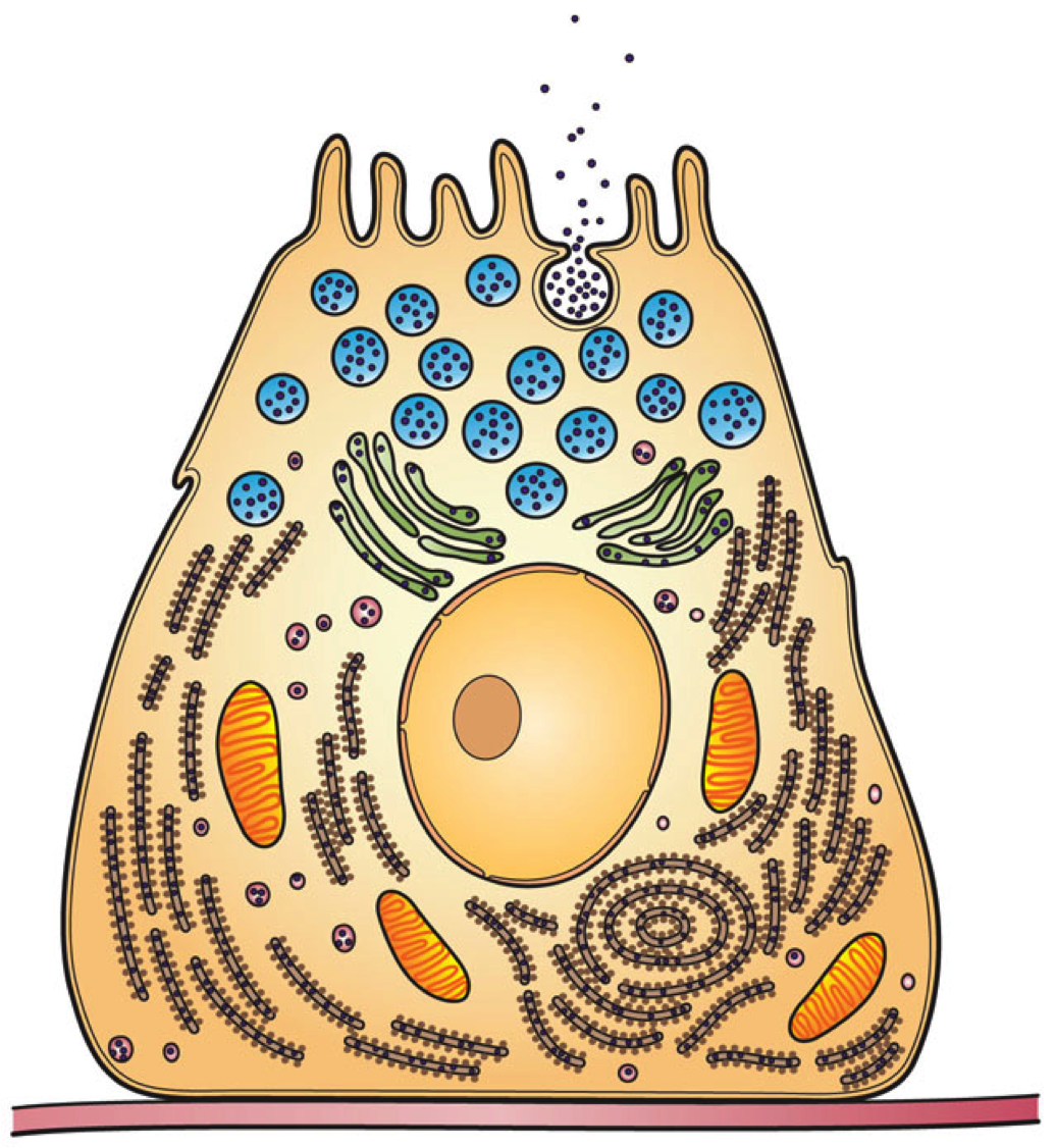
In eukaryotic cells, exoenzymes are manufactured like any other enzyme via protein synthesis, and are transported via the secretory pathway. After moving through the rough endoplasmic reticulum, they are processed through the Golgi apparatus, where they are packaged in vesicles and released out of the cell.[12] In humans, a majority of such exoenzymes can be found in the digestive system and are used for metabolic breakdown of macronutrients via hydrolysis. Breakdown of these nutrients allows for their incorporation into other metabolic pathways.[13]

Examples of exoenzymes as virulence factors

[edit]

Source:[3]

Microscopic view of necrotizing fasciitis as caused by Streptococcus pyogenes

Necrotizing enzymes

[edit]

Necrotizing enzymes destroy cells and tissue. One of the best known examples is an exoenzyme produced by Streptococcus pyogenes that causes necrotizing fasciitis in humans.

Coagulase

[edit]

By binding to prothrombin, coagulase facilitates clotting in a cell by ultimately converting fibrinogen to fibrin. Bacteria such as Staphylococcus aureus use the enzyme to form a layer of fibrin around their cell to protect against host defense mechanisms.

Fibrin layer formed by Staphylococcus aureus

Kinases

[edit]

The opposite of coagulase, kinases can dissolve clots. S. aureus can also produce staphylokinase, allowing them to dissolve the clots they form, to rapidly diffuse into the host at the correct time.[14]

Hyaluronidase

[edit]

Similar to collagenase, hyaluronidase enables a pathogen to penetrate deep into tissues. Bacteria such as Clostridium do so by using the enzyme to dissolve collagen and hyaluronic acid, the protein and saccharides, respectively, that hold tissues together.

Hemolysins

[edit]

Hemolysins target erythrocytes, a.k.a. red blood cells. Attacking and lysing these cells harms the host organism, and provides the microorganism, such as the fungus Candida albicans, with a source of iron from the lysed hemoglobin.[15] Organisms can either by alpha-hemolytic, beta-hemolytic, or gamma-hemolytic (non-hemolytic).

Examples of digestive exoenzymes

[edit]

Amylases

[edit]
Pancreatic alpha-amylase 1HNY

Amylases are a group of extracellular enzymes (glycoside hydrolases) that catalyze the hydrolysis of starch into maltose. These enzymes are grouped into three classes based on their amino acid sequences, mechanism of reaction, method of catalysis and their structure.[16] The different classes of amylases are α-amylases, β-amylases, and glucoamylases. The α-amylases hydrolyze starch by randomly cleaving the 1,4-a-D-glucosidic linkages between glucose units, β-amylases cleave non-reducing chain ends of components of starch such as amylose, and glucoamylases hydrolyze glucose molecules from the ends of amylose and amylopectin.[17] Amylases are critically important extracellular enzymes and are found in plants, animals, and microorganisms. In humans, amylases are secreted by the pancreas and salivary glands, with both sources of the enzyme required for complete starch hydrolysis.[18]

Lipoprotein lipase

[edit]

Lipoprotein lipase (LPL) is a type of digestive enzyme that helps regulate the uptake of triacylglycerols from chylomicrons and other low-density lipoproteins from fatty tissues in the body.[19] The exoenzymatic function allows it to break down the triacylglycerol into two free fatty acids and one molecule of monoacylglycerol. LPL can be found in endothelial cells in fatty tissues, such as adipose, cardiac, and muscle.[19] Lipoprotein lipase is downregulated by high levels of insulin,[20] and upregulated by high levels of glucagon and adrenaline.[19]

Pectinase

[edit]

Pectinases, also called pectolytic enzymes, are a class of exoenzymes that are involved in the breakdown of pectic substances, most notably pectin.[21] Pectinases can be classified into two different groups based on their action against the galacturonan backbone of pectin: de-esterifying and depolymerizing.[22] These exoenzymes can be found in both plants and microbial organisms including fungi and bacteria.[23] Pectinases are most often used to break down the pectic elements found in plants and plant-derived products.

Pepsin

[edit]

Discovered in 1836, pepsin was one of the first enzymes to be classified as an exoenzyme.[8] The enzyme is first made in the inactive form, pepsinogen by chief cells in the lining of the stomach.[24] With an impulse from the vagus nerve, pepsinogen is secreted into the stomach, where it mixes with hydrochloric acid to form pepsin.[25] Once active, pepsin works to break down proteins in foods such as dairy, meat, and eggs.[24] Pepsin works best at the pH of gastric acid, 1.5 to 2.5, and is deactivated when the acid is neutralized to a pH of 7.[24]

Trypsin

[edit]

Also one of the first exoenzymes to be discovered, trypsin was named in 1876, forty years after pepsin.[26] This enzyme is responsible for the breakdown of large globular proteins and its activity is specific to cleaving the C-terminal sides of arginine and lysine amino acid residues.[26] It is the derivative of trypsinogen, an inactive precursor that is produced in the pancreas.[27] When secreted into the small intestine, it mixes with enterokinase to form active trypsin. Due to its role in the small intestine, trypsin works at an optimal pH of 8.0.[28]

Bacterial assays

[edit]
Amylase test results
Lipase test results
Results of bacterial assays. Left:amylase bacterial assay on a starch medium. A indicates a positive result, D indicates a negative result. Right: lipase bacterial assay on an olive oil medium. 1 shows a positive result, 3 shows a negative result

The production of a particular digestive exoenzyme by a bacterial cell can be assessed using plate assays. Bacteria are streaked across the agar, and are left to incubate. The release of the enzyme into the surroundings of the cell cause the breakdown of the macromolecule on the plate. If a reaction does not occur, this means that the bacteria does not create an exoenzyme capable of interacting with the surroundings. If a reaction does occur, it becomes clear that the bacteria does possess an exoenzyme, and which macromolecule is hydrolyzed determines its identity.[2]

Amylase

[edit]

Amylase breaks down carbohydrates into mono- and disaccharides, so a starch agar must be used for this assay. Once the bacteria is streaked on the agar, the plate is flooded with iodine. Since iodine binds to starch but not its digested by-products, a clear area will appear where the amylase reaction has occurred. Bacillus subtilis is a bacterium that results in a positive assay as shown in the picture.[2]

Lipase

[edit]

Lipase assays are done using a lipid agar with a spirit blue dye. If the bacteria has lipase, a clear streak will form in the agar, and the dye will fill the gap, creating a dark blue halo around the cleared area. Staphylococcus epidermidis results in a positive lipase assay.[2]

Biotechnological and industrial applications

[edit]

Microbiological sources of exoenzymes including amylases, proteases, pectinases, lipases, xylanases, and cellulases are used for a wide range of biotechnological and industrial uses including biofuel generation, food production, paper manufacturing, detergents and textile production.[4] Optimizing the production of biofuels has been a focus of researchers in recent years and is centered around the use of microorganisms to convert biomass into ethanol. The enzymes that are of particular interest in ethanol production are cellobiohydrolase which solubilizes crystalline cellulose and xylanase that hydrolyzes xylan into xylose.[29] One model of biofuel production is the use of a mixed population of bacterial strains or a consortium that work to facilitate the breakdown of cellulose materials into ethanol by secreting exoenzymes such as cellulases and laccases.[29] In addition to the important role it plays in biofuel production, xylanase is utilized in a number of other industrial and biotechnology applications due to its ability to hydrolyze cellulose and hemicellulose. These applications include the breakdown of agricultural and forestry wastes, working as a feed additive to facilitate greater nutrient uptake by livestock, and as an ingredient in bread making to improve the rise and texture of the bread.[30]

Generic Biodiesel Reaction. Lipases can serve as a biocatalyst in this reaction

Lipases are one of the most used exoenzymes in biotechnology and industrial applications. Lipases make ideal enzymes for these applications because they are highly selective in their activity, they are readily produced and secreted by bacteria and fungi, their crystal structure is well characterized, they do not require cofactors for their enzymatic activity, and they do not catalyze side reactions.[31] The range of uses of lipases encompasses production of biopolymers, generation of cosmetics, use as a herbicide, and as an effective solvent.[31] However, perhaps the most well known use of lipases in this field is its use in the production of biodiesel fuel. In this role, lipases are used to convert vegetable oil to methyl- and other short-chain alcohol esters by a single transesterification reaction.[32]

Cellulases, hemicellulases and pectinases are different exoenzymes that are involved in a wide variety of biotechnological and industrial applications. In the food industry these exoenzymes are used in the production of fruit juices, fruit nectars, fruit purees and in the extraction of olive oil among many others.[33] The role these enzymes play in these food applications is to partially breakdown the plant cell walls and pectin. In addition to the role they play in food production, cellulases are used in the textile industry to remove excess dye from denim, soften cotton fabrics, and restore the color brightness of cotton fabrics.[33] Cellulases and hemicellulases (including xylanases) are also used in the paper and pulp industry to de-ink recycled fibers, modify coarse mechanical pulp, and for the partial or complete hydrolysis of pulp fibers.[33] Cellulases and hemicellulases are used in these industrial applications due to their ability to hydrolyze the cellulose and hemicellulose components found in these materials.

Bioremediation applications

[edit]
Water pollution from runoff of soil and fertilizer

Bioremediation is a process in which pollutants or contaminants in the environment are removed through the use of biological organisms or their products. The removal of these often hazardous pollutants is mostly carried out by naturally occurring or purposely introduced microorganisms that are capable of breaking down or absorbing the desired pollutant. The types of pollutants that are often the targets of bioremediation strategies are petroleum products (including oil and solvents) and pesticides.[34] In addition to the microorganisms ability to digest and absorb the pollutants, their secreted exoenzymes play an important role in many bioremediation strategies.[35]

Fungi have been shown to be viable organisms to conduct bioremediation and have been used to aid in the decontamination of a number of pollutants including polycyclic aromatic hydrocarbons (PAHs), pesticides, synthetic dyes, chlorophenols, explosives, crude oil, and many others.[36] While fungi can breakdown many of these contaminants intracellularly, they also secrete numerous oxidative exoenzymes that work extracellularly. One critical aspect of fungi in regards to bioremediation is that they secrete these oxidative exoenzymes from their ever elongating hyphal tips.[36] Laccases are an important oxidative enzyme that fungi secrete and use oxygen to oxidize many pollutants. Some of the pollutants that laccases have been used to treat include dye-containing effluents from the textile industry, wastewater pollutants (chlorophenols, PAHs, etc.), and sulfur-containing compounds from coal processing.[36]

Exocytic vesicles move along actin microfilaments toward the fungal hyphal tip where they release their contents including exoenzymes

Bacteria are also a viable source of exoenzymes capable of facilitating the bioremediation of the environment. There are many examples of the use of bacteria for this purpose and their exoenzymes encompass many different classes of bacterial enzymes. Of particular interest in this field are bacterial hydrolases as they have an intrinsic low substrate specificity and can be used for numerous pollutants including solid wastes.[37] Plastic wastes including polyurethanes are particularly hard to degrade, but an exoenzyme has been identified in a Gram-negative bacterium, Comamonas acidovorans, that was capable of degrading polyurethane waste in the environment.[37] Cell-free use of microbial exoenzymes as agents of bioremediation is also possible although their activity is often not as robust and introducing the enzymes into certain environments such as soil has been challenging.[37] In addition to terrestrial based microorganisms, marine based bacteria and their exoenzymes show potential as candidates in the field of bioremediation. Marine based bacteria have been utilized in the removal of heavy metals, petroleum/diesel degradation and in the removal of polyaromatic hydrocarbons among others.[38]

References

[edit]
Revisions and contributorsEdit on WikipediaRead on Wikipedia
from Grokipedia
Exoenzymes are extracellular enzymes secreted by microorganisms, primarily and fungi, that function outside the cell to break down complex organic substrates into forms that can be assimilated, often through of high-molecular-weight polymers such as , proteins, and into simpler monomers. These enzymes enable microbes to access in environments where substrates are too large or insoluble to enter cells directly, playing a pivotal role in microbial , cycling, and processes in soils, aquatic systems, and host tissues. Unlike endoenzymes, which operate intracellularly, exoenzymes are released into the periplasmic space or external milieu, often via specialized systems in (e.g., Type I–VI systems) or through hyphal tips in fungi. In , exoenzymes are essential for both saprophytic and pathogenic lifestyles, where hydrolytic types aid in breaking down and virulence factors contribute to host tissue and immune evasion. Beyond their biological roles, exoenzymes have applications in and . Their activity is influenced by environmental factors like , temperature, and substrate availability, with kinetic properties varying across depths to optimize microbial fitness.

Fundamentals

Definition

Exoenzymes are extracellular enzymes secreted by cells, primarily microorganisms such as and fungi, but also by plants and animals, that catalyze the breakdown of large, insoluble substrates in the external environment into smaller, diffusible molecules that can be taken up by the cell for and . These enzymes enable organisms to access complex that cannot cross cell membranes directly, playing a crucial role in . Key characteristics of exoenzymes include their localization and outside the cell, where they typically operate in the periplasmic space of or the broader external environment, performing hydrolytic or oxidative reactions on polymeric substrates like proteins, , and . Unlike endoenzymes, which function intracellularly within the cell that produces them, exoenzymes are released to act remotely on substrates. Exoenzymes occur widely across diverse organisms, with high prevalence in bacteria such as and species, fungi that excrete them at hyphal tips to degrade substrates, and eukaryotic cells including pancreatic acinar cells in animals, which secrete them into the digestive tract. Common types include amylases, which target , and proteases, which break down proteins, illustrating their role in processing essential biomolecules.

Classification

Exoenzymes are classified functionally according to the standard Enzyme Commission (EC) system, which categorizes based on the reactions they catalyze, with many exoenzymes falling into the class (EC 3) due to their role in breaking down complex polymers. include glycoside hydrolases (EC 3.2), such as cellulases that degrade (e.g., EC 3.2.1.4), and peptidases (EC 3.4), such as elastases that cleave peptide bonds (e.g., EC 3.4.21.37). Other structural classes represented among exoenzymes are oxidoreductases (EC 1), exemplified by laccases that oxidize (e.g., EC 1.10.3.2 in fungal systems), and lyases (EC 4), such as pectate lyases that cleave pectate (e.g., EC 4.2.2.2 in bacterial pathogens). Functionally, exoenzymes are often grouped by their substrate specificity, reflecting their adaptation to extracellular degradation of insoluble macromolecules. Carbohydrases target , including cellulases that hydrolyze β-1,4-glycosidic bonds in and amylases that break down . Proteases degrade proteins, with examples like elastases that target in host tissues and collagenases such as ColH that disrupt matrices. Lipases hydrolyze lipids, such as LipA in species that cleaves esters. In comparison to endoenzymes, which operate intracellularly on soluble substrates to support metabolic processes, exoenzymes function extracellularly to depolymerize insoluble substrates into absorbable monomers, with no primary overlap in their operational locations. Exoenzymes can be further subdivided by localization and oligomeric state. Periplasmic subtypes, common in , reside in the periplasmic space between inner and outer , such as certain pectate lyase isoenzymes (e.g., PLb in Erwinia carotovora). Truly extracellular subtypes are secreted beyond the outer into the environment, often via type II or type V systems, as seen in fungal cellulases. Regarding structure, many exoenzymes are monomeric, but some form multimeric complexes, such as ExoS in , which aggregates into structures exceeding 300 kDa facilitated by a localization domain.

Historical Development

Early Observations

The foundational observations leading to the understanding of exoenzymes emerged in the late 19th century through studies on microbial and . Louis Pasteur's experiments in the 1850s and 1860s on alcoholic and established the role of living microorganisms in these processes, laying groundwork for later enzymology by challenging vitalistic views. These findings highlighted microbial nutrient acquisition but did not identify extracellular agents. Eduard Buchner's landmark 1897 work on yeast extracts demonstrated cell-free fermentation, where lysed yeast cells released capable of converting sugar to alcohol and without intact living cells. This established enzymes as non-living catalysts functioning outside cells, advancing the general enzyme concept, though the enzymes were intracellular extracts rather than secreted products. Key early experiments in the early 20th century focused on isolating and characterizing extracellular enzymes from microbes. A notable example is Alexander Fleming's 1922 discovery of , an antibacterial exoenzyme secreted by bacteria and found in nasal secretions, which lysed bacterial cell walls and confirmed active secretion by intact cells. In 1894, Japanese chemist Jokichi Takamine isolated and patented takadiastase, an exoenzyme secreted by the fungus during koji fermentation, marking the first commercial production of a microbial exoenzyme for in industrial and digestive applications. Building on this, Japanese researchers in the early 1900s identified bacterial amylases from soil microbes, such as species, through cultivation and activity assays on substrates, demonstrating the role of these exoenzymes in degrading polymers in natural environments like soil. These studies emphasized the diversity of microbial sources and the practical utility of exoenzymes in , with assays showing enzymatic activity in cell-free culture filtrates. Early observations sometimes attributed extracellular activity to leakage from damaged cells, but studies in the early , including viability assays, supported active by intact microbes.

Key Milestones

In the mid-, significant progress was made in the purification and early of exoenzymes, exemplified by Carlsberg, a bacterial secreted by (now Bacillus subtilis subsp. licheniformis), isolated and crystallized around 1948–1957 at the Carlsberg Laboratory by researchers including Martin Ottesen. This achievement advanced understanding of the biochemical properties of extracellular proteases, enabling studies on their stability and catalytic mechanisms; high-resolution structures followed in the . During this era, research also highlighted the role of exoenzymes in microbial , including their production during sporulation in species, which coincided with and contributed to industrial interest in these enzymes for bioprocessing applications. The 1970s and 1980s ushered in the revolution, transforming exoenzyme production through . A landmark example was the cloning and of the Rhizomucor miehei lipase gene in 1989, which allowed high-yield production in and facilitated its commercial use in food and detergent industries. By the 1990s, this technology extended to other exoenzymes, such as fungal cellulases and bacterial amylases, enabling scalable manufacturing and reducing reliance on native microbial . Concurrently, studies in the mid-1980s elucidated the contributions of staphylococcal exoenzymes, including proteases and nucleases, to bacterial by demonstrating their ability to degrade host tissues and evade immune responses in infections. Entering the 2000s, advanced exoenzyme research through high-resolution techniques like , providing insights into active sites and substrate interactions. For instance, in the 2010s, the of bacterial from Streptomyces koganeiensis, resolved at 1.55 Å in 2016, revealed unique catalytic domains that informed inhibitor design for therapeutic applications. The advent of CRISPR-Cas9 post-2012 further revolutionized exoenzyme engineering, enabling precise genome edits in microbial hosts to enhance secretion yields and modify specificity for biotechnological uses, such as improved cellulases for biofuel production. In the 2020s, metagenomic approaches have uncovered novel exoenzymes from uncultured microbes, expanding the repertoire available for environmental and industrial applications. High-throughput sequencing of and microbiomes has identified diverse extracellular hydrolases, including lignin-degrading enzymes from uncultured , which play roles in carbon cycling and offer potential for mitigating through enhanced breakdown. These discoveries, driven by advances in bioinformatics and functional screening, underscore ' impact on revealing exoenzyme diversity in complex ecosystems, with implications for sustainable strategies.

Mechanism and Function

Secretion Process

In , exoenzymes are primarily secreted via the Type II secretion system (T2SS), also known as the general secretory pathway (GSP), which exports folded proteins from the across the outer membrane into the extracellular environment. This two-step process begins with translocation across the inner membrane in an unfolded state using the Sec pathway or, less commonly, the (SRP) pathway, guided by cleavable N-terminal signal peptides on the preproteins. In the , the proteins fold with assistance from chaperones before T2SS components—a pseudopilus, inner membrane platform, and outer membrane secretin channel—facilitate their export, powered by a cytoplasmic . Examples include proteases like LasB in and pullulanase in . In , which lack an outer , exoenzymes are secreted directly into the extracellular environment primarily via the Sec or twin-arginine translocation (Tat) pathways. The Sec pathway translocates unfolded proteins across the cytoplasmic in a post-translational manner, dependent on signal peptides and chaperones like PrsA to prevent aggregation in the wall matrix. The Tat pathway, used for folded proteins, ensures cofactor assembly before export and is crucial for enzymes like those in , such as protease. Secretion is often regulated by environmental signals, with the thick layer posing a diffusion barrier that specialized mechanisms help overcome. Another key bacterial pathway is the Type V secretion system, exemplified by autotransporters, which enables autonomous export of exoenzymes without additional dedicated machinery beyond the Sec system. Autotransporter proteins feature an N-terminal that directs Sec-dependent translocation across the inner membrane to the , where periplasmic chaperones such as Skp, DegP, and SurA maintain the polypeptide in a secretion-competent, unfolded state, often with bond formation aided by DsbA. The C-terminal β-barrel domain then inserts into the outer membrane as a pore (typically 12-14 antiparallel strands, sometimes oligomeric), translocating the N-terminal passenger domain—the functional exoenzyme—extracellularly, where it folds into its active form. Relevant examples include the EatA in enterotoxigenic Escherichia coli and IgA1 protease in . In microbial eukaryotes such as fungi, exoenzyme follows the classical (ER)-Golgi pathway, involving synthesis, modification, and regulated release, as seen in filamentous fungi like where hydrolytic enzymes such as glucoamylase are produced. Translation occurs on rough ER ribosomes, with hydrophobic N-terminal signal peptides directing nascent polypeptides into the ER lumen for folding and initial ; inactive pro-enzymes are then transported to the Golgi apparatus via vesicles. In the Golgi, further posttranslational modifications, including , occur before concentration and packaging into secretory vesicles at the trans-Golgi network. These vesicles are directed to the hyphal tips or cell surface for , often in a polarized manner to facilitate acquisition in substrates. The secretion of exoenzymes is tightly regulated, often induced by the presence of substrates or environmental cues, with quorum sensing (QS) playing a central role in bacteria to coordinate population-level responses. In species like Pseudomonas aeruginosa, QS via N-acylhomoserine lactone signals (e.g., through LasR/LasI and RhlR/RhlI systems) upregulates expression of T2SS-secreted exoenzymes such as elastases LasA and LasB when substrates like elastin are detected or at high cell densities, ensuring efficient resource utilization. Similarly, in Vibrio cholerae, QS-regulated HapR activates T6SS components for effector secretion under nutrient-limiting conditions induced by chitin substrates. These processes are energy-intensive, relying on ATP hydrolysis for translocation—such as by the T2SS cytoplasmic ATPase or ABC transporters in Type I systems—imposing metabolic costs that QS helps balance by timing secretion to favorable conditions. Evolutionarily, bacterial exoenzyme machinery has co-evolved with the s encoding the enzymes themselves, often within operons or clusters that promote coordinated expression and horizontal transfer. In Type V systems, autotransporter clusters integrate the passenger (exoenzyme) and translocator domains, with evidence of modular through co-option of outer β-barrel proteins, facilitating adaptation to diverse niches like . For T2SS, genomic analyses reveal conserved clusters across , where apparatus s (e.g., encoding secretins and pseudopilins) have co-evolved with exoenzyme loci, enhancing fitness in environments requiring extracellular , as seen in the diversification of protease-secreting strains. This co-evolution underscores the selective pressure for efficient export mechanisms in nutrient-scarce or host-associated habitats.

Catalytic Activity

Exoenzymes catalyze the extracellular of complex into soluble monomers or oligomers, enabling acquisition by microorganisms and other organisms. This process typically involves the enzyme binding to the substrate's surface or integrating into its structure, where the positions water molecules for nucleophilic attack on specific bonds, such as glycosidic, , or linkages. For example, amylases facilitate the of α-1,4-glycosidic bonds in , cleaving the as follows: (14)α-D-glucan+H2Oglucose oligomers (e.g., maltose)(1 \to 4)-\alpha\text{-D-glucan} + \text{H}_2\text{O} \to \text{glucose oligomers (e.g., maltose)} This reaction proceeds via a double-displacement mechanism in many glycoside hydrolases, involving a covalent enzyme-substrate intermediate stabilized by acidic residues in the active site. The kinetics of exoenzyme catalysis deviate from classical Michaelis-Menten models due to the insolubility of many substrates, requiring adaptations that incorporate surface-binding steps. Enzymes adsorb onto the substrate via carbohydrate-binding modules, forming an enzyme-substrate complex where the catalytic domain accesses exposed bonds; the effective KmK_m reflects both binding affinity and substrate accessibility, often modeled as Km=(k1+k2)/k1K_m = (k_{-1} + k_2)/k_1, with k1k_1 as the association rate to surface sites. For cellulases, this surface erosion model predicts that degradation rates depend on the density of attack sites (Γ\Gamma), linking molar substrate concentration to mass load and enabling steady-state hydrolysis even as the substrate diminishes. Such models highlight how non-productive binding can limit turnover, particularly for heterogeneous substrates like lignocellulose. Environmental factors profoundly influence exoenzyme activity, with optimal and varying by enzyme origin to match extracellular conditions. Acidic environments favor enzymes like fungal aspartic proteases (e.g., from ), which exhibit peak activity at 2–4 due to of catalytic aspartates that enhance nucleophilic attack on bonds. In contrast, bacterial often operate optimally at neutral around 8, where serine-histidine-aspartate triads maintain hydrolytic efficiency on substrates. optima typically range from 20–40°C for and microbial exoenzymes, with activity increasing via enhanced molecular collisions up to the point of denaturation; beyond this, Q_{10} values of 1.4–1.8 indicate moderate sensitivity. Product inhibition commonly arises as oligomers or monomers competitively bind the , elevating apparent KmK_m and reducing VmaxV_{max}, a feedback mechanism that regulates extracellular degradation in nutrient-limited settings. In multi-enzyme systems, exoenzymes from bacterial consortia exhibit synergistic during lignocellulose degradation, where complementary activities amplify overall rates. Endohydrolytic enzymes create nicks in cellulose chains, exposing ends for exohydrolases like cellobiohydrolases to release , which β-glucosidases then convert to glucose, preventing product inhibition and boosting efficiency by up to 18-fold in co-cultures such as and Sphingobacterium multivorum. This division of labor, driven by metabolic cross-feeding, ensures sequential bond cleavage and minimizes unproductive adsorption, optimizing polymer breakdown in complex matrices.

Biological Roles

In Nutrient Acquisition and Digestion

Exoenzymes play a crucial role in microbial acquisition by degrading complex environmental polymers into simpler monomers that can be transported and utilized for carbon and nitrogen sources. In and fungi, extracellular chitinases hydrolyze , a major component of fungal cell walls and exoskeletons, enabling microbes to access nitrogen-rich units under nutrient-scarce conditions. Similarly, pectinases secreted by and -associated microbes break down in plant cell walls, releasing galacturonic acid and other sugars as carbon sources, which supports microbial growth in terrestrial environments. These degradative processes are essential for heterotrophic microbes that lack the ability to synthesize all required nutrients de novo. Symbiotic gut further augment host nutrient acquisition through the production of exoenzymes that target indigestible dietary components. These microbes, primarily in the colon, secrete hydrolases and polysaccharide lyases to ferment complex plant like and , generating and monosaccharides that the host can absorb for energy. This mutualistic interaction expands the host's nutritional niche, particularly for fiber-rich diets, by converting otherwise inaccessible substrates into bioavailable forms. Biofilm formation enhances the efficiency of exoenzyme-mediated acquisition by increasing the effective surface area for enzymatic reactions and substrate contact. In microbial communities embedded in , exoenzymes are concentrated within the , promoting localized degradation of polymers and reducing limitations, which can significantly improve scavenging compared to planktonic cells. Under limitation, such as , microbes upregulate exoenzyme as an adaptive response; for instance, marine increase activity during carbon or deprivation to mine refractory , sustaining minimal metabolic functions. Ecologically, microbial exoenzymes drive carbon cycling in soils and oceans by remineralizing recalcitrant . In soils, they hydrolyze lignocellulosic inputs from , releasing bioavailable carbon that fuels microbial respiration and supports turnover, with enzymatic being a major driver of terrestrial carbon . In oceanic environments, bacterioplankton exoenzymes process from exudates and sinking particles, facilitating rapid recycling in the surface and influencing the biological pump's efficiency.

In Pathogenesis and Virulence

Exoenzymes contribute to bacterial by enabling tissue invasion through the action of spreading factors that degrade components of the host extracellular matrix, such as , thereby reducing tissue viscosity and facilitating dissemination during infection. These enzymes also support delivery by hydrolyzing surrounding host barriers, allowing other factors to penetrate deeper into tissues and exacerbate damage. Furthermore, exoenzymes promote nutrient scavenging from host tissues via the breakdown of complex biomolecules like proteins and nucleic acids, which not only supplies the with resources but simultaneously inflicts destructive effects on host structures, undermining tissue integrity. In host-pathogen dynamics, exoenzymes function as secreted effectors that modulate interactions in both Gram-positive and Gram-negative bacteria, often exported through dedicated secretion systems that enhance their extracellular activity. In Gram-negative pathogens, type III secretion systems deliver enzymatic effectors directly to host cells, disrupting cellular functions and aiding infection progression, while Gram-positive bacteria rely on general secretion pathways to release these enzymes into the extracellular milieu. Exoenzymes further contribute to chronic infections by participating in biofilm dynamics, where they degrade polymeric matrix components to promote biofilm maturation, dispersal, and persistence, enabling pathogens to evade host defenses and establish long-term colonization in tissues. The evolutionary adaptation of exoenzymes in pathogens is driven by , particularly in , where genes encoding these enzymes are frequently acquired via like bacteriophages and plasmids, allowing rapid dissemination of capabilities across bacterial populations and enhancing adaptability to host environments. Clinically, exoenzymes confer resistance through direct degradation of antimicrobial drugs; for instance, secreted beta-lactamases hydrolyze the beta-lactam ring in penicillins, rendering these agents ineffective and promoting the survival of resistant strains in infected hosts.

Examples of Exoenzymes

Digestive Exoenzymes

Digestive exoenzymes are extracellular hydrolases secreted by organisms to break down complex dietary macromolecules into simpler, absorbable forms during nutrient acquisition. In animals, these enzymes primarily target carbohydrates, proteins, and in the alimentary , facilitating efficient . Microbial digestive exoenzymes, often produced by fungi and in symbiotic or fermentative contexts, similarly degrade plant-based substrates, enabling breakdown of structural . Amylases represent a key class of digestive exoenzymes that hydrolyze , a major dietary , into and other oligosaccharides. In humans, salivary α-amylase, secreted by the parotid glands, initiates in the oral cavity by cleaving internal α-1,4-glycosidic bonds, producing , , and dextrins. This process begins the conversion of granules into fermentable sugars, with optimal activity at neutral pH around 6.7. Pancreatic α-amylase, released into the , continues this under slightly alkaline conditions (pH 6.7–7.0), further degrading the partially digested products into and limit dextrins for subsequent action by brush-border . Microbial sources, such as the fungus , produce α-amylase that performs analogous , breaking down to in fermentative processes like production, with the exhibiting up to 60°C. Proteases, another essential group of digestive exoenzymes, target peptide bonds in dietary proteins to yield and peptides. , the primary gastric in vertebrates, is secreted as the inactive pepsinogen by chief cells in the and undergoes autocatalytic activation at low pH (1.5–2.5) due to , which cleaves the proenzyme to form the active form. Active preferentially hydrolyzes bonds involving aromatic or hydrophobic , initiating protein denaturation and partial digestion in the acidic environment. In the , serves as a major , secreted as from the and activated by enterokinase; it specifically cleaves peptide bonds on the carboxyl side of and residues, generating smaller peptides for further degradation by other peptidases. Lipases hydrolyze ester bonds in triglycerides, releasing fatty acids and monoglycerides for absorption. Pancreatic lipase, secreted by acinar cells into the , acts on emulsified dietary fats, where salts from the liver reduce size, increasing the surface area for enzymatic access and enabling efficient at the oil-water interface. This enzyme requires colipase for optimal activity in the presence of acids, producing free fatty acids and 2-monoacylglycerols that form micelles for intestinal uptake. , anchored on the endothelial surfaces of blood vessels in adipose and muscle tissues, hydrolyzes triglycerides in circulating chylomicrons and very-low-density lipoproteins (VLDL), liberating non-esterified fatty acids for local storage or energy use, with activity modulated by as a cofactor. In microbial digestion of plant material, pectinases degrade , a complex heteropolysaccharide in primary cell walls, facilitating access to and . Produced by gut-associated and fungi in herbivores or ruminants, these exoenzymes, including polygalacturonases and pectin lyases, cleave α-1,4-galactosiduronic linkages, solubilizing the gel-like pectin matrix and enabling comprehensive breakdown of for into short-chain fatty acids.

Virulence Exoenzymes

Virulence exoenzymes are secreted by to facilitate host tissue invasion, immune evasion, and nutrient acquisition during . These enzymes often degrade host barriers or modulate physiological processes to promote bacterial survival and dissemination. In particular, certain exoenzymes contribute to the destructive of infections by enabling tissue necrosis, clot formation, proteolytic spread, breakdown, and for iron release. DNases and RNases from (group A Streptococcus) act as necrotizing enzymes by degrading nucleic acids from host cells and neutrophils, which disrupts (NETs) and liquefies to aid bacterial spread in tissues. This enzymatic activity contributes to the rapid tissue necrosis observed in severe infections like , where multiple DNases such as Sda1 and Spd1 are upregulated to enhance . Coagulase from is a key exoenzyme that non-proteolytically activates host prothrombin, leading to the conversion of fibrinogen to and the formation of protective barriers around bacterial . This shield encapsulates staphylococcal communities, shielding them from and promoting persistent in skin and soft tissues. S. aureus produces two coagulases—staphylocoagulase (Coa) and von Willebrand factor-binding protein (vWbp)—that synergistically generate distinct networks, both essential for development and bacterial survival . Streptokinase, a (kinase) secreted by , binds host to form a complex that generates , a that degrades clots and components. This activity facilitates bacterial dissemination from local infection sites into the bloodstream, enhancing systemic spread and invasion in models of infection. Streptokinase-mediated activation is a critical mechanism, as mutants lacking this show reduced tissue invasion and bloodstream persistence compared to wild-type strains. (mu-toxin) from hydrolyzes , a major component of the in connective tissues, thereby increasing tissue permeability and enabling bacterial invasion and spread. This exoenzyme is an established in and other clostridial infections, where it degrades host barriers to promote deeper tissue penetration and toxin dissemination. Recent studies confirm that C. perfringens hyaluronate lyase variants, such as NagH, function as intrinsic hyaluronan-degrading enzymes essential for invasion in opportunistic infections. Hemolysins, exemplified by α-hemolysin (HlyA) from uropathogenic Escherichia coli, are pore-forming exoenzymes that lyse red blood cells by inserting transmembrane pores, releasing hemoglobin as a source of iron for bacterial growth during infection. This lytic activity supports iron acquisition in iron-limited host environments, such as the urinary tract or bloodstream, and enhances overall virulence by damaging host epithelia and promoting bacterial persistence. HlyA mutants exhibit attenuated virulence in murine models of urinary tract infection, underscoring its role as an alternative iron-uptake mechanism alongside siderophores like aerobactin.

Detection and Assays

Methods for

Laboratory techniques for detecting and quantifying activity, particularly in bacterial samples, rely on the enzyme's ability to hydrolyze into reducing sugars, allowing for qualitative and quantitative assessments. These methods are essential for screening microbial isolates producing exoenzymes like , which functions in nutrient acquisition by breaking down complex carbohydrates. Plate assays provide a simple qualitative approach for initial screening of amylase-producing . In the starch-agar method, bacterial colonies are grown on plates supplemented with soluble as the substrate; after incubation, the plates are flooded with iodine solution, which stains intact blue-black, while zones of clearing around colonies indicate hydrolysis by secreted . This technique is widely used for isolating amylase-positive strains from environmental samples, with the diameter of the clear zone correlating to enzyme activity levels. For quantitative measurement, spectrophotometric assays such as the dinitrosalicylic acid (DNS) method are standard. In this procedure, hydrolyzes to produce reducing sugars like , which react with DNS reagent to form a colored product measured at 540 nm ; one unit of activity is typically defined as the amount of enzyme releasing 1 μmol of reducing sugar per minute under specified conditions. This method offers high sensitivity and is suitable for crude extracts from bacterial cultures, though it requires a standard curve with for accurate quantification. Commercial kits enhance throughput and specificity for detection in settings. Fluorometric kits, such as the EnzChek Ultra Assay, employ chromogenic or fluorogenic substrates like DQ-starch, where relieves and produces measurable , enabling detection of low activity levels (down to 10 mU/mL) in 96-well formats for of bacterial supernatants. Chromogenic kits, like the EnzyChrom α- Assay, use similar principles but with colorimetric readouts at 570 nm, providing linear detection ranges from 0.3 to 50 U/L. To ensure specificity, assays differentiate from endoglucanases by substrate selectivity; targets α-1,4-glycosidic bonds in , while endoglucanases act on β-1,4 linkages in derivatives like carboxymethylcellulose, allowing parallel plate tests with distinct substrates to confirm activity without cross-reactivity.

Methods for

Assays for exoenzymes, which hydrolyze into free fatty acids and , require careful handling of substrates due to their insolubility in water. These methods are essential for detecting and quantifying extracellular lipase production in and fungi, often in the context of nutrient acquisition or . Common approaches include plate-based qualitative tests and quantitative titrimetric or fluorometric techniques that measure products. The is a widely used qualitative plate method for screening bacterial lipase activity. In this test, , a short-chain , is incorporated into an medium, creating an opaque suspension. Bacterial colonies producing exolipase hydrolyze the , releasing and forming a clear halo around the due to the breakdown of the emulsified oil droplets. The appearance of these halos after incubation indicates positive lipase activity, with halo size correlating to enzyme production levels. This is simple, cost-effective, and suitable for initial screening of microbial isolates. Titrimetric methods, particularly , provide a precise quantitative measure of activity by monitoring the release of free fatty acids from substrates. The principle relies on the proton-liberating of (e.g., triolein) at a constant , where an automatic titrator adds base (such as NaOH) to neutralize the acids produced, and the rate of base addition directly reflects activity. The setup involves emulsifying the substrate in a reaction vessel maintained at optimal (typically 7-8) and , with continuous stirring to ensure interfacial contact. This method is highly sensitive, applicable to crude preparations, and distinguishes initial products, making it a reference standard for specificity studies. Rhodamine-based fluorescence assays offer sensitive detection of , particularly in emulsified substrates, and can be adapted for both plate and liquid formats. In plate versions, media containing triglycerides (e.g., trioleoylglycerol) and are inoculated with ; releases fatty acids that interact with the , producing orange fluorescent halos visible under UV (excitation at ~350 nm), allowing quantification via halo diameter (linear from 1-30 nkat). Liquid-state high-throughput variants use in emulsions of natural substrates like ; action frees fatty acids, enhancing intensity, which is measured in multi-well plates for rapid screening. These assays are automatable, stable for months at 4°C, and effective across and ranges, though they require UV equipment. Lipase assays face challenges related to substrate emulsification and potential inhibition. Lipases act at lipid-water interfaces, necessitating stable emulsions (e.g., via mechanical stirring or emulsifiers) to provide sufficient surface area; variations in droplet size (e.g., 5-37% efficiency) or stability can alter results, with reducing access and slowing activity. salts, often present in physiological or assay mimics, can inhibit by displacing the enzyme from interfaces or forming inhibitory micelles, particularly without colipase, complicating quantification in complex samples.

Applications

Biotechnological Uses

Exoenzymes play a pivotal role in the , where microbial-derived amylases are extensively utilized for liquefaction in and processes. Alpha-amylases, secreted extracellularly by such as species, hydrolyze into fermentable sugars, improving handling, volume, and filtration efficiency. Similarly, pectinases facilitate juice clarification by breaking down in fruit cell walls, enhancing extraction yields and reducing viscosity in products like apple and juices. These enzymes, often from , enable clearer, more stable beverages while minimizing the need for chemical treatments. In pharmaceutical and related industrial applications, exoenzymes such as and offer targeted functionalities. , a from , is engineered for use in detergents, where it enhances stain removal by hydrolyzing protein-based soils under alkaline conditions, comprising a significant portion of commercial formulations. , an exoglycosidase, serves as a spreading agent in subcutaneous injections, depolymerizing in the to improve the dispersion and absorption of co-administered drugs like anesthetics or antibiotics. Genetic engineering has advanced exoenzyme production through overexpression in heterologous hosts like Pichia pastoris, a methylotrophic favored for its high-density and secretion capabilities. This system enables scalable production of such as lipases and proteases by integrating strong promoters like AOX1, yielding gram-per-liter titers for commercial applications. techniques, prominent in the , have further optimized ; for instance, error-prone PCR on Bacillus subtilis lipases introduced mutations that increased half-life at 60°C by over 10-fold, facilitating use in high-temperature processes like synthesis. The biotechnological significance of exoenzymes is underscored by their dominance in the global industrial enzyme market, estimated at USD 8.76 billion as of 2025 with a of approximately 6.8% through 2035, driven by demand in food, pharmaceuticals, and detergents. Hydrolase-class exoenzymes, including amylases, proteases, and lipases, account for nearly 75% of industrial enzyme production, reflecting their versatility and economic impact.

Bioremediation Uses

Exoenzymes, also known as extracellular enzymes, play a pivotal role in by catalyzing the breakdown of environmental pollutants outside microbial cells, transforming complex xenobiotics into less toxic or mineralizable forms. These enzymes, primarily produced by and fungi, facilitate the degradation of organic contaminants such as polycyclic aromatic hydrocarbons (PAHs), synthetic dyes, pesticides, and pharmaceuticals in , , and . Their extracellular nature allows them to act directly on insoluble substrates in harsh environments, overcoming limitations of intracellular processes and enabling applications in cell-free systems for enhanced stability and targeted remediation. In bacterial systems, exoenzymes like monooxygenases and laccases are key for oxidizing persistent pollutants. For instance, enzymes from Pseudomonas putida and Bacillus megaterium hydroxylate PAHs such as and fluoranthene, converting them into or quinones that are further metabolized in contaminated soils. Laccases from Streptomyces cyaneus and Pseudomonas putida oxidize phenolic compounds and synthetic dyes like Reactive Black 5, with bacterial laccases, such as from Bacillus sp., achieving efficiencies exceeding 90% for (BPA) breakdown to less harmful metabolites. Hydrolases, including lipases and proteases from various soil bacteria, hydrolyze residues like organophosphates, contributing to the remediation of agricultural runoff. White-rot fungi represent a major source of potent exoenzymes for bioremediation, particularly ligninolytic enzymes that mimic natural wood decay to target lignin-like pollutants. Laccases, manganese peroxidases (MnPs), and lignin peroxidases (LiPs) from species such as Phanerochaete chrysosporium and Trametes versicolor degrade PAHs like pyrene and phenanthrene, with P. chrysosporium achieving 99.55% phenanthrene removal in 60 days through oxidative cleavage. These enzymes also decolorize azo dyes such as Congo Red (up to 97%) and Remazol Brilliant Blue R (up to 96%), with some dyes reaching nearly 100% efficiency in fungal cultures supplemented with mediators. For emerging contaminants, versatile peroxidases (VPs) and dye-decolorizing peroxidases (DyPs) from white-rot fungi such as Trametes versicolor (94% diclofenac removal in 1 hour) and Pleurotus ostreatus and Ganoderma lucidum remove perfluoroalkyl substances (PFAS) such as PFOA (50% in 157 days), reducing toxicity in polluted ecosystems. Applications of these exoenzymes extend to immobilized systems for scalable , where enzymes from white-rot fungi are fixed on supports to treat industrial effluents, enhancing reusability and activity in high-toxicity settings. For example, immobilization from Trametes versicolor has demonstrated sustained degradation of 2,4-dichlorophenol (2,4-DCP) in soils in combined enzymatic-microbial setups. Overall, exoenzyme-based strategies offer eco-friendly alternatives to chemical treatments, though challenges like enzyme stability under field conditions persist, driving ongoing into bioengineering for improved performance. As of 2025, advancements include CRISPR-modified for enhanced PFAS degradation in wastewater.

References

Add your contribution
Related Hubs
User Avatar
No comments yet.