Hubbry Logo
BronchusBronchusMain
Open search
Bronchus
Community hub
Bronchus
logo
8 pages, 0 posts
0 subscribers
Be the first to start a discussion here.
Bronchus
Bronchus
from Wikipedia
Bronchus
The bronchi are conducting passages for air into the lungs.
The bronchi form part of the lower respiratory tract
Details
SystemRespiratory system
ArteryBronchial artery
VeinBronchial vein
NervePulmonary branches of vagus nerve
Identifiers
Latinbronchus
Greekβρόγχος
MeSHD001980
TA98A06.4.01.001
A06.3.01.008
TA23226
FMA7409
Anatomical terminology

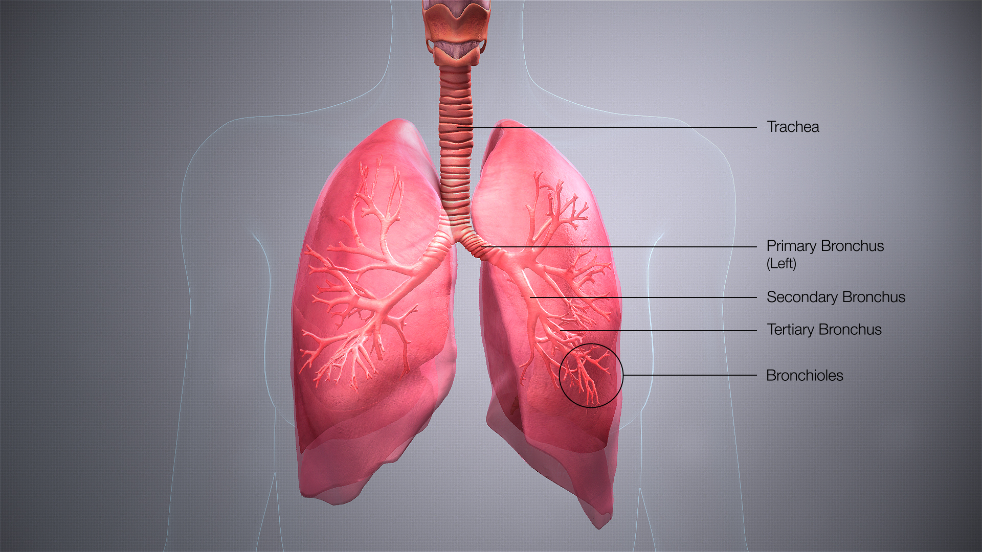
A bronchus (/ˈbrɒŋkəs/ BRONG-kəs; pl.: bronchi, /ˈbrɒŋk/ BRONG-ky) is a passage or airway in the lower respiratory tract that conducts air into the lungs. The first or primary bronchi to branch from the trachea at the carina are the right main bronchus and the left main bronchus. These are the widest bronchi, and enter the right lung, and the left lung at each hilum. The main bronchi branch into narrower secondary bronchi or lobar bronchi, and these branch into narrower tertiary bronchi or segmental bronchi. Further divisions of the segmental bronchi are known as 4th order, 5th order, and 6th order segmental bronchi, or grouped together as subsegmental bronchi.[1][2] The bronchi, when too narrow to be supported by cartilage, are known as bronchioles. No gas exchange takes place in the bronchi.

Structure

[edit]

The trachea (windpipe) divides at the carina into two main or primary bronchi, the left bronchus and the right bronchus. The carina of the trachea is located at the level of the sternal angle and the fifth thoracic vertebra (at rest).

The right main bronchus is wider, shorter, and more vertical than the left main bronchus,[3] its mean length is 1.09 cm.[4] It enters the root of the right lung at approximately the fifth thoracic vertebra. The right main bronchus subdivides into three secondary bronchi (also known as lobar bronchi), which deliver oxygen to the three lobes of the right lung—the superior, middle and inferior lobe. The azygos vein arches over it from behind; and the right pulmonary artery lies at first below and then in front of it. About 2 cm from its commencement it gives off a branch to the superior lobe of the right lung, which is also called the eparterial bronchus. Eparterial refers to its position above the right pulmonary artery. The right bronchus now passes below the artery, and is known as the hyparterial branch which divides into the two lobar bronchi to the middle and lower lobes.

The left main bronchus is smaller in caliber but longer than the right, being 5 cm long. It enters the root of the left lung opposite the sixth thoracic vertebra. It passes beneath the aortic arch, crosses in front of the esophagus, the thoracic duct, and the descending aorta, and has the left pulmonary artery lying at first above, and then in front of it. The left bronchus has no eparterial branch, and therefore it has been supposed by some that there is no upper lobe to the left lung, but that the so-called upper lobe corresponds to the middle lobe of the right lung. The left main bronchus divides into two secondary bronchi or lobar bronchi, to deliver air to the two lobes of the left lung—the superior and the inferior lobe.

The secondary bronchi divide further into tertiary bronchi, (also known as segmental bronchi), each of which supplies a bronchopulmonary segment. A bronchopulmonary segment is a division of a lung separated from the rest of the lung by a septum of connective tissue. This property allows a bronchopulmonary segment to be surgically removed without affecting other segments. Initially, there are ten segments in each lung, but during development with the left lung having just two lobes, two pairs of segments fuse to give eight, four for each lobe. The tertiary bronchi divide further in another three branchings known as 4th order, 5th order and 6th order segmental bronchi which are also referred to as subsegmental bronchi. These branch into many smaller bronchioles which divide into terminal bronchioles, each of which then gives rise to several respiratory bronchioles, which go on to divide into two to eleven alveolar ducts. There are five or six alveolar sacs associated with each alveolar duct. The alveolus is the basic anatomical unit of gas exchange in the lung.

The main bronchi have relatively large lumens that are lined by respiratory epithelium. This cellular lining has cilia departing towards the mouth which removes dust and other small particles. There is a smooth muscle layer below the epithelium arranged as two ribbons of muscle that spiral in opposite directions. This smooth muscle layer contains seromucous glands, which secrete mucus, in its wall. Hyaline cartilage is present in the bronchi, surrounding the smooth muscle layer. In the main bronchi, the cartilage forms C-shaped rings like those in the trachea, while in the smaller bronchi, hyaline cartilage is present in irregularly arranged crescent-shaped plates and islands. These plates give structural support to the bronchi and keep the airway open.[5]

The bronchial wall normally has a thickness of 10% to 20% of the total bronchial diameter.[6]

Microanatomy

[edit]
Cilia and much smaller microvilli on non-ciliated bronchiolar epithelium

The cartilage and mucous membrane of the main bronchus (primary bronchi) are similar to those in the trachea. They are lined with respiratory epithelium, which is classified as ciliated pseudostratified columnar epithelium.[7] The epithelium in the main bronchi contains goblet cells, which are glandular, modified simple columnar epithelial cells that produce mucins, the main component of mucus. Mucus plays an important role in keeping the airways clear in the mucociliary clearance process.

As branching continues through the bronchial tree, the amount of hyaline cartilage in the walls decreases until it is absent in the bronchioles. As the cartilage decreases, the amount of smooth muscle increases. The mucous membrane also undergoes a transition from ciliated pseudostratified columnar epithelium, to simple ciliated cuboidal epithelium, to simple squamous epithelium in the alveolar ducts and alveoli[7][8]

Variation

[edit]

In 0.1 to 5% of people there is a right superior lobe bronchus arising from the main stem bronchus prior to the carina. This is known as a tracheal bronchus, and seen as an anatomical variation.[9] It can have multiple variations and, although usually asymptomatic, it can be the root cause of pulmonary disease such as a recurrent infection. In such cases resection is often curative.[10] [11]

The cardiac bronchus has a prevalence of ≈0.3% and presents as an accessory bronchus arising from the bronchus intermedius between the upper lobar bronchus and the origin of the middle and lower lobar bronchi of the right main bronchus.[12]

An accessory cardiac bronchus is usually an asymptomatic condition but may be associated with persistent infection or hemoptysis.[13][14] In about half of observed cases the cardiac bronchus presents as a short dead-ending bronchial stump, in the remainder the bronchus may exhibit branching and associated aerated lung parenchyma.

Function

[edit]

The bronchi function to carry air that is breathed in through to the functional tissues of the lungs, called alveoli. Exchange of gases between the air in the lungs and the blood in the capillaries occurs across the walls of the alveolar ducts and alveoli. The alveolar ducts and alveoli consist primarily of simple squamous epithelium, which permits rapid diffusion of oxygen and carbon dioxide.

Clinical significance

[edit]
Bronchial wall thickness (T) and bronchial diameter (D).

Bronchial wall thickening, as can be seen on CT scan, generally (but not always) implies inflammation of the bronchi (bronchitis).[15] Normally, the ratio of the bronchial wall thickness and the bronchial diameter is between 0.17 and 0.23.[16]

Bronchitis

[edit]

Bronchitis is defined as inflammation of the bronchi, which can either be acute or chronic. Acute bronchitis is usually caused by viral or bacterial infections. Many sufferers of chronic bronchitis also suffer from chronic obstructive pulmonary disease (COPD), and this is usually associated with smoking or long-term exposure to irritants.

Aspiration

[edit]

The left main bronchus departs from the trachea at a greater angle than that of the right main bronchus. The right bronchus is also wider than the left and these differences predispose the right lung to aspirational problems. If food, liquids, or foreign bodies are aspirated, they will tend to lodge in the right main bronchus. Bacterial pneumonia and aspiration pneumonia may result.

If a tracheal tube used for intubation is inserted too far, it will usually lodge in the right bronchus, allowing ventilation only of the right lung.

Asthma

[edit]

Asthma is marked by hyperresponsiveness of the bronchi with an inflammatory component, often in response to allergens.

In asthma, the constriction of the bronchi can result in difficulty in breathing giving shortness of breath; this can lead to a lack of oxygen reaching the body for cellular processes. In this case, an inhaler can be used to rectify the problem. The inhaler administers a bronchodilator, which serves to soothe the constricted bronchi and to re-expand the airways. This effect occurs quite quickly.

Bronchial atresia

[edit]

Bronchial atresia is a rare congenital disorder that can have a varied appearance. A bronchial atresia is a defect in the development of the bronchi, affecting one or more bronchi – usually segmental bronchi and sometimes lobar. The defect takes the form of a blind-ended bronchus. The surrounding tissue secretes mucus normally but builds up and becomes distended.[17] This can lead to regional emphysema.[18]

The collected mucus may form a mucoid impaction or a bronchocele, or both. A pectus excavatum may accompany a bronchial atresia.[17]

Additional images

[edit]

Citations

[edit]
  1. ^ Netter, Frank H. (2014). Atlas of Human Anatomy Including Student Consult Interactive Ancillaries and Guides (6th ed.). Philadelphia, Penn.: W B Saunders Co. p. 200. ISBN 978-1-4557-0418-7.
  2. ^ Maton, Anthea; Jean Hopkins; Charles William McLaughlin; Susan Johnson; Maryanna Quon Warner; David LaHart; Jill D. Wright (1993). Human Biology and Health. wood Cliffs, New Jersey, USA: Prentice Hall. ISBN 0-13-981176-1.[page needed]
  3. ^ Brodsky, JB; Lemmens, JM (2003). "Left Double-Lumen Tubes: Clinical Experience With 1,170 Patients" (PDF). Journal of Cardiothoracic and Vascular Anesthesia. 17 (3): 289–98. doi:10.1016/S1053-0770(03)00046-6. PMID 12827573. Archived from the original (PDF) on 2017-03-12. Alt URL
  4. ^ Robinson, CL; Müller, NL; Essery, C (January 1989). "Clinical significance and measurement of the length of the right main bronchus". Canadian Journal of Surgery. 32 (1): 27–8. PMID 2642720.
  5. ^ Saladin, K (2012). Anatomy & physiology : the unity of form and function (6th ed.). McGraw-Hill. p. 862. ISBN 9780073378251.
  6. ^ Section SA6-PA4 ("Airway Inflammation with Wall Thickening") in: Brett M. Elicker, W. Richard Webb (2012). Fundamentals of High-Resolution Lung CT: Common Findings, Common Patterns, Common Diseases, and Differential Diagnosis. Lippincott Williams & Wilkins. ISBN 9781469824796.
  7. ^ a b Marieb, Elaine N.; Hoehn, Katja (2012). Human Anatomy & Physiology (9th ed.). Pearson. ISBN 978-0321852120.
  8. ^ "Bronchi, Bronchial Tree & Lungs". nih.gov. Retrieved 18 September 2019.
  9. ^ Weerakkody, Yuranga. "Tracheal bronchus | Radiology Reference Article | Radiopaedia.org". Radiopaedia. Retrieved 21 November 2021.
  10. ^ Shih, Fu-Chieh; Wei-Jing Lee; Hung-Jung Lin (2009-03-31). "Tracheal bronchus". Canadian Medical Association Journal. 180 (7): 783. doi:10.1503/cmaj.080280. ISSN 0820-3946. PMC 2659830. PMID 19332762.
  11. ^ Barat, Michael; Horst R. Konrad (1987-03-04). "Tracheal bronchus". American Journal of Otolaryngology. 8 (2): 118–122. doi:10.1016/S0196-0709(87)80034-0. ISSN 0196-0709. PMID 3592078.
  12. ^ "Cardiac bronchus". Radiopedia. Archived from the original on 2015-11-15.
  13. ^ Parker MS, Christenson ML, Abbott GF. Teaching atlas of chest imaging. 2006, ISBN 3131390212
  14. ^ McGuinness G, Naidich DP, Garay SM, Davis AL, Boyd AD, Mizrachi HH (1993). "Accessory cardiac bronchus: CT features and clinical significance". Radiology. 189 (2): 563–6. doi:10.1148/radiology.189.2.8210391. PMID 8210391.
  15. ^ Weerakkody, Yuranga (2021-01-13). "Bronchial wall thickening". Radiopaedia. Retrieved 2018-01-05.
  16. ^ Page 112 in: David P. Naidich (2005). Imaging of the Airways: Functional and Radiologic Correlations. Lippincott Williams & Wilkins. ISBN 9780781757683.
  17. ^ a b Traibi, A.; Seguin-Givelet, A.; Grigoroiu, M.; Brian, E.; Gossot, D. (2017). "Congenital bronchial atresia in adults: Thoracoscopic resection". Journal of Visualized Surgery. 3: 174. doi:10.21037/jovs.2017.10.15. PMC 5730535. PMID 29302450.
  18. ^ Van Klaveren, R. J.; Morshuis, W. J.; Lacquet, L. K.; Cox, A. L.; Festen, J.; Heystraten, F. M. (1992). "Congenital bronchial atresia with regional emphysema associated with pectus excavatum". Thorax. 47 (12): 1082–3. doi:10.1136/thx.47.12.1082. PMC 1021111. PMID 1494776.

Sources

[edit]
  • Moore, Keith L. and Arthur F. Dalley. Clinically Oriented Anatomy, 4th ed. (1999). ISBN 0-7817-5936-6.
[edit]
Revisions and contributorsEdit on WikipediaRead on Wikipedia
from Grokipedia
The bronchus (plural: bronchi) is a principal airway in the lower that extends from the trachea into the lungs, serving as the primary conduit for air to reach the site of . These tubes branch from the tracheal bifurcation at the level of the (carina), dividing into a right and left main bronchus that distribute air to the respective lungs. Structurally, the bronchi are reinforced by cartilaginous rings or plates that provide rigidity and prevent , surrounded by layers that regulate , and lined with a featuring pseudostratified ciliated columnar and goblet cells for production. The right main bronchus is shorter, wider, and more vertical than the left, which is narrower, longer, and positioned more horizontally under the , influencing the pathway of inhaled foreign bodies. As the bronchi progress distally, the diminishes, transitioning into smaller secondary (lobar) and tertiary (segmental) bronchi that supply specific lobes and bronchopulmonary segments—three secondary bronchi on the right serving the three lobes, and two on the left for the two lobes. The primary function of the bronchi is to transport inhaled air to the lungs while conditioning it—warming, humidifying, and filtering particles through the mucociliary escalator, where cilia propel mucus-trapped debris upward for expulsion. Innervated by parasympathetic fibers from the (cranial nerve X), the bronchi also contribute to or dilation in response to autonomic signals, aiding in airflow regulation during respiration. Blood supply derives from the bronchial arteries, branching from the , ensuring nourishment to the airway walls. Embryologically, the bronchi originate from the respiratory diverticulum of the around the fourth week of , with branching completing by the 16th week to form the tracheobronchial tree. This foundational structure underscores the bronchi's critical role in pulmonary ventilation, with disruptions leading to conditions such as or aspiration risks.

Anatomy

Gross anatomy

The bronchi represent the principal conduits of the , bifurcating from the trachea at the carina—a cartilaginous located at the level of the and approximately the fourth thoracic —to form the right and left primary (main) bronchi, which convey air into their respective . The right main bronchus is characteristically shorter, measuring about 2.5 cm in length, wider with a of approximately 1.5–1.6 cm, and more vertically oriented, facilitating a straighter path from the trachea; it gives rise to three secondary (lobar) bronchi that supply the three lobes of the right (upper, middle, and lower). In contrast, the left main bronchus is longer at around 5 cm, narrower with a of about 1.2–1.3 cm, and more horizontally angled; it branches into two secondary (lobar) bronchi serving the two lobes of the left (upper and lower). From the primary bronchi, the airway tree progresses through a hierarchical branching pattern: secondary bronchi divide into tertiary (segmental) bronchi—typically 10 on the right and 8–10 on the left—each supplying a , followed by further subdivisions into smaller bronchi over 16–23 generations until reaching the terminal bronchioles. Anatomically, the main bronchi reside within the and enter the lungs at the hila, with the left main bronchus positioned inferior to the and anterior to the , , and , while the right relates superiorly to the arch and inferiorly to the ; laterally, both are adjacent to pulmonary vessels, and they are enveloped by hilar lymph nodes.

Microscopic anatomy

The microscopic anatomy of the bronchus reveals a structured wall composed of four primary layers: the mucosa, , muscularis, and , which collectively support respiratory conduction and protection. The mucosa forms the innermost layer, consisting of a resting on a and supported by the , a thin layer of containing elastic fibers and lymphoid elements. This epithelial layer is characterized by multiple cell types that facilitate and secretion. The epithelial cells include ciliated cells, which comprise over 50% of the population and bear 200–300 cilia per cell to propel upward; , which secrete granules for entrapment; basal cells, cuboidal progenitors anchored to the that differentiate into other epithelial types; brush cells, sparse elements possibly functioning as chemoreceptors; and neuroendocrine cells, accounting for about 3% and releasing substances like serotonin and calcitonin. density decreases distally along the bronchial tree. Beneath the mucosa lies the submucosa, a layer rich in and , housing submucosal seromucinous glands composed of mucous and serous cells akin to salivary glands; these glands produce and enzymes via ducts that open into the epithelial surface. The muscularis consists of spirally arranged bundles that regulate airway diameter and decrease in prominence toward smaller bronchi. The outermost is a fibrous sheath that incorporates irregular plates of , providing structural rigidity; these plates transition from C-shaped in main bronchi to smaller, discontinuous fragments in lobar and segmental bronchi, and are absent in bronchioles. In contrast to cartilaginous bronchi, non-cartilaginous bronchioles exhibit a simpler wall lacking glands and , with their shifting to simple columnar or cuboidal cells, including club cells that replace goblet cells for .

Anatomical variations

Anatomical variations in the bronchial tree deviate from the typical branching pattern and can influence respiratory function and clinical interventions. These variations include asymmetries between the right and left main bronchi, as well as accessory or anomalous branches such as the tracheal bronchus and cardiac bronchus. Such deviations occur in a notable portion of the and are often but may pose challenges during procedures. The right main bronchus exhibits inherent asymmetries compared to the left, being shorter (approximately 2.5 cm versus 5 cm), wider (about 1.6 cm versus 1.3 cm in ), and oriented more vertically at an of roughly 25° from the trachea, while the left forms a more horizontal 45° . This configuration optimizes airflow but predisposes the right side to and contributes to variations in segmental branching. Segmental bronchi may also show absent or supernumerary patterns, with the highest frequency in the right lower lobe (up to 25.4% of cases), particularly involving the subsuperior bronchus (B*) at 19.4%. Common accessory variations include the tracheal bronchus, an anomalous bronchus arising directly from the trachea to supply part or all of the right upper lobe, with a ranging from 0.1% to 5%, more frequently on the right side (0.1–2%) than left (0.3–1%). The cardiac bronchus, a rarer variant with around 0.08–0.5%, originates from the medial wall of the right intermediate or main bronchus and supplies an accessory segment of the right lower lobe, often ending in a blind pouch or rudimentary . Slight differences in bronchial exist across demographics, with studies reporting gender-related variations in branching angles and dimensions; for instance, adult females tend to have larger tracheal bifurcation angles (75.8° ± 14.7°) compared to males (71.9° ± 12.9°), alongside smaller cross-sectional areas (160.7 mm² versus 275.7 mm² in males). Racial differences are less pronounced, though multiethnic analyses indicate comparable frequencies of lower-lobe segmental variants across groups. These variations hold clinical relevance, particularly in procedures like endotracheal intubation, where the right bronchus's vertical orientation may lead to inadvertent right mainstem placement, and anomalies like tracheal bronchus can cause uneven ventilation or complicate fiberoptic bronchoscopy navigation. In resection surgeries, unrecognized supernumerary bronchi increase risks of incomplete resection or postoperative complications. Detection primarily relies on multidetector computed (MDCT) with for precise visualization of branching patterns, supplemented by virtual bronchoscopy; traditional bronchography is less common due to its invasiveness.

Development and physiology

Embryological development

The embryological development of the bronchus begins during the fourth week of gestation, when the emerges as an outpocketing from the ventral wall of the , posterior to the pharyngeal pouches. This bud elongates to form the of the trachea and principal bronchi, establishing the initial separation from the digestive system. By the end of this embryonic phase, the diverticulum bifurcates into right and left primary bronchial buds, marking the onset of lung-specific differentiation. Branching morphogenesis of the bronchial tree occurs primarily during the pseudoglandular stage, spanning weeks 5 to 17 of , characterized by successive dichotomous bifurcations that generate the airways up to the terminal bronchioles. This process is driven by 10 (FGF10) signaling, where FGF10 expressed in the distal induces epithelial outgrowth and budding, ensuring iterative branching patterns. Key milestones include the formation of the carina, the tracheal bifurcation point, around week 6, followed by the emergence of lobar bronchi and the initial 10 segmental bronchi by week 7. Asymmetry in bronchial development is established early, with the right primary bronchus undergoing more rapid branching to form three lobar bronchi (upper, middle, and lower), corresponding to the trilobed right , while the left forms two (upper and lower), resulting in a bilobed left . This differential timing arises from inherent signaling gradients that favor earlier and more extensive bifurcations on the right side. Molecular regulators play crucial roles in patterning the bronchial tree; sonic hedgehog (Shh), secreted by the endodermal , particularly at bud tips, represses mesenchymal to fine-tune branching sites while promoting overall lung bud outgrowth. Bone morphogenetic protein 4 (BMP4), expressed in the surrounding prospective bronchial regions, antagonizes FGF signaling to restrict excessive budding and establish proximal-distal polarity along the airways. These interactions form a feedback loop essential for the precise architecture of the bronchial tree.

Postnatal changes and growth

Following birth, the bronchial tree undergoes significant postnatal maturation, building upon the branching patterns established during fetal development. In infancy and , rapid growth occurs in conjunction with alveolar and volume expansion, with airways doubling or tripling in both length and from birth to adulthood. This expansion is particularly pronounced in the first few years, as larger bronchi grow faster than smaller ones, leading to an increase in the diameter ratio from approximately 1.35 in early infancy to 1.45 by 13 months of age. By around age 5, bronchial length has roughly doubled, supporting the overall increase in capacity driven by body size changes. During , further elongation and widening of the bronchi take place, influenced by hormonal surges that accentuate sex differences in airway dimensions. Males typically develop airways 20-30% larger in diameter and length compared to females by late , a divergence not evident in prepubertal children but emerging around ages 11-13 due to and testosterone effects on growth. These changes ensure proportional scaling with body size, maintaining a stable length-to-diameter ratio across generations of bronchi. In adulthood and aging, bronchial continues to evolve, with quantitative growth stabilizing but showing progressive alterations by the sixth . Bronchi enlarge proportionally to body height and weight, with diameters increasing approximately 1 mm per on average through early adulthood, though this tapers off. By age 60 and beyond, in the tracheobronchial walls calcifies in up to 40% of individuals, contributing to rigidity; undergoes and , reducing elasticity and increasing stiffness. These age-related shifts impair airway compliance without altering overall branching patterns. Environmental factors, such as exposure to and respiratory infections during postnatal periods, can influence bronchial remodeling by promoting wall thickening and altered growth trajectories, particularly in when lungs are most vulnerable. Prenatal foundations of branching may modulate susceptibility, but postnatal insults like fine particulate matter accelerate structural changes, potentially leading to persistent modifications in airway caliber.

Respiratory function

The bronchi form a low-resistance conduit for the transport of air from the trachea to the alveoli, minimizing energy expenditure during ventilation. In the larger bronchi, airflow is predominantly turbulent, characterized by a Reynolds number exceeding 2000, which arises due to the relatively high velocities and diameters of these airways. This turbulent regime facilitates rapid mixing and distribution of air but contributes to a portion of the overall frictional losses in the proximal respiratory tract. The architecture of the bronchial tree, with its progressively branching diameters, promotes uniform ventilation distribution across segments, ensuring that tidal air reaches various regions proportionally to their volume. Bronchial tone, modulated by contraction, further influences this distribution by altering airway caliber and resistance, thereby optimizing regional during both inspiration and expiration. In supporting gas exchange, the bronchi condition inhaled air through warming to body temperature, humidification to near 100% relative humidity, and initial filtration of particulates via the epithelial lining and associated mucus layer. These processes protect the delicate alveolar epithelium and maintain optimal conditions for diffusion-based oxygen uptake and carbon dioxide elimination in the distal respiratory zones. Regulation of bronchial diameter occurs primarily through autonomic inputs: sympathetic stimulation induces bronchodilation via β-adrenergic receptors on , increasing airway patency, while parasympathetic activation promotes through muscarinic receptors, fine-tuning in response to physiological demands. Biomechanically, bronchial compliance allows elastic deformation during cycles, while resistance in the smaller branches follows Poiseuille's for , where resistance RR is inversely proportional to the fourth power of the radius (R1r4R \propto \frac{1}{r^4}), underscoring the profound impact of even minor changes in airway caliber on overall ventilatory efficiency.

Protective mechanisms

The bronchi are equipped with multiple innate protective mechanisms to defend against inhaled pathogens, irritants, and particulates, primarily through physical clearance, chemical barriers, and immune oversight. The mucociliary escalator serves as the primary defense, consisting of coordinated ciliary beating on the epithelial surface that propels a mucus layer containing trapped debris toward the pharynx for expulsion. Cilia in the bronchial epithelium beat at a frequency of 10-20 Hz in metachronal waves, generating a propulsion speed of 5-20 mm/min for the mucus gel layer. This continuous transport removes the majority of inhaled particles and microbes before they can penetrate deeper into the lung parenchyma. The mucus layer itself acts as a sticky trap, composed of approximately 95% water, with the remaining solids including gel-forming mucins such as , produced by goblet cells in the proximal airways. forms homotypic polymers that contribute to the viscoelastic properties of the gel, enabling effective entrapment of pathogens. Embedded within this matrix are , including β-defensins (e.g., β-defensin-1 and -2) secreted by airway epithelial cells, which exhibit microbicidal activity against and viruses at micromolar concentrations under physiological salt conditions. These components collectively form a biochemical barrier that neutralizes or immobilizes invaders during transit. In cases where mucociliary clearance is insufficient, the cough reflex provides a rapid expulsion mechanism, initiated by irritant sensors such as rapidly adapting receptors in the bronchial walls that detect mechanical or chemical stimuli via vagal afferents. This reflex generates high intrathoracic pressures exceeding 300 mm Hg, propelling mucus-laden air at velocities over 200 m/s (equivalent to more than 500 mph) to dislodge and eject secretions from the airways. Complementing these physical defenses, immune surveillance is maintained by resident alveolar macrophages and dendritic cells in the bronchial submucosa, which phagocytose trapped particles and present antigens to initiate adaptive responses while suppressing excessive inflammation through cytokine regulation like IL-10. Finally, the airway surface liquid (ASL) overlying the is regulated for optimal defense, with and balance maintained primarily through CFTR channels that facilitate and secretion in ciliated cells and ionocytes. CFTR-mediated transport ensures ASL remains near neutral (around 7.0-7.4), which is essential for unfolding, ciliary function, and peptide efficacy; disruptions, as in CFTR dysfunction, lead to acidification and impaired clearance. This ionoregulatory system integrates with the mucociliary apparatus to sustain a hydrated, hostile environment for pathogens.

Blood supply and innervation

Vascular supply

The bronchial arteries provide the primary arterial supply to the walls of the bronchi, delivering oxygenated to the mucosa, , and supporting structures. Typically, two left bronchial arteries arise directly from the descending at the level of the T5-T6 vertebrae, while the right bronchial artery originates from the third or fourth posterior intercostal artery or occasionally directly from the . These vessels enter the at the hilum and follow the branching pattern of the bronchial tree, accounting for approximately 1% of the total to nourish non-respiratory lung tissues. Venous drainage from the bronchial walls occurs primarily through the bronchial veins, which empty into the systemic circulation: the right bronchial veins drain into the , and the left into the accessory hemiazygos or left superior intercostal vein. A portion of the , particularly from the visceral pleura and distal bronchial regions, recirculates into the pulmonary veins, contributing to a mixed drainage pattern. Within the bronchial walls, capillary networks are densely distributed in the mucosa to facilitate nutrient exchange, gas diffusion, and support for epithelial secretion, forming an extensive submucosal plexus. In contrast, the cartilage plates are avascular, with sparse or absent capillaries, relying on diffusion from surrounding perichondrium for nourishment. Anastomoses between the bronchial and pulmonary circulations occur distally, where bronchial artery branches connect with pulmonary arterioles; these connections play a critical role in cases of hemorrhage by providing alternative pathways for blood flow. Regional variations exist along the bronchial tree, with proximal bronchi showing greater reliance on the bronchial arteries for their blood supply compared to distal segments, where pulmonary circulation contributes more significantly.

Lymphatic drainage

The lymphatic drainage of the bronchi originates from a deep subepithelial located in the submucosal and peribronchial tissues, which collects excess to maintain and support immune surveillance. This drains centripetally into intrapulmonary lymph nodes within the , followed by bronchopulmonary (hilar) nodes at the hilum. From the hilar nodes, flows to tracheobronchial nodes and then to mediastinal nodes, eventually converging into the bronchomediastinal trunks that empty into the venous system via the on the left or the right lymphatic duct on the right. The drainage pathway exhibits regional specificity in node groups, with the upper bronchi primarily directing lymph to superior tracheobronchial nodes located above the tracheal bifurcation, while the lower bronchi drain to inferior tracheobronchial nodes, including subcarinal nodes below the carina. Right-left is evident, as lymph from the left lower lobe may cross to the right superior tracheobronchial nodes, facilitating bilateral drainage patterns. These nodes play a crucial role in filtering before it reaches broader mediastinal stations. Bronchial lymphatic vessels are thin-walled, one-way conduits featuring endothelial flaps that form semilunar valves to prevent and ensure unidirectional flow toward the hilum. Under normal physiological conditions, the flow rate through pulmonary lymphatics, including those from the bronchi, is low at approximately 10-20 mL per hour, reflecting steady-state fluid filtration; this rate increases substantially during to enhance clearance of pathogens and debris. In clinical contexts, these lymphatics represent the primary route for early in bronchogenic carcinomas, with tumor cells often first disseminating to hilar and tracheobronchial nodes, influencing staging and .

Nerve supply

The nerve supply to the bronchi is provided primarily through the , a network of autonomic and sensory fibers located at the hilum, formed by contributions from the and the . This distributes along the bronchial tree, modulating tone, glandular secretion, and sensory responses to maintain airway patency and protect against irritants. Parasympathetic innervation arises from the pulmonary branches of the (cranial nerve X), which in intramural ganglia within the bronchial wall. These postganglionic fibers release , acting on muscarinic receptors to induce and stimulate from submucosal glands, thereby facilitating airway clearance during rest. Sympathetic innervation originates from the upper thoracic sympathetic chain (T1-T5 segments), with postganglionic fibers traveling via the pulmonary plexus to the bronchi. These fibers release norepinephrine, which binds to beta-2 adrenergic receptors on airway , promoting bronchodilation and reducing glandular to enhance airflow during sympathetic activation, such as in response to stress or exercise. Sensory innervation is mediated by vagal afferent fibers, including unmyelinated C-fibers and myelinated rapidly adapting receptors (RARs), which detect mechanical stretch, irritants, and chemical stimuli in the bronchial mucosa. C-fibers, sensitive to and inflammatory mediators, contribute to and irritant reflexes, while RARs respond to rapid airflow changes, triggering protective coughing to expel foreign material. The pulmonary plexus also contains non-adrenergic non-cholinergic (NANC) neurons, which form an inhibitory component of the autonomic control. These neurons release (VIP) and (NO), acting on to induce bronchodilation independent of adrenergic or cholinergic pathways, with NO serving as the primary mediator in humans. Innervation density is greater in the proximal bronchi, where autonomic and sensory fibers are abundant around and glands, becoming sparser in distal bronchioles to allow finer regulation of in smaller airways. Some autonomic fibers shared with the vascular supply influence both bronchial tone and adjacent pulmonary vessels.

Clinical significance

Inflammatory and infectious conditions

Acute bronchitis represents an acute of the trachea and bronchi, predominantly triggered by viral infections that account for 90% to 95% of cases in otherwise healthy adults, with common etiologic agents including , influenza viruses, adenovirus, , and coronaviruses. Symptoms typically manifest as a persistent , which may be dry or productive of clear to purulent , accompanied by , low-grade fever, chest discomfort, and occasional wheezing; these features usually develop following an . The condition is self-limiting in most instances, resolving within 1 to 3 weeks, although the can persist for up to 3 weeks or longer in some patients. In contrast, chronic bronchitis is characterized by a long-term inflammatory process in the airways, clinically defined as a productive cough with lasting at least 3 months per year for 2 consecutive years, in the absence of other identifiable causes, and it constitutes a major of (COPD). The primary risk factor is cigarette smoking, which drives the condition in approximately 80% to 90% of cases through chronic exposure to irritants that provoke sustained airway inflammation and mucus hypersecretion. Other contributors include long-term environmental exposures such as , occupational dusts, and biomass fuel combustion, particularly in non-smokers where prevalence ranges from 4% to 22%. Infectious agents play a variable role in both acute and chronic forms, with bacteria such as , , and implicated in up to 10% of acute cases and more frequently in exacerbations of chronic bronchitis, where they colonize damaged airways and provoke acute worsening. Fungal pathogens, including species, are less common but can cause bronchitis-like syndromes such as in immunocompromised hosts, leading to invasive or allergic bronchial involvement. The of these conditions centers on initial epithelial damage from infectious or irritant insults, which disrupts the bronchial mucosa and triggers the release of proinflammatory cytokines such as interleukin-8 (IL-8), promoting recruitment and influx into the airways. This inflammatory cascade results in hyperplasia, excessive production, and bronchial , exacerbating airflow limitation and in chronic cases through sustained activation and release of mediators like tumor necrosis factor-alpha (TNF-α). Diagnosis of inflammatory and infectious bronchial conditions relies primarily on clinical and examination, with identified by the presence of without evidence of on chest , while chronic bronchitis is confirmed by the temporal criteria of persistent production. and Gram staining may be employed in suspected bacterial cases or exacerbations to guide therapy, though purulent alone does not reliably indicate bacterial , as it occurs in up to 50% of viral infections. Treatment for acute bronchitis is predominantly supportive, emphasizing rest, hydration, and symptomatic relief with antitussives or expectorants, as the condition resolves spontaneously without antibiotics in the vast majority of viral cases. Antibiotics such as amoxicillin or are reserved for confirmed bacterial infections, such as pertussis, or in patients with comorbidities, where they may shorten duration by about 0.6 days but do not alter overall recovery. For chronic bronchitis, management focuses on to halt progression, alongside bronchodilators and inhaled corticosteroids for control, with antibiotics used judiciously during bacterial exacerbations to target pathogens like H. influenzae.

Obstructive diseases

Obstructive diseases of the bronchi encompass a range of chronic conditions that impair airflow through the bronchial tree, leading to significant respiratory morbidity. These disorders are characterized by increased resistance to airflow due to narrowing or blockage of the airways. typically relies on pulmonary function tests, such as , where obstructive patterns show a forced expiratory volume in one second to forced ratio (FEV1/FVC) below 70% after administration. Asthma is a common obstructive airway disease marked by reversible and chronic primarily affecting the bronchi. The involves type 2 helper T-cell (Th2) cytokines, such as interleukin-4 and interleukin-13, which promote recruitment and IgE-mediated responses, leading to . Common triggers include allergens, exercise, and irritants, which induce episodic wheezing, dyspnea, and . In severe cases, persistent can cause airway remodeling with subepithelial fibrosis and smooth muscle hypertrophy. Bronchiectasis represents an irreversible obstructive condition characterized by permanent bronchial dilation resulting from recurrent infections and impaired . This leads to mucostasis, where mucus accumulates and fosters chronic bacterial colonization, perpetuating a cycle of inflammation and tissue damage. (HRCT) reveals characteristic bronchial dilatations, including cylindrical, varicose, or cystic forms, often with a "signet ring" sign where the dilated bronchus exceeds the adjacent diameter. Unlike reversible obstructions, this structural change causes persistent airflow limitation and recurrent exacerbations. Chronic obstructive pulmonary disease (COPD), including its emphysematous subtype, often overlaps with bronchial obstruction due to chronic exposure to irritants like cigarette smoke. In the bronchi, this manifests as small airway disease with mucus hypersecretion, metaplasia, and peribronchiolar , contributing to fixed airflow limitation. involves alveolar destruction, but bronchial involvement exacerbates obstruction through loss of radial traction on airways. A key genetic factor is , where insufficient protease inhibition allows unchecked elastase activity, accelerating in susceptible individuals, particularly smokers. The protease-antiprotease imbalance underlies much of the destructive pathology. Cystic fibrosis (CF) is a genetic obstructive disorder primarily affecting the bronchi due to mutations in the (CFTR) gene, which encodes a essential for hydration. Defective CFTR leads to dehydrated, viscous that obstructs bronchial lumens, impairing clearance and predisposing to recurrent infections like colonization. This results in progressive and inflammation. Genotype-phenotype correlations exist, with severe mutations (e.g., ΔF508 homozygosity) associating with earlier onset and worse pulmonary function, while residual CFTR function in certain genotypes correlates with milder disease. Management of these bronchial obstructive diseases focuses on symptom relief, inflammation control, and preserving lung function. Bronchodilators, particularly short- and long-acting beta-2 agonists like albuterol and salmeterol, relax bronchial to improve , serving as first-line therapy in and COPD. Inhaled corticosteroids, such as fluticasone, reduce inflammation in and moderate-to-severe COPD. For CF, CFTR modulators such as (Trikafta) target ΔF508 mutations by combining correctors and potentiators to improve CFTR folding, trafficking, and function, enhancing clearance. Recent modulators, such as vanzacaftor/tezacaftor/deutivacaftor (Alyftrek, approved 2024), further enhance outcomes for a broader range of genotypes, significantly improving FEV1 and reducing exacerbations as of 2025. severity is often staged using metrics like the Global Initiative for Chronic Obstructive Lung Disease (GOLD) criteria for COPD, which classify based on FEV1 percentage predicted (e.g., GOLD 1: mild, FEV1 ≥80%; GOLD 4: very severe, FEV1 <30%) and symptom/exacerbation risk to guide therapy escalation. Multidisciplinary approaches, including and airway clearance techniques, are integral to slowing progression.

Congenital and structural anomalies

Congenital anomalies of the bronchus encompass a range of developmental defects that alter bronchial structure from birth, potentially leading to impaired ventilation or clearance mechanisms. These conditions are typically rare and may remain undetected until or symptomatic presentation. Structural anomalies, including acquired changes like post-intubation narrowing, can further compromise bronchial patency, distinguishing them from functional disorders. Bronchial atresia represents a focal obliteration of a proximal bronchial segment, resulting in a blind-ending bronchus while the distal receives collateral ventilation through and canals of Lambert. This congenital malformation is often asymptomatic and discovered incidentally on chest imaging, though symptomatic cases may involve recurrent respiratory infections, dyspnea, or cough due to mucostasis in the affected segment. Detection commonly occurs via computed tomography (CT), which reveals a and of the involved lobe. The condition exhibits a predominance, with an estimated prevalence of 1.2 cases per 100,000 s. Tracheobronchomegaly, also known as Mounier-Kuhn syndrome, is a rare congenital disorder characterized by marked dilation of the trachea and major bronchi, attributed to atrophy or absence of elastic fibers and in the airway wall. This structural weakness leads to ineffective , predisposing individuals to recurrent bronchopulmonary infections and . Diagnosis is confirmed by CT demonstrating tracheal diameters exceeding 3 cm in men or 2.5 cm in women, often with redundant mucosal folds. The syndrome predominantly affects males and typically manifests in early adulthood. Foreign body aspiration constitutes an acute structural compromise of the bronchus, particularly prevalent in children under 3 years due to exploratory behaviors and immature airway protective reflexes. Inhaled objects, such as particles or small , lodge in the right main bronchus more frequently owing to its straighter angle from the trachea, causing partial or complete obstruction. Immediate symptoms include , paroxysmal coughing, and , progressing to unilateral wheezing and if unresolved. is the gold standard for confirmation and removal. Bronchial stenosis involves congenital or acquired narrowing of the bronchial lumen, defined as greater than 50% reduction in , which can impede . Congenital forms often arise from extrinsic compression by vascular rings, such as a right encircling and indenting the bronchus, leading to symptoms like or recurrent infections in infancy. Acquired stenosis frequently follows prolonged in neonates or adults, resulting from ischemic mucosal injury and fibrotic scarring. Symptomatic cases warrant evaluation for surgical resection to restore patency.

Diagnostic and interventional procedures

Diagnostic procedures for bronchial pathologies primarily involve direct visualization, imaging, and functional assessments to identify abnormalities such as obstructions, infections, or malignancies. Bronchoscopy, utilizing flexible or rigid scopes, enables real-time visualization of the tracheobronchial tree, allowing for biopsy sampling of suspicious lesions and performance of bronchoalveolar lavage (BAL) to collect cellular and microbial samples from the airways. During BAL, 100-150 mL of sterile saline is typically instilled in aliquots to retrieve representative alveolar fluid, aiding in the diagnosis of inflammatory or infectious conditions. Flexible bronchoscopes, inserted via the nose or mouth, are preferred for their maneuverability in distal bronchi, while rigid scopes facilitate larger biopsies and therapeutic interventions in proximal airways. Imaging modalities complement bronchoscopy by providing non-invasive structural and functional insights. (HRCT) serves as the gold standard for detecting , revealing bronchial dilation, wall thickening, and mucus plugging with superior sensitivity compared to plain . (MRI) excels in evaluating involvement in bronchial tumors, offering detailed assessment of peribronchial structures without . (PET), often combined with CT, is essential for staging bronchial malignancies by identifying metabolically active lesions and metastatic spread. Pulmonary function tests, particularly with flow-volume loops, provide non-invasive evaluation of bronchial airflow dynamics. A reduced forced expiratory volume in one second (FEV1) to forced (FVC) ratio indicates obstructive patterns, while a characteristic "scooped" appearance in the expiratory limb of the flow-volume loop suggests variable intrathoracic airway obstruction, as seen in conditions like . Interventional procedures address bronchial , tumors, and obstructions through endoscopic techniques. Bronchial stenting involves placement of self-expanding metallic or prostheses to maintain airway patency in cases of malignant or benign , improving ventilation and . , using neodymium-doped yttrium aluminum garnet (Nd:YAG) or other lasers delivered via bronchoscope, vaporizes endobronchial tumors to restore lumen patency, particularly effective for central airway obstructions. applies extreme cold to freeze and necrose tumor tissue or mucus plugs, facilitating their removal and recanalization of narrowed bronchi with minimal thermal damage to surrounding structures. Common complications of these procedures include minor bleeding, occurring in approximately 1-2% of bronchoscopies, and , which arises in up to 0.5% of cases involving transbronchial biopsies. Advances such as endobronchial ultrasound (EBUS), developed in the early 2000s, enhance diagnostic precision by enabling real-time ultrasound-guided needle aspiration of peribronchial lymph nodes for staging and .

References

Add your contribution
Related Hubs
User Avatar
No comments yet.