

Community hub
Recent from talks
Contribute something
Nothing was collected or created yet.
Smallpox
View on Wikipedia
| Smallpox | |
|---|---|
| Other names | variola,[1] variola vera,[2] pox,[3] red plague[4] |
 | |
| A child with smallpox in Bangladesh in 1973. The bumps filled with thick fluid and a depression or dimple in the center are characteristic. | |
| Specialty | Infectious disease |
| Symptoms | |
| Complications | Scarring of the skin, blindness[6] |
| Usual onset | 1 to 3 weeks following exposure[5] |
| Duration | About 4 weeks[5] |
| Causes | variola major virus, variola minor virus (spread between people)[6][7] |
| Diagnostic method | Based on symptoms and confirmed by PCR[8] |
| Differential diagnosis | Chickenpox, impetigo, molluscum contagiosum, mpox[8] |
| Prevention | Smallpox vaccine[9] |
| Treatment | Supportive care[10] |
| Medication | Brincidofovir |
| Prognosis | 30% risk of death[5] |
| Frequency | Eradicated (last naturally occurring case in 1977) |
Smallpox was an infectious disease caused by Variola virus (often called Smallpox virus), which belongs to the genus Orthopoxvirus.[7][11] The last naturally occurring case was diagnosed in October 1977, and the World Health Organization (WHO) certified the global eradication of the disease in 1980,[10] making smallpox the only human disease to have been eradicated to date.[12]
The initial symptoms of the disease included fever and vomiting.[5] This was followed by formation of ulcers in the mouth and a skin rash.[5] Over a number of days, the skin rash turned into the characteristic fluid-filled blisters with a dent in the center.[5] The bumps then scabbed over and fell off, leaving scars.[5] The disease was transmitted from one person to another primarily through prolonged face-to-face contact with an infected person or rarely via contaminated objects.[6][13][14] Prevention was achieved mainly through the smallpox vaccine.[9] Once the disease had developed, certain antiviral medications could potentially have helped, but such medications did not become available until after the disease was eradicated.[9] The risk of death was about 30%, with higher rates among babies.[6][15] Often, those who survived had extensive scarring of their skin, and some were left blind.[6]
The earliest evidence of the disease dates to around 1500 BCE in Egyptian mummies.[16][17] The disease historically occurred in outbreaks.[10] It was one of several diseases introduced by the Columbian exchange to the New World, resulting in large swathes of Native Americans dying. In 18th-century Europe, it is estimated that 400,000 people died from the disease per year, and that one-third of all cases of blindness were due to smallpox.[10][18] Smallpox is estimated to have killed up to 300 million people in the 20th century[19][20] and around 500 million people in the last 100 years of its existence.[21] Earlier deaths included six European monarchs, including Louis XV of France in 1774.[10][18] As recently as 1967, 15 million cases occurred a year.[10] The final known fatal case occurred in 1978 in a laboratory in the United Kingdom.
Inoculation for smallpox appears to have started in China around the 1500s.[22][23] Europe adopted this practice from Asia in the first half of the 18th century.[24] In 1796, Edward Jenner introduced the modern smallpox vaccine.[25][26] In 1967, the WHO intensified efforts to eliminate the disease.[10] Smallpox is one of two infectious diseases to have been eradicated, the other being rinderpest (a disease of even-toed ungulates) in 2011.[27][28] The term "smallpox" was first used in England in the 16th century to distinguish the disease from syphilis, which was then known as the "great pox".[29][30] Other historical names for the disease include pox, speckled monster, and red plague.[3][4][30]
The United States and Russia retain samples of variola virus in laboratories, which has sparked debates over safety.
Classification
[edit]| Type of disease | Case fatality rate (%) | Frequency (%) | ||
|---|---|---|---|---|
| Unvac. | Vac. | Unvac. | Vac. | |
| Ordinary discrete | 9.3 | 0.7 | 42.1 | 58.4 |
| Ordinary confluent | 62 | 26.3 | 22.8 | 4.6 |
| Ordinary semiconfluent | 37 | 8.4 | 23.9 | 7 |
| Modified | 0 | 0 | 2.1 | 25.3 |
| Malignant aka Flat | 96.5 | 66.7 | 6.7 | 1.3 |
| Early hemorrhagic | 100 | 100 | 0.7 | 1.4 |
| Late hemorrhagic | 96.8 | 89.8 | 1.7 | 2.0 |
There are two forms of the smallpox. Variola major is the severe and most common form, with a more extensive rash and higher fever. Variola minor is a less common presentation, causing less severe disease, typically discrete smallpox, with historical death rates of 1% or less.[32] Subclinical (asymptomatic) infections with variola virus were noted but were not common.[33] In addition, a form called variola sine eruptione (smallpox without rash) was seen generally in vaccinated persons. This form was marked by a fever that occurred after the usual incubation period and could be confirmed only by antibody studies or, rarely, by viral culture.[33] In addition, there were two very rare and fulminating types of smallpox, the malignant (flat) and hemorrhagic forms, which were usually fatal.
Signs and symptoms
[edit]| External videos | |
|---|---|
The initial symptoms were similar to other viral diseases that are still extant, such as influenza and the common cold: fever of at least 38.3 °C (101 °F), muscle pain, malaise, headache and fatigue. As the digestive tract was commonly involved, nausea, vomiting, and backache often occurred. The early prodromal stage usually lasted 2–4 days. By days 12–15, the first visible lesions – small reddish spots called enanthem – appeared on mucous membranes of the mouth, tongue, palate, and throat, and the temperature fell to near-normal. These lesions rapidly enlarged and ruptured, releasing large amounts of virus into the saliva.[34]
Variola virus tended to attack skin cells, causing the characteristic pimples, or macules, associated with the disease. A rash developed on the skin 24 to 48 hours after lesions on the mucous membranes appeared. Typically the macules first appeared on the forehead, then rapidly spread to the whole face, proximal portions of extremities, the trunk, and lastly to distal portions of extremities. The process took no more than 24 to 36 hours, after which no new lesions appeared.[34] At this point, variola major disease could take several very different courses, which resulted in four types of smallpox disease based on the Rao classification:[35] ordinary, modified, malignant (or flat), and hemorrhagic smallpox. Historically, ordinary smallpox had an overall fatality rate of about 30%, and the malignant and hemorrhagic forms were usually fatal. The modified form was almost never fatal. In early hemorrhagic cases, hemorrhages occurred before any skin lesions developed.[36] The incubation period between contraction and the first obvious symptoms of the disease was 7–14 days.[37]
Ordinary
[edit]
At least 90% of smallpox cases among unvaccinated persons were of the ordinary type.[33] In this form of the disease, by the second day of the rash the macules had become raised papules. By the third or fourth day, the papules had filled with an opalescent fluid to become vesicles. This fluid became opaque and turbid within 24–48 hours, resulting in pustules.
By the sixth or seventh day, all the skin lesions had become pustules. Between seven and ten days the pustules had matured and reached their maximum size. The pustules were sharply raised, typically round, tense, and firm to the touch. The pustules were deeply embedded in the dermis, giving them the feel of a small bead in the skin. Fluid slowly leaked from the pustules, and by the end of the second week, the pustules had deflated and began to dry up, forming crusts or scabs. By day 16–20 scabs had formed over all of the lesions, which had started to flake off, leaving depigmented scars.[38]
Ordinary smallpox generally produced a discrete rash, in which the pustules stood out on the skin separately. The distribution of the rash was most dense on the face, denser on the extremities than on the trunk, and denser on the distal parts of the extremities than on the proximal. The palms of the hands and soles of the feet were involved in most cases.[33]
Confluent
[edit]Sometimes, the blisters merged into sheets, forming a confluent rash, which began to detach the outer layers of skin from the underlying flesh. Patients with confluent smallpox often remained ill even after scabs had formed over all the lesions. In one case series, the case-fatality rate in confluent smallpox was 62%.[33]
Modified
[edit]
Referring to the character of the eruption and the rapidity of its development, modified smallpox occurred mostly in previously vaccinated people. It was rare in unvaccinated people, with one case study showing 1–2% of modified cases compared to around 25% in vaccinated people. In this form, the prodromal illness still occurred but may have been less severe than in the ordinary type. There was usually no fever during the evolution of the rash. The skin lesions tended to be fewer and evolved more quickly, were more superficial, and may not have shown the uniform characteristic of more typical smallpox.[38] Modified smallpox was rarely, if ever, fatal. This form of variola major was more easily confused with chickenpox.[33]
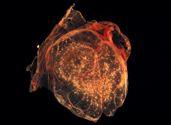
Malignant
[edit].jpg)
In malignant-type smallpox (also called flat smallpox) the lesions remained almost flush with the skin at the time when raised vesicles would have formed in the ordinary type. It is unknown why some people developed this type. Historically, it accounted for 5–10% of cases, and most (72%) were children.[3] Malignant smallpox was accompanied by a severe prodromal phase that lasted 3–4 days, prolonged high fever, and severe symptoms of viremia. The prodromal symptoms continued even after the onset of the rash.[3] The rash on the mucous membranes (enanthem) was extensive. Skin lesions matured slowly, were typically confluent or semi-confluent, and by the seventh or eighth day, they were flat and appeared to be buried in the skin. Unlike ordinary-type smallpox, the vesicles contained little fluid, were soft and velvety to the touch, and may have contained hemorrhages. Malignant smallpox was nearly always fatal and death usually occurred between the 8th and 12th day of illness. Often, a day or two before death, the lesions turned ashen gray, which, along with abdominal distension, was a bad prognostic sign.[3] This form is thought to be caused by deficient cell-mediated immunity to smallpox. If the person recovered, the lesions gradually faded and did not form scars or scabs.[39]
Hemorrhagic
[edit]Hemorrhagic smallpox is a severe form accompanied by extensive bleeding into the skin, mucous membranes, gastrointestinal tract, and viscera. This form develops in approximately 2% of infections and occurs mostly in adults.[33] Pustules do not typically form in hemorrhagic smallpox. Instead, bleeding occurs under the skin, making it look charred and black,[33] hence this form of the disease is also referred to as variola nigra or "black pox".[40] Hemorrhagic smallpox has very rarely been caused by variola minor virus.[41] While bleeding may occur in mild cases and not affect outcomes,[42] hemorrhagic smallpox is typically fatal.[43] Vaccination does not appear to provide any immunity to either form of hemorrhagic smallpox and some cases even occurred among people that were revaccinated shortly before. It has two forms.[3]
Early
[edit].jpg)
The early or fulminant form of hemorrhagic smallpox (referred to as purpura variolosa) begins with a prodromal phase characterized by a high fever, severe headache, and abdominal pain.[39] The skin becomes dusky and erythematous, and this is rapidly followed by the development of petechiae and bleeding in the skin, conjunctiva and mucous membranes. Death often occurs suddenly between the fifth and seventh days of illness, when only a few insignificant skin lesions are present. Some people survive a few days longer, during which time the skin detaches and fluid accumulates under it, rupturing at the slightest injury. People are usually conscious until death or shortly before.[43] Autopsy reveals petechiae and bleeding in the spleen, kidney, serous membranes, skeletal muscles, pericardium, liver, gonads and bladder.[41] Historically, this condition was frequently misdiagnosed, with the correct diagnosis made only at autopsy.[41] This form is more likely to occur in pregnant women than in the general population (approximately 16% of cases in unvaccinated pregnant women were early hemorrhagic smallpox, versus roughly 1% in nonpregnant women and adult males).[43] The case fatality rate of early hemorrhagic smallpox approaches 100%.[43]
Late
[edit]There is also a later form of hemorrhagic smallpox (referred to late hemorrhagic smallpox, or variolosa pustula hemorrhagica). The prodrome is severe and similar to that observed in early hemorrhagic smallpox, and the fever persists throughout the course of the disease.[3] Bleeding appears in the early eruptive period (but later than that seen in purpura variolosa), and the rash is often flat and does not progress beyond the vesicular stage. Hemorrhages in the mucous membranes appear to occur less often than in the early hemorrhagic form.[33] Sometimes the rash forms pustules which bleed at the base and then undergo the same process as in ordinary smallpox. This form of the disease is characterized by a decrease in all of the elements of the coagulation cascade and an increase in circulating antithrombin.[34] This form of smallpox occurs anywhere from 3% to 25% of fatal cases, depending on the virulence of the smallpox strain.[36] Most people with the late-stage form die within eight to 10 days of illness. Among the few who recover, the hemorrhagic lesions gradually disappear after a long period of convalescence.[3] The case fatality rate for late hemorrhagic smallpox is around 90–95%.[35] Pregnant women are slightly more likely to experience this form of the disease, though not as much as early hemorrhagic smallpox.[3]
Cause
[edit]| Variola virus | |
|---|---|

| |
| This transmission electron micrograph depicts a number of smallpox virions. The "dumbbell-shaped" structure inside the virion is the viral core, which contains the viral DNA; Mag. = ~370,000× | |
| Virus classification | |
| (unranked): | Virus |
| Realm: | Varidnaviria |
| Kingdom: | Bamfordvirae |
| Phylum: | Nucleocytoviricota |
| Class: | Pokkesviricetes |
| Order: | Chitovirales |
| Family: | Poxviridae |
| Genus: | Orthopoxvirus |
| Species: | †Orthopoxvirus variola |
Smallpox is caused by infection with Variola virus, which belongs to the family Poxviridae, subfamily Chordopoxvirinae, genus Orthopoxvirus.
Evolution
[edit]The date of the appearance of smallpox is not settled. It most probably evolved from a terrestrial African rodent virus between 68,000 and 16,000 years ago.[44] The wide range of dates is due to the different records used to calibrate the molecular clock. One clade was the variola major strains (the more clinically severe form of smallpox) which spread from Asia between 400 and 1,600 years ago. A second clade included both alastrim (a phenotypically mild smallpox) described from the American continents and isolates from West Africa which diverged from an ancestral strain between 1,400 and 6,300 years before present. This clade further diverged into two subclades at least 800 years ago.[45]
A second estimate has placed the separation of variola virus from Taterapox (an Orthopoxvirus of some African rodents including gerbils) at 3,000 to 4,000 years ago.[46] This is consistent with archaeological and historical evidence regarding the appearance of smallpox as a human disease which suggests a relatively recent origin. If the mutation rate is assumed to be similar to that of the herpesviruses, the divergence date of variola virus from Taterapox has been estimated to be 50,000 years ago.[46] While this is consistent with the other published estimates, it suggests that the archaeological and historical evidence is very incomplete. Better estimates of mutation rates in these viruses are needed.
Examination of a strain that dates from c. 1650 found that this strain was basal to the other presently sequenced strains.[47] The mutation rate of this virus is well modeled by a molecular clock. Diversification of strains only occurred in the 18th and 19th centuries.
Virology
[edit]Variola virus is large and brick-shaped and is approximately 302 to 350 nanometers by 244 to 270 nm,[48] with a single linear double stranded DNA genome 186 kilobase pairs (kbp) in size and containing a hairpin loop at each end.[49][50]
Four orthopoxviruses cause infection in humans: variola, vaccinia, cowpox, and monkeypox. Variola virus infects only humans in nature, although primates and other animals have been infected in an experimental setting. Vaccinia, cowpox, and monkeypox viruses can infect both humans and other animals in nature.[33]
The life cycle of poxviruses is complicated by having multiple infectious forms, with differing mechanisms of cell entry. Poxviruses are unique among human DNA viruses in that they replicate in the cytoplasm of the cell rather than in the nucleus. To replicate, poxviruses produce a variety of specialized proteins not produced by other DNA viruses, the most important of which is a viral-associated DNA-dependent RNA polymerase.
Both enveloped and unenveloped virions are infectious. The viral envelope is made of modified Golgi membranes containing viral-specific polypeptides, including hemagglutinin.[49] Infection with either variola major virus or variola minor virus confers immunity against the other.[34]
Variola major
[edit]The more common, infectious form of the disease was caused by the variola major virus strain, known for its significantly higher mortality rate compared to its counterpart, variola minor. Variola major had a fatality rate of around 30%, while variola minor’s mortality rate was about 1%. Throughout the 18th century, variola major was responsible for around 400,000 deaths annually in Europe alone. Survivors of the disease often faced lifelong consequences, such as blindness and severe scarring, which were nearly universal among those who recovered.[51]
In the first half of the 20th century, variola major was the primary cause of smallpox outbreaks across Asia and most of Africa. Meanwhile, variola minor was more commonly found in regions of Europe, North America, South America, and certain parts of Africa.[52]
Variola minor
[edit]Variola minor virus, also called alastrim, was a less common form of the virus, and much less deadly. Although variola minor had the same incubation period and pathogenetic stages as smallpox, it is believed to have had a mortality rate of less than 1%, as compared to variola major's 30%. Like variola major, variola minor was spread through inhalation of the virus in the air, which could occur through face-to-face contact or through fomites. Infection with variola minor virus conferred immunity against the more dangerous variola major virus.
Because variola minor was a less debilitating disease than smallpox, people were more frequently ambulant and thus able to infect others more rapidly. As such, variola minor swept through the United States, Great Britain, and South Africa in the early 20th century, becoming the dominant form of the disease in those areas and thus rapidly decreasing mortality rates. Along with variola major, the minor form has now been totally eradicated from the globe. The last case of indigenous variola minor was reported in a Somali cook, Ali Maow Maalin, in October 1977, and smallpox was officially declared eradicated worldwide in May 1980.[16] Variola minor was also called white pox, kaffir pox, Cuban itch, West Indian pox, milk pox, and pseudovariola.
Genome composition
[edit]The genome of variola major virus is about 186,000 base pairs in length.[53] It is made from linear double stranded DNA and contains the coding sequence for about 200 genes.[54] The genes are usually not overlapping and typically occur in blocks that point towards the closer terminal region of the genome.[55] The coding sequence of the central region of the genome is highly consistent across orthopoxviruses, and the arrangement of genes is consistent across chordopoxviruses.[54][55]
The center of the variola virus genome contains the majority of the essential viral genes, including the genes for structural proteins, DNA replication, transcription, and mRNA synthesis.[54] The ends of the genome vary more across strains and species of orthopoxviruses.[54] These regions contain proteins that modulate the hosts' immune systems, and are primarily responsible for the variability in virulence across the orthopoxvirus family.[54] These terminal regions in poxviruses are inverted terminal repetitions (ITR) sequences.[55] These sequences are identical but oppositely oriented on either end of the genome, leading to the genome being a continuous loop of DNA.[55] Components of the ITR sequences include an incompletely base paired A/T rich hairpin loop, a region of roughly 100 base pairs necessary for resolving concatomeric DNA (a stretch of DNA containing multiple copies of the same sequence), a few open reading frames, and short tandemly repeating sequences of varying number and length.[55] The ITRs of poxviridae vary in length across strains and species.[55] The coding sequence for most of the viral proteins in variola major virus have at least 90% similarity with the genome of vaccinia, a related virus used for vaccination against smallpox.[55]
Gene expression
[edit]Gene expression of variola virus occurs entirely within the cytoplasm of the host cell, and follows a distinct progression during infection.[55] After entry of an infectious virion into a host cell, synthesis of viral mRNA can be detected within 20 minutes.[55] About half of the viral genome is transcribed prior to the replication of viral DNA.[55] The first set of expressed genes are transcribed by pre-existing viral machinery packaged within the infecting virion.[55] These genes encode the factors necessary for viral DNA synthesis and for transcription of the next set of expressed genes.[55] Unlike most DNA viruses, DNA replication in variola virus and other poxviruses takes place within the cytoplasm of the infected cell.[55] The exact timing of DNA replication after infection of a host cell varies across the poxviridae.[55] Recombination of the genome occurs within actively infected cells.[55] Following the onset of viral DNA replication, an intermediate set of genes codes for transcription factors of late gene expression.[55] The products of the later genes include transcription factors necessary for transcribing the early genes for new virions, as well as viral RNA polymerase and other essential enzymes for new viral particles.[55] These proteins are then packaged into new infectious virions capable of infecting other cells.[55]
Research
[edit]Two live samples of variola major virus remain, one in the United States at the CDC in Atlanta, and one at the Vector Institute in Koltsovo, Russia.[56] Research with the remaining virus samples is tightly controlled, and each research proposal must be approved by the WHO and the World Health Assembly (WHA).[56] Most research on poxviruses is performed using the closely related Vaccinia virus as a model organism.[55] Vaccinia virus, which is used to vaccinate for smallpox, is also under research as a viral vector for vaccines for unrelated diseases.[57]
The genome of variola major virus was first sequenced in its entirety in the 1990s.[54] The complete coding sequence is publicly available online. The current reference sequence for variola major virus was sequenced from a strain that circulated in India in 1967. In addition, there are sequences for samples of other strains that were collected during the WHO eradication campaign.[54] A genome browser for a complete database of annotated sequences of variola virus and other poxviruses is publicly available through the Viral Bioinformatics Resource Center.[58]
Genetic engineering
[edit]The WHO currently bans genetic engineering of the variola virus.[59] However, in 2004, a committee advisory to the WHO voted in favor of allowing editing of the genome of the two remaining samples of variola major virus to add a marker gene.[59] This gene, called GFP, or green fluorescent protein, would cause live samples of the virus to glow green under fluorescent light.[60] The insertion of this gene, which would not influence the virulence of the virus, would be the only allowed modification of the genome.[60] The committee stated the proposed modification would aid in research of treatments by making it easier to assess whether a potential treatment was effective in killing viral samples.[60] The recommendation could only take effect if approved by the WHA.[60] When the WHA discussed the proposal in 2005, it refrained from taking a formal vote on the proposal, stating that it would review individual research proposals one at a time.[61] Addition of the GFP gene to the Vaccinia genome is routinely performed during research on the closely related vaccinia virus.[62]
Controversies
[edit]The public availability of the variola virus complete sequence has raised concerns about the possibility of illicit synthesis of infectious virus.[63] Vaccinia, a cousin of the variola virus, was artificially synthesized in 2002 by NIH scientists.[64] They used a previously established method that involved using a recombinant viral genome to create a self-replicating bacterial plasmid that produced viral particles.[64]
In 2016, another group synthesized the horsepox virus using publicly available sequence data for horsepox.[65] The researchers argued that their work would be beneficial to creating a safer and more effective vaccine for smallpox, although an effective vaccine is already available.[65] The horsepox virus had previously seemed to have gone extinct, raising concern about potential revival of variola major and causing other scientists to question their motives.[63] Critics found it especially concerning that the group was able to recreate viable virus in a short time frame with relatively little cost or effort.[65] Although the WHO bans individual laboratories from synthesizing more than 20% of the genome at a time, and purchases of smallpox genome fragments are monitored and regulated, a group with malicious intentions could compile, from multiple sources, the full synthetic genome necessary to produce viable virus.[65]
Transmission
[edit]Smallpox was highly contagious, but generally spread more slowly and less widely than some other viral diseases, perhaps because transmission required close contact and occurred after the onset of the rash. The overall rate of infection was also affected by the short duration of the infectious stage. In temperate areas, the number of smallpox infections was highest during the winter and spring. In tropical areas, seasonal variation was less evident and the disease was present throughout the year.[33] Age distribution of smallpox infections depended on acquired immunity. Vaccination immunity declined over time and was probably lost within thirty years.[34] Smallpox was not known to be transmitted by insects or animals and there was no asymptomatic carrier state.[33]
Transmission occurred through inhalation of airborne variola virus, usually droplets expressed from the oral, nasal, or pharyngeal mucosa of an infected person. It was transmitted from one person to another primarily through prolonged face-to-face contact with an infected person.[14]
Some infections of laundry workers with smallpox after handling contaminated bedding suggested that smallpox could be spread through direct contact with contaminated objects (fomites), but this was found to be rare.[14][35] Also rarely, smallpox was spread by virus carried in the air in enclosed settings such as buildings, buses, and trains.[32] The virus can cross the placenta, but the incidence of congenital smallpox was relatively low.[34] Smallpox was not notably infectious in the prodromal period and viral shedding was usually delayed until the appearance of the rash, which was often accompanied by lesions in the mouth and pharynx. The virus can be transmitted throughout the course of the illness, but this happened most frequently during the first week of the rash when most of the skin lesions were intact.[33] Infectivity waned in 7 to 10 days when scabs formed over the lesions, but the infected person was contagious until the last smallpox scab fell off.[66]
Concern about possible use of smallpox for biological warfare led in 2002 to Donald K. Milton's detailed review of existing research on its transmission and of then-current recommendations for controlling its spread. He agreed, citing Rao, Fenner and others, that "careful epidemiologic investigation rarely implicated fomites as a source of infection"; noted that "Current recommendations for control of secondary smallpox infections emphasize transmission 'by expelled droplets to close contacts (those within 6–7 feet)'"; but warned that the "emphasis on spread via large droplets may reduce the vigilance with which more difficult airborne precautions [i.e. against finer droplets capable of traveling longer distances and penetrating deeply into the lower respiratory tract] are maintained".[67]
Mechanism
[edit]Once inhaled, the variola virus invaded the mucous membranes of the mouth, throat, and respiratory tract. From there, it migrated to regional lymph nodes and began to multiply. In the initial growth phase, the virus seemed to move from cell to cell, but by around the 12th day, widespread lysis of infected cells occurred and the virus could be found in the bloodstream in large numbers, a condition known as viremia. This resulted in the second wave of multiplication in the spleen, bone marrow, and lymph nodes.
Diagnosis
[edit]The clinical definition of ordinary smallpox is an illness with acute onset of fever equal to or greater than 38.3 °C (101 °F) followed by a rash characterized by firm, deep-seated vesicles or pustules in the same stage of development without other apparent cause.[33] When a clinical case was observed, smallpox was confirmed using laboratory tests.
Microscopically, poxviruses produce characteristic cytoplasmic inclusion bodies, the most important of which are known as Guarnieri bodies, and are the sites of viral replication. Guarnieri bodies are readily identified in skin biopsies stained with hematoxylin and eosin, and appear as pink blobs. They are found in virtually all poxvirus infections but the absence of Guarnieri bodies could not be used to rule out smallpox.[68] The diagnosis of an orthopoxvirus infection can also be made rapidly by electron microscopic examination of pustular fluid or scabs. All orthopoxviruses exhibit identical brick-shaped virions by electron microscopy.[34] If particles with the characteristic morphology of herpesviruses are seen this will eliminate smallpox and other orthopoxvirus infections.
Definitive laboratory identification of variola virus involved growing the virus on chorioallantoic membrane (part of a chicken embryo) and examining the resulting pock lesions under defined temperature conditions.[69] Strains were characterized by polymerase chain reaction (PCR) and restriction fragment length polymorphism (RFLP) analysis. Serologic tests and enzyme linked immunosorbent assays (ELISA), which measured variola virus-specific immunoglobulin and antigen were also developed to assist in the diagnosis of infection.[70]
Chickenpox was commonly confused with smallpox in the immediate post-eradication era. Chickenpox and smallpox could be distinguished by several methods. Unlike smallpox, chickenpox does not usually affect the palms and soles. Additionally, chickenpox pustules are of varying size due to variations in the timing of pustule eruption: smallpox pustules are all very nearly the same size since the viral effect progresses more uniformly. A variety of laboratory methods were available for detecting chickenpox in the evaluation of suspected smallpox cases.[33]
-
 Smallpox virus lesions on the chorioallantoic membrane of a developing chick
 -
 In contrast to the rash in smallpox, the rash in chickenpox occurs mostly on the torso, spreading less to the limbs.
 -
 An Italian female smallpox patient whose skin displayed the characteristics of late-stage confluent maculopapular scarring, 1965


Prevention
[edit].jpg)
The earliest procedure used to prevent smallpox was inoculation with variola minor virus (a method later known as variolation after the introduction of smallpox vaccine to avoid possible confusion), which likely occurred in India, Africa, and China well before the practice arrived in Europe.[15] The idea that inoculation originated in India has been challenged, as few of the ancient Sanskrit medical texts described the process of inoculation.[71] Accounts of inoculation against smallpox in China can be found as early as the late 10th century, and the procedure was widely practiced by the 16th century, during the Ming dynasty.[72] If successful, inoculation produced lasting immunity to smallpox. Because the person was infected with variola virus, a severe infection could result, and the person could transmit smallpox to others. Variolation had a 0.5–2 percent mortality rate, considerably less than the 20–30 percent mortality rate of smallpox.[33] Two reports on the Chinese practice of inoculation were received by the Royal Society in London in 1700; one by Dr. Martin Lister who received a report by an employee of the East India Company stationed in China and another by Clopton Havers.[73]
Lady Mary Wortley Montagu observed smallpox inoculation during her stay in the Ottoman Empire, writing detailed accounts of the practice in her letters, and enthusiastically promoted the procedure in England upon her return in 1718.[74] According to Voltaire (1742), the Turks derived their use of inoculation from neighbouring Circassia. Voltaire does not speculate on where the Circassians derived their technique from, though he reports that the Chinese have practiced it "these hundred years".[75] In 1721, Cotton Mather and colleagues provoked controversy in Boston by inoculating hundreds. After publishing The present method of inoculating for the small-pox in 1767, Dr Thomas Dimsdale was invited to Russia to variolate the Empress Catherine the Great of Russia and her son, Grand Duke Paul, which he successfully did in 1768. In 1796, Edward Jenner, a doctor in Berkeley, Gloucestershire, rural England, discovered that immunity to smallpox could be produced by inoculating a person with material from a cowpox lesion. Cowpox is a poxvirus in the same family as variola. Jenner called the material used for inoculation vaccine from the root word vacca, which is Latin for cow. The procedure was much safer than variolation and did not involve a risk of smallpox transmission. Vaccination to prevent smallpox was soon practiced all over the world. During the 19th century, the cowpox virus used for smallpox vaccination was replaced by the vaccinia virus. Vaccinia is in the same family as cowpox and variola virus but is genetically distinct from both. The origin of the vaccinia virus and how it came to be in the vaccine are not known.[33]

The current formulation of the smallpox vaccine is a live virus preparation of the infectious vaccinia virus. The vaccine is given using a bifurcated (two-pronged) needle that is dipped into the vaccine solution. The needle is used to prick the skin (usually the upper arm) several times in a few seconds. If successful, a red and itchy bump develops at the vaccine site in three or four days. In the first week, the bump becomes a large blister (called a "Jennerian vesicle") which fills with pus and begins to drain. During the second week, the blister begins to dry up, and a scab forms. The scab falls off in the third week, leaving a small scar.[76]
The antibodies induced by the vaccinia vaccine are cross-protective for other orthopoxviruses, such as monkeypox, cowpox, and variola (smallpox) viruses. Neutralizing antibodies are detectable 10 days after first-time vaccination and seven days after revaccination. Historically, the vaccine has been effective in preventing smallpox infection in 95 percent of those vaccinated.[77] Smallpox vaccination provides a high level of immunity for three to five years and decreasing immunity thereafter. If a person is vaccinated again later, the immunity lasts even longer. Studies of smallpox cases in Europe in the 1950s and 1960s demonstrated that the fatality rate among persons vaccinated less than 10 years before exposure was 1.3 percent; it was 7 percent among those vaccinated 11 to 20 years prior, and 11 percent among those vaccinated 20 or more years before infection. By contrast, 52 percent of unvaccinated persons died.[78]

There are side effects and risks associated with the smallpox vaccine. In the past, about 1 out of 1,000 people vaccinated for the first time experienced serious, but non-life-threatening, reactions, including toxic or allergic reaction at the site of the vaccination (erythema multiforme), spread of the vaccinia virus to other parts of the body, and spread to other individuals. Potentially life-threatening reactions occurred in 14 to 500 people out of every 1 million people vaccinated for the first time. Based on past experience, it is estimated that 1 or 2 people in 1 million (0.000198 percent) who receive the vaccine may die as a result, most often the result of postvaccinial encephalitis or severe necrosis in the area of vaccination (called progressive vaccinia).[77]
Given these risks, as smallpox became effectively eradicated and the number of naturally occurring cases fell below the number of vaccine-induced illnesses and deaths, routine childhood vaccination was discontinued in the United States in 1972 and was abandoned in most European countries in the early 1970s.[10][79] Routine vaccination of health care workers was discontinued in the U.S. in 1976, and among military recruits in 1990 (although military personnel deploying to the Middle East and Korea still receive the vaccination[80]). By 1986, routine vaccination had ceased in all countries.[10] It is now primarily recommended for laboratory workers at risk for occupational exposure.[33] However, the possibility of variola virus being used as a biological weapon has rekindled interest in the development of newer vaccines.[81] The smallpox vaccine is also effective in, and therefore administered for, the prevention of mpox.[82]
ACAM2000 is a smallpox vaccine developed by Acambis, approved for use in the United States by the U.S. FDA on August 31, 2007. It contains live vaccinia virus, cloned from the same strain used in an earlier vaccine, Dryvax. While the Dryvax virus was cultured in the skin of calves and freeze-dried, ACAM2000s virus is cultured in kidney epithelial cells (Vero cells) from an African green monkey. Efficacy and adverse reaction incidence are similar to Dryvax.[81] The vaccine is not routinely available to the US public; it is, however, used in the military and maintained in the Strategic National Stockpile.[83]
Treatment
[edit]Smallpox vaccination within three days of exposure will prevent or significantly lessen the severity of smallpox symptoms in the vast majority of people. Vaccination four to seven days after exposure can offer some protection from disease or may modify the severity of the disease.[77] Other than vaccination, treatment of smallpox is primarily supportive, such as wound care and infection control, fluid therapy, and possible ventilator assistance. Flat and hemorrhagic types of smallpox are treated with the same therapies used to treat shock, such as fluid resuscitation. People with semi-confluent and confluent types of smallpox may have therapeutic issues similar to patients with extensive skin burns.[84]
Antiviral treatments have improved since the last large smallpox epidemics, and as of 2004, studies suggested that the antiviral drug cidofovir might be useful as a therapeutic agent. The drug must be administered intravenously, and may cause serious kidney toxicity.[85] In July 2018, the Food and Drug Administration approved tecovirimat, the first drug approved for treatment of smallpox.[86] However, during treatment viral mutations causing resistance have been known to occur, especially since its use in the 2022–2023 mpox outbreak which jeopardize its effectiveness for smallpox biothreat preparedness.[87]
In June 2021, brincidofovir was approved for medical use in the United States for the treatment of human smallpox disease caused by variola virus.[88][89]
Prognosis
[edit].jpg)
The mortality rate from variola minor is approximately 1%, while the mortality rate from variola major is approximately 30%.[90]
Ordinary type-confluent is fatal about 50–75% of the time, ordinary-type semi-confluent about 25–50% of the time, in cases where the rash is discrete the case-fatality rate is less than 10%. The overall fatality rate for children younger than 1 year of age is 40–50%. Hemorrhagic and flat types have the highest fatality rates. The fatality rate for flat or late hemorrhagic type smallpox is 90% or greater and nearly 100% is observed in cases of early hemorrhagic smallpox.[43] The case-fatality rate for variola minor is 1% or less.[38] There is no evidence of chronic or recurrent infection with variola virus.[38] In cases of flat smallpox in vaccinated people, the condition was extremely rare but less lethal, with one case series showing a 67% death rate.[3]
In fatal cases of ordinary smallpox, death usually occurs between days 10–16 of the illness. The cause of death from smallpox is not clear, but the infection is now known to involve multiple organs. Circulating immune complexes, overwhelming viremia, or an uncontrolled immune response may be contributing factors.[33] In early hemorrhagic smallpox, death occurs suddenly about six days after the fever develops. The cause of death in early hemorrhagic cases is commonly due to heart failure and pulmonary edema. In late hemorrhagic cases, high and sustained viremia, severe platelet loss and poor immune response were often cited as causes of death.[3] In flat smallpox modes of death are similar to those in burns, with loss of fluid, protein and electrolytes, and fulminating sepsis.[84]
Complications
[edit]Complications of smallpox arise most commonly in the respiratory system and range from simple bronchitis to fatal pneumonia. Respiratory complications tend to develop on about the eighth day of the illness and can be either viral or bacterial in origin. Secondary bacterial infection of the skin is a relatively uncommon complication of smallpox. When this occurs, the fever usually remains elevated.[33]
Other complications include encephalitis (1 in 500 patients), which is more common in adults and may cause temporary disability; permanent pitted scars, most notably on the face; and complications involving the eyes (2% of all cases). Pustules can form on the eyelid, conjunctiva, and cornea, leading to complications such as conjunctivitis, keratitis, corneal ulcer, iritis, iridocyclitis, and atrophy of the optic nerve. Blindness results in approximately 35–40% of eyes affected with keratitis and corneal ulcer. Hemorrhagic smallpox can cause subconjunctival and retinal hemorrhages. In 2–5% of young children with smallpox, virions reach the joints and bone, causing osteomyelitis variolosa. Bony lesions are symmetrical, most common in the elbows, legs, and characteristically cause separation of the epiphysis and marked periosteal reactions. Swollen joints limit movement, and arthritis may lead to limb deformities, ankylosis, malformed bones, flail joints, and stubby fingers.[34]
Between 65 and 80% of survivors are marked with deep pitted scars (pockmarks), most prominent on the face.
History
[edit]Disease emergence
[edit]
The earliest credible clinical evidence of smallpox is found in the descriptions of smallpox-like disease in medical writings from ancient India (as early as 1500 BCE),[91][92] and China (1122 BCE),[93] as well as a study of the Egyptian mummy of Ramses V (died 1145 BCE).[92][94] It has been speculated that Egyptian traders brought smallpox to India during the 1st millennium BCE, where it remained as an endemic human disease for at least 2000 years. Smallpox was probably introduced into China during the 1st century CE from the southwest, and in the 6th century was carried from China to Japan.[3] In Japan, the epidemic of 735–737 is believed to have killed as much as one-third of the population.[18][95] At least seven religious deities have been specifically dedicated to smallpox, such as the god Sopona in the Yoruba religion in West Africa. In India, the Hindu goddess of smallpox, Shitala, was worshipped in temples throughout the country.[96]
A different viewpoint is that smallpox emerged 1588 CE and the earlier reported cases were incorrectly identified as smallpox.[97][47]
The timing of the arrival of smallpox in Europe and south-western Asia is less clear. Smallpox is not clearly described in either the Old or New Testaments of the Bible or in the literature of the Greeks or Romans. While some have identified the Plague of Athens – which was said to have originated in "Ethiopia" and Egypt – or the plague that lifted Carthage's 396 BCE siege of Syracuse – with smallpox,[3] many scholars agree it is very unlikely such a serious disease as variola major would have escaped being described by Hippocrates if it had existed in the Mediterranean region during his lifetime.[42]
While the Antonine Plague that swept through the Roman Empire in 165–180 CE may have been caused by smallpox,[98] Saint Nicasius of Rheims became the patron saint of smallpox victims for having supposedly survived a bout in 450,[3] and Saint Gregory of Tours recorded a similar outbreak in France and Italy in 580, the first use of the term variola.[3] Other historians speculate that Arab armies first carried smallpox from Africa into Southwestern Europe during the 7th and 8th centuries.[3] In the 9th century the Persian physician, Rhazes, provided one of the most definitive descriptions of smallpox and was the first to differentiate smallpox from measles and chickenpox in his Kitab fi al-jadari wa-al-hasbah (The Book of Smallpox and Measles).[99] During the Middle Ages several smallpox outbreaks occurred in Europe. However, smallpox had not become established there until the population growth and mobility marked by the Crusades allowed it to do so. By the 16th century, smallpox had become entrenched across most of Europe,[3] where it had a mortality rate as high as 30 percent. This endemic occurrence of smallpox in Europe is of particular historical importance, as successive exploration and colonization by Europeans tended to spread the disease to other nations. By the 16th century, smallpox had become a predominant cause of morbidity and mortality throughout much of the world.[3]

There were no credible descriptions of smallpox-like disease in the Americas before the westward exploration by Europeans in the 15th century CE.[45] Smallpox was introduced into the Caribbean island of Hispaniola in 1507, and into the mainland in 1520, when Spanish settlers from Hispaniola arrived in Mexico, inadvertently carrying smallpox with them. Because the native Amerindian population had no acquired immunity to this new disease, their peoples were decimated by epidemics. Such disruption and population losses were an important factor in the Spanish achieving conquest of the Aztecs and the Incas.[3] Similarly, English settlement of the east coast of North America in 1633 in Plymouth, Massachusetts was accompanied by devastating outbreaks of smallpox among Native American populations,[100] and subsequently among the native-born colonists.[101] Case fatality rates during outbreaks in Native American populations were as high as 90%.[102] Smallpox was introduced into Australia in 1789 and again in 1829,[3] though colonial surgeons, who by 1829 were attempting to distinguish between smallpox and chickenpox (which could be almost equally fatal to Aboriginal Australians), were divided as to whether the 1829–1830 epidemic was chickenpox or smallpox.[103] Although smallpox was never endemic on the continent,[3] it has been described as the principal cause of death in Aboriginal populations between 1780 and 1870.[104]
.svg)

By the mid-18th century, smallpox was a major endemic disease everywhere in the world except in Australia and small islands untouched by outside exploration. In 18th century Europe, smallpox was a leading cause of death, killing an estimated 400,000 Europeans each year.[105] Up to 10 percent of Swedish infants died of smallpox each year,[18] and the death rate of infants in Russia might have been even higher.[93] The widespread use of variolation in a few countries, notably Great Britain, its North American colonies, and China, somewhat reduced the impact of smallpox among the wealthy classes during the latter part of the 18th century, but a real reduction in its incidence did not occur until vaccination became a common practice toward the end of the 19th century. Improved vaccines and the practice of re-vaccination led to a substantial reduction in cases in Europe and North America, but smallpox remained almost unchecked everywhere else in the world. By the mid-20th century, variola minor occurred along with variola major, in varying proportions, in many parts of Africa. Patients with variola minor experience only a mild systemic illness, are often ambulant throughout the course of the disease, and are therefore able to more easily spread disease. Infection with variola minor virus induces immunity against the more deadly variola major form. Thus, as variola minor spread all over the US, into Canada, the South American countries, and Great Britain, it became the dominant form of smallpox, further reducing mortality rates.[3]
Eradication
[edit]

The first clear reference to smallpox inoculation was made by the Chinese author Wan Quan (1499–1582) in his Dòuzhěn xīnfǎ (痘疹心法, "Pox Rash Teachings") published in 1549,[106] with earliest hints of the practice in China during the 10th century.[107] In China, powdered smallpox scabs were blown up the noses of the healthy. People would then develop a mild case of the disease and from then on were immune to it. The technique did have a 0.5–2.0% mortality rate, but that was considerably less than the 20–30% mortality rate of the disease itself. Two reports on the Chinese practice of inoculation were received by the Royal Society in London in 1700: one by Dr. Martin Lister who received a report by an employee of the East India Company stationed in China and another by Clopton Havers.[108] Voltaire (1742) reports that the Chinese had practiced smallpox inoculation "these hundred years".[75] Variolation had also been witnessed in Turkey by Lady Mary Wortley Montagu, who later introduced it in the UK.[109]
An early mention of the possibility of smallpox's eradication was made in reference to the work of Johnnie Notions, a self-taught inoculator from Shetland, Scotland. Notions found success in treating people from at least the late 1780s through a method devised by himself despite having no formal medical background.[110][111] His method involved exposing smallpox pus to peat smoke, burying it in the ground with camphor for up to 8 years, and then inserting the matter into a person's skin using a knife, and covering the incision with a cabbage leaf.[112] He was reputed not to have lost a single patient.[112] Arthur Edmondston, in writings on Notions' technique that were published in 1809, stated, "Had every practitioner been as uniformly successful in the disease as he was, the small-pox might have been banished from the face of the earth, without injuring the system, or leaving any doubt as to the fact."[113]

The English physician Edward Jenner demonstrated the effectiveness of cowpox to protect humans from smallpox in 1796, after which various attempts were made to eliminate smallpox on a regional scale. In Russia in 1796, the first child to receive this treatment was bestowed the name "Vaccinov" by Catherine the Great, and was educated at the expense of the nation.[114]
The introduction of the vaccine to the New World took place in Trinity, Newfoundland in 1800 by Dr. John Clinch, boyhood friend and medical colleague of Jenner.[115] As early as 1803, the Spanish Crown organized the Balmis expedition to transport the vaccine to the Spanish colonies in the Americas and the Philippines, and establish mass vaccination programs there.[116] The U.S. Congress passed the Vaccine Act of 1813 to ensure that safe smallpox vaccine would be available to the American public. By about 1817, a robust state vaccination program existed in the Dutch East Indies.[117]
On March 26, 1806, the Swiss canton Thurgau became the first state in the world to introduce compulsory smallpox vaccinations, by order of the cantonal councillor Jakob Christoph Scherb.[118][119] Half a year later, Elisa Bonaparte issued a corresponding order for her Principality of Lucca and Piombino.[120] Baden followed in 1809, Prussia in 1815, Württemberg in 1818, Sweden in 1816 and the German Empire in 1874 through the Reichs Vaccination Act.[121][122] In Lutheran Sweden, the Protestant clergy played a pioneering role in voluntary smallpox vaccination as early as 1800.[123] The first vaccination was carried out in Liechtenstein in 1801, and from 1812 it was mandatory to vaccinate.[124]
In British India a program was launched to propagate smallpox vaccination, through Indian vaccinators, under the supervision of European officials.[125] Nevertheless, British vaccination efforts in India, and in Burma in particular, were hampered by indigenous preference for inoculation and distrust of vaccination, despite tough legislation, improvements in the local efficacy of the vaccine and vaccine preservative, and education efforts.[126] By 1832, the federal government of the United States established a smallpox vaccination program for Native Americans.[127] In 1842, the United Kingdom banned inoculation (variolation), later progressing to mandatory vaccination. The British government introduced compulsory smallpox vaccination by an Act of Parliament in 1853.[128] An epidemic in Sheffield in 1887/88 demonstrated that, in addition to the vaccine, several other factors such as extensive isolation measures contributed to the control of the disease.[129]
In the United States, from 1843 to 1855, first Massachusetts and then other states required smallpox vaccination. Although some disliked these measures,[93] coordinated efforts against smallpox went on, and the disease continued to diminish in the wealthy countries. In Northern Europe a number of countries had eliminated smallpox by 1900, and by 1914, the incidence in most industrialized countries had decreased to comparatively low levels.
Vaccination continued in industrialized countries as protection against reintroduction until the mid to late 1970s. Australia and New Zealand are two notable exceptions; neither experienced endemic smallpox and never vaccinated widely, relying instead on protection by distance and strict quarantines.[130]

The first hemisphere-wide effort to eradicate smallpox was made in 1950 by the Pan American Health Organization.[131] The campaign was successful in eliminating smallpox from all countries of the Americas except Argentina, Brazil, Colombia, and Ecuador.[130] In 1958 Professor Viktor Zhdanov, Deputy Minister of Health for the USSR, called on the World Health Assembly to undertake a global initiative to eradicate smallpox.[132] The proposal (Resolution WHA11.54) was accepted in 1959.[132] At this point, 2 million people were dying from smallpox every year. Overall, the progress towards eradication was disappointing, especially in Africa and in the Indian subcontinent. In 1966 an international team, the Smallpox Eradication Unit, was formed under the leadership of an American, Donald Henderson.[132] In 1967, the World Health Organization intensified the global smallpox eradication by contributing $2.4 million annually to the effort, and adopted the new disease surveillance method promoted by Czech epidemiologist Karel Raška.[133]

In the early 1950s, an estimated 50 million cases of smallpox occurred in the world each year.[10] To eradicate smallpox, each outbreak had to be stopped from spreading, by isolation of cases and vaccination of everyone who lived close by.[134] This process is known as "ring vaccination". The key to this strategy was the monitoring of cases in a community (known as surveillance) and containment.
The initial problem the WHO team faced was inadequate reporting of smallpox cases, as many cases did not come to the attention of the authorities. The fact that humans are the only reservoir for smallpox infection (the virus only infected humans and not other animals) and that carriers did not exist played a significant role in the eradication of smallpox. The WHO established a network of consultants who assisted countries in setting up surveillance and containment activities. Early on, donations of vaccine were provided primarily by the Soviet Union and the United States, but by 1973, more than 80 percent of all vaccines were produced in developing countries.[130] The Soviet Union provided one and a half billion doses between 1958 and 1979, as well as the medical staff.[135]
The last major European outbreak of smallpox was in 1972 in Yugoslavia, after a pilgrim from Kosovo returned from the Middle East, where he had contracted the virus. The epidemic infected 175 people, causing 35 deaths. Authorities declared martial law, enforced quarantine, and undertook widespread re-vaccination of the population, enlisting the help of the WHO. In two months, the outbreak was over.[136] Prior to this, there had been a smallpox outbreak in May–July 1963 in Stockholm, Sweden, brought from the Far East by a Swedish sailor; this had been dealt with by quarantine measures and vaccination of the local population.[137]

By the end of 1975, smallpox persisted only in the Horn of Africa. Conditions were very difficult in Ethiopia and Somalia, where there were few roads. Civil war, famine, and refugees made the task even more difficult. An intensive surveillance, containment, and vaccination program was undertaken in these countries in early and mid-1977, under the direction of Australian microbiologist Frank Fenner. As the campaign neared its goal, Fenner and his team played an important role in verifying eradication.[139] The last naturally occurring case of indigenous smallpox (Variola minor) was diagnosed in Ali Maow Maalin, a hospital cook in Merca, Somalia, on 26 October 1977.[33] The last naturally occurring case of the more deadly Variola major had been detected in October 1975 in a three-year-old Bangladeshi girl, Rahima Banu.[40]
The global eradication of smallpox was certified, based on intense verification activities, by a commission of eminent scientists on 9 December 1979 and subsequently endorsed by the World Health Assembly on 8 May 1980.[10][140] The first two sentences of the resolution read:
Having considered the development and results of the global program on smallpox eradication initiated by WHO in 1958 and intensified since 1967 ... Declares solemnly that the world and its peoples have won freedom from smallpox, which was a most devastating disease sweeping in epidemic form through many countries since earliest time, leaving death, blindness and disfigurement in its wake and which only a decade ago was rampant in Africa, Asia and South America.[141]
The World Health Organization records of the smallpox eradication programme, from 1948 to 1987, were added to the UNESCO Memory of the World register in 2017.[142]
Costs and benefits
[edit]The cost of the eradication effort, from 1967 to 1979, was roughly US$300 million. Roughly a third came from the developed world, which had largely eradicated smallpox decades earlier. The United States, the largest contributor to the program, has reportedly recouped that investment every 26 days since in money not spent on vaccinations and the costs of incidence.[143]
Since eradication
[edit]
The last case of smallpox in the world occurred in an outbreak in the United Kingdom in 1978.[144] A medical photographer, Janet Parker, contracted the disease at the University of Birmingham Medical School and died on 11 September 1978. Although it has remained unclear how Parker became infected, the source of the infection was established to be the variola virus grown for research purposes at the Medical School laboratory.[145][146] All known stocks of smallpox worldwide were subsequently destroyed or transferred to two WHO-designated reference laboratories with BSL-4 facilities – the United States' Centers for Disease Control and Prevention (CDC) and the Soviet Union's (now Russia's) State Research Center of Virology and Biotechnology VECTOR.[147]
WHO first recommended destruction of the virus in 1986 and later set the date of destruction to be 30 December 1993. This was postponed to 30 June 1999.[148] Due to resistance from the U.S. and Russia, in 2002 the World Health Assembly agreed to permit the temporary retention of the virus stocks for specific research purposes.[149] Destroying existing stocks would reduce the risk involved with ongoing smallpox research; the stocks are not needed to respond to a smallpox outbreak.[150] Some scientists have argued that the stocks may be useful in developing new vaccines, antiviral drugs, and diagnostic tests;[151] a 2010 review by a team of public health experts appointed by WHO concluded that no essential public health purpose is served by the U.S. and Russia continuing to retain virus stocks.[152] The latter view is frequently supported in the scientific community, particularly among veterans of the WHO Smallpox Eradication Program.[153]
On March 31, 2003, smallpox scabs were found inside an envelope in an 1888 book on Civil War medicine in Santa Fe, New Mexico.[154] The envelope was labeled as containing scabs from a vaccination and gave scientists at the CDC an opportunity to study the history of smallpox vaccination in the United States.
On July 1, 2014, six sealed glass vials of smallpox dated 1954, along with sample vials of other pathogens, were discovered in a cold storage room in an FDA laboratory at the National Institutes of Health location in Bethesda, Maryland. The smallpox vials were subsequently transferred to the custody of the CDC in Atlanta, where virus taken from at least two vials proved viable in culture.[155][156] After studies were conducted, the CDC destroyed the virus under WHO observation on February 24, 2015.[157]
In 2017, scientists at the University of Alberta recreated an extinct horse pox virus to demonstrate that the variola virus can be recreated in a small lab at a cost of about $100,000, by a team of scientists without specialist knowledge.[158] Although the scientists performed the research to help development of new vaccines as well as trace smallpox's history, the possibility of the techniques being used for nefarious purposes was immediately recognized, raising questions on dual use research and regulations.[159][160]
In September 2019, the Russian lab housing smallpox samples experienced a gas explosion that injured one worker. It did not occur near the virus storage area, and no samples were compromised, but the incident prompted a review of risks to containment.[161]
Society and culture
[edit]Biological warfare
[edit]In 1763, Pontiac's War broke out as a Native American confederacy led by Pontiac attempted to counter British control over the Great Lakes region.[162][163][164] A group of Native American warriors laid siege to British-held Fort Pitt on June 22.[165] In response, Henry Bouquet, the commander of the fort, ordered his subordinate Simeon Ecuyer to give smallpox-infested blankets from the infirmary to a Delaware delegation outside the fort. Bouquet had discussed this with his superior, Sir Jeffrey Amherst, who wrote to Bouquet stating: "Could it not be contrived to send the small pox among the disaffected tribes of Indians? We must on this occasion use every stratagem in our power to reduce them." Bouquet agreed with the proposal, writing back that "I will try to inocculate [sic] the Indians by means of Blankets that may fall in their hands".[166] On 24 June 1763, William Trent, a local trader and commander of the Fort Pitt militia, wrote, "Out of our regard for them, we gave them two Blankets and an Handkerchief out of the Small Pox Hospital. I hope it will have the desired effect."[167][162] The effectiveness of this effort to broadcast the disease is unknown. There are also accounts that smallpox was used as a weapon during the American Revolutionary War (1775–1783).[168][169]
According to a theory put forward in Journal of Australian Studies (JAS) by independent researcher Christopher Warren, Royal Marines used smallpox in 1789 against indigenous tribes in New South Wales.[170] This theory was also considered earlier in Bulletin of the History of Medicine[171] and by David Day.[172] However it is disputed by some medical academics, including Professor Jack Carmody, who in 2010 claimed that the rapid spread of the outbreak in question was more likely indicative of chickenpox – a more infectious disease which, at the time, was often confused, even by surgeons, with smallpox, and may have been comparably deadly to Aboriginals and other peoples without natural immunity to it.[173] Carmody noted that in the 8-month voyage of the First Fleet and the following 14 months there were no reports of smallpox amongst the colonists and that, since smallpox has an incubation period of 10–12 days, it is unlikely it was present in the First Fleet; however, Warren argued in the JAS article that the likely source was bottles of variola virus possessed by First Fleet surgeons. Ian and Jennifer Glynn, in The life and death of smallpox, confirm that bottles of "variolous matter" were carried to Australia for use as a vaccine, but think it unlikely the virus could have survived till 1789.[104] In 2007, Christopher Warren offered evidence that the British smallpox may have been still viable.[174] However, the only non-Aboriginal reported to have died in this outbreak was a seaman called Joseph Jeffries, who was recorded as being of "American Indian" origin.[175]
W. S. Carus, an expert in biological weapons, has written that there is circumstantial evidence that smallpox was deliberately introduced to the Aboriginal population.[176] However Carmody and the Australian National University's Boyd Hunter continue to support the chickenpox hypothesis.[177] In a 2013 lecture at the Australian National University,[178] Carmody pointed out that chickenpox, unlike smallpox, was known to be present in the Sydney Cove colony. He also suggested that all c. 18th century (and earlier) identifications of smallpox outbreaks were dubious because: "surgeons ... would have been unaware of the distinction between smallpox and chickenpox – the latter having traditionally been considered a milder form of smallpox."[179]
During World War II, scientists from the United Kingdom, United States, and Japan (Unit 731 of the Imperial Japanese Army) were involved in research into producing a biological weapon from smallpox.[180] Plans of large scale production were never carried through as they considered that the weapon would not be very effective due to the wide-scale availability of a vaccine.[168]
In 1947, the Soviet Union established a smallpox weapons factory in the Scientific Research Institute of Medicine of the Ministry of Defense in Sergiyev Posad in Zagorsk, 75 km to the northeast of Moscow.[181] An outbreak of weaponized smallpox occurred during testing at a facility on an island in the Aral Sea in 1971. General Prof. Peter Burgasov, former Chief Sanitary Physician of the Soviet Army and a senior researcher within the Soviet program of biological weapons, described the incident:
On Vozrozhdeniya Island in the Aral Sea, the strongest recipes of smallpox were tested. Suddenly I was informed that there were mysterious cases of mortalities in Aralsk. A research ship of the Aral fleet came to within 15 km of the island (it was forbidden to come any closer than 40 km). The lab technician of this ship took samples of plankton twice a day from the top deck. The smallpox formulation – 400 gr. of which was exploded on the island – "got her" and she became infected. After returning home to Aralsk, she infected several people including children. All of them died. I suspected the reason for this and called the Chief of General Staff of the Ministry of Defense and requested to forbid the stop of the Alma-Ata–Moscow train in Aralsk. As a result, the epidemic around the country was prevented. I called Andropov, who at that time was Chief of KGB, and informed him of the exclusive recipe of smallpox obtained on Vozrazhdenie Island.[182][183]
Others contend that the first patient may have contracted the disease while visiting Uyaly or Komsomolsk-on-Ustyurt, two cities where the boat docked.[184][185]
Responding to international pressures, in 1991 the Soviet government allowed a joint U.S.–British inspection team to tour four of its main weapons facilities at Biopreparat. The inspectors were met with evasion and denials from the Soviet scientists and were eventually ordered out of the facility.[186] In 1992, Soviet defector Ken Alibek alleged that the Soviet bioweapons program at Zagorsk had produced a large stockpile – as much as twenty tons – of weaponized smallpox (possibly engineered to resist vaccines, Alibek further alleged), along with refrigerated warheads to deliver it. Alibek's stories about the former Soviet program's smallpox activities have never been independently verified.
In 1997, the Russian government announced that all of its remaining smallpox samples would be moved to the Vector Institute in Koltsovo.[186] With the breakup of the Soviet Union and unemployment of many of the weapons program's scientists, U.S. government officials have expressed concern that smallpox and the expertise to weaponize it may have become available to other governments or terrorist groups who might wish to use virus as means of biological warfare.[187] Specific allegations made against Iraq in this respect proved to be false.[188]
Notable cases
[edit]
Famous historical figures who contracted smallpox include Lakota Chief Sitting Bull, Ramses V,[189] the Kangxi Emperor (survived), Shunzhi Emperor and Tongzhi Emperor of China, Emperor Komei of Japan (died of smallpox in 1867), and Date Masamune of Japan (who lost an eye to the disease). Cuitláhuac, the 10th tlatoani (ruler) of the Aztec city of Tenochtitlan, died of smallpox in 1520, shortly after its introduction to the Americas, and the Incan emperor Huayna Capac died of it in 1527 (causing a civil war of succession in the Inca empire and the eventual conquest by the Spaniards). More recent public figures include Guru Har Krishan, 8th Guru of the Sikhs, in 1664, Louis I of Spain in 1724 (died), Peter II of Russia in 1730 (died),[190] George Washington (survived), Louis XV of France in 1774 (died), and Maximilian III Joseph of Bavaria in 1777 (died).
Prominent families throughout the world often had several people infected by and/or perish from the disease. For example, several relatives of Henry VIII of England survived the disease but were scarred by it. These include his sister Margaret, his wife Anne of Cleves, and his two daughters: Mary I in 1527 and Elizabeth I in 1562. Elizabeth tried to disguise the pockmarks with heavy makeup. Mary, Queen of Scots, contracted the disease as a child but had no visible scarring.
In Europe, deaths from smallpox often changed dynastic succession. Louis XV of France succeeded his great-grandfather Louis XIV through a series of deaths of smallpox or measles among those higher in the succession line. He himself died of the disease in 1774. Peter II of Russia died of the disease at 14 years of age. Also, before becoming emperor, Peter III of Russia caught the virus and suffered greatly from it.[citation needed] He was left scarred and disfigured. His wife, Catherine the Great, was spared but fear of the virus clearly had its effects on her. She feared for the safety of her son, Paul, so much that she made sure that large crowds were kept at bay and sought to isolate him. Eventually, she decided to have herself inoculated by a British doctor, Thomas Dimsdale. While this was considered a controversial method at the time, she succeeded. Paul was later inoculated as well. Catherine then sought to have inoculations throughout her empire stating: "My objective was, through my example, to save from death the multitude of my subjects who, not knowing the value of this technique, and frightened of it, were left in danger." By 1800, approximately two million inoculations had been administered in the Russian Empire.[191]
In China, the Qing dynasty had extensive protocols to protect Manchus from Peking's endemic smallpox.
U.S. Presidents George Washington, Andrew Jackson, and Abraham Lincoln all contracted and recovered from the disease. Washington became infected with smallpox on a visit to Barbados in 1751.[192] Jackson developed the illness after being taken prisoner by the British during the American Revolution, and though he recovered, his brother Robert did not.[192] Lincoln contracted the disease during his presidency, possibly from his son Tad, and was quarantined shortly after giving the Gettysburg address in 1863.[192]
The famous theologian Jonathan Edwards died of smallpox in 1758 following an inoculation.[193]
Soviet leader Joseph Stalin fell ill with smallpox at the age of seven. His face was badly scarred by the disease. He later had photographs retouched to make his pockmarks less apparent.[194]
Hungarian poet Ferenc Kölcsey, who wrote the Hungarian national anthem, lost his right eye to smallpox.[195]
Tradition and religion
[edit]
In the face of the devastation of smallpox, various smallpox gods and goddesses have been worshipped throughout parts of the Old World, for example in China and India. In China, the smallpox goddess was referred to as T'ou-Shen Niang-Niang (Chinese: 痘疹娘娘).[196] Chinese believers actively worked to appease the goddess and pray for her mercy, by such measures as referring to smallpox pustules as "beautiful flowers" as a euphemism intended to avert offending the goddess, for example (the Chinese word for smallpox is 天花, literally "heaven flower").[197] In a related New Year's Eve custom it was prescribed that the children of the house wear ugly masks while sleeping, so as to conceal any beauty and thereby avoid attracting the goddess, who would be passing through sometime that night.[197] If a case of smallpox did occur, shrines would be set up in the homes of the victims, to be worshipped and offered to as the disease ran its course. If the victim recovered, the shrines were removed and carried away in a special paper chair or boat for burning. If the patient did not recover, the shrine was destroyed and cursed, to expel the goddess from the house.[196]
In the Yoruba language smallpox is known as ṣọpọná, but it was also written as shakpanna, shopona, ṣhapana, and ṣọpọnọ. The word is a combination of 3 words, the verb ṣán, meaning to cover or plaster (referring to the pustules characteristic of smallpox), kpa or pa, meaning to kill, and enia, meaning human. Roughly translated, it means One who kills a person by covering them with pustules.[198] Among the Yorùbá people of West Africa, and also in Dahomean religion, Trinidad, and in Brazil, The deity Sopona, also known as Obaluaye, is the deity of smallpox and other deadly diseases (like leprosy, HIV/AIDS, and fevers). One of the most feared deities of the orisha pantheon, smallpox was seen as a form of punishment from Shopona.[199] Worship of Shopona was highly controlled by his priests, and it was believed that priests could also spread smallpox when angered.[199] However, Shopona was also seen as a healer who could cure the diseases he inflicted, and he was often called upon by his victims to heal them.[200] The British government banned the worship of the god because it was believed his priests were purposely spreading smallpox to their opponents.[200][199]
India's first records of smallpox can be found in a medical book that dates back to before 400 CE. This book describes a disease that sounds exceptionally like smallpox.[201] India, like China and the Yorùbá, created a goddess in response to its exposure to smallpox. The Hindu goddess Shitala was both worshipped and feared during her reign. It was believed that this goddess was both evil and kind and had the ability to inflict victims when angered, as well as calm the fevers of the already affected.[202][93] Portraits of the goddess show her holding a broom in her right hand to continue to move the disease and a pot of cool water in the other hand in an attempt to soothe patients.[197] Shrines were created where many Indian natives, both healthy and not, went to worship and attempt to protect themselves from this disease. Some Indian women, in an attempt to ward off Shitala, placed plates of cooling foods and pots of water on the roofs of their homes.[203]
In cultures that did not recognise a smallpox deity, there was often nonetheless a belief in smallpox demons, who were accordingly blamed for the disease. Such beliefs were prominent in Japan, Europe, Africa, and other parts of the world. Nearly all cultures who believed in the demon also believed that it was afraid of the colour red. This led to the invention of the so-called red treatment, where patients and their rooms would be decorated in red. The practise spread to Europe in the 12th century and was practised by (among others) Charles V of France and Elizabeth I of England.[3] Afforded scientific credibility through the studies by Niels Ryberg Finsen showing that red light reduced scarring,[3] this belief persisted even until the 1930s.
See also
[edit]References
[edit]- ^ Barton LL, Friedman NR (2008). The Neurological Manifestations of Pediatric Infectious Diseases and Immunodeficiency Syndromes. Springer Science & Business Media. p. 151. ISBN 978-1-59745-391-2.
- ^ Schaller KF (2012). Colour Atlas of Tropical Dermatology and Venerology. Springer Science & Business Media. p. Chapter 1. ISBN 978-3-642-76200-0.
- ^ a b c d e f g h i j k l m n o p q r s t u v w x Fenner F, Henderson DA, Arita I, Ježek Z, Ladnyi ID (1988). "The History of Smallpox and its Spread Around the World" (PDF). Smallpox and its eradication. History of International Public Health. Vol. 6. Geneva: World Health Organization. pp. 209–44. hdl:10665/39485. ISBN 978-92-4-156110-5. Archived (PDF) from the original on 12 December 2019. Retrieved 14 December 2017.
- ^ a b Medicine: The Definitive Illustrated History. Pengui. 2016. p. 100. ISBN 978-1-4654-5893-3.
- ^ a b c d e f g h i "Signs and Symptoms". CDC. 7 June 2016. Archived from the original on 7 June 2020. Retrieved 14 December 2017.
- ^ a b c d e "What is Smallpox?". CDC. 7 June 2016. Archived from the original on 7 June 2020. Retrieved 14 December 2017.
- ^ a b Ryan KJ, Ray CG, eds. (2004). Sherris Medical Microbiology (4th ed.). McGraw Hill. pp. 525–28. ISBN 978-0-8385-8529-0.
- ^ a b "Diagnosis & Evaluation". CDC. 25 July 2017. Archived from the original on 9 December 2017. Retrieved 14 December 2017.
- ^ a b c "Prevention and Treatment". CDC. 13 December 2017. Archived from the original on 15 December 2017. Retrieved 14 December 2017.
- ^ a b c d e f g h i j k "Smallpox". WHO Factsheet. Archived from the original on 21 September 2007.
- ^ Babkin, I, Babkina, I (March 2015). "The Origin of the variola Virus". Viruses. 7 (3): 1100–12. doi:10.3390/v7031100. ISSN 1999-4915. PMC 4379562. PMID 25763864.
- ^ "Smallpox – Symptoms and causes". Mayo Clinic. Archived from the original on 20 April 2022. Retrieved 27 April 2022.
- ^ Lebwohl MG, Heymann WR, Berth-Jones J, Coulson I (2013). Treatment of Skin Disease E-Book: Comprehensive Therapeutic Strategies. Elsevier Health Sciences. p. 89. ISBN 978-0-7020-5236-1.
- ^ a b c Donald K. Milton (29 November 2012). "What was the primary mode of smallpox transmission? Implications for biodefense". Front Cell Infect Microbiol. 2 (150): 150. doi:10.3389/fcimb.2012.00150. PMC 3509329. PMID 23226686.
the rarity of smallpox transmission via fomites suggests that mucosal exposure was not the primary means of transmission and is consistent with a preference for infection via the lower respiratory tract. The rarity of transmission on crowded buses and trains could be evidence that airborne transmission was not important. However, Fenner et al. (1988) state that transmission on public transport was rare because patients seldom traveled after becoming ill.
- ^ a b Riedel S (January 2005). "Edward Jenner and the history of smallpox and vaccination". Proceedings. 18 (1): 21–25. doi:10.1080/08998280.2005.11928028. PMC 1200696. PMID 16200144.
- ^ a b "History of Smallpox". CDC. 25 July 2017. Archived from the original on 14 June 2020. Retrieved 14 December 2017.
- ^ Thèves C, Crubézy E, Biagini P (2016). "History of Smallpox and Its Spread in Human Populations". Microbiology Spectrum. 4 (4). doi:10.1128/microbiolspec.PoH-0004-2014. ISSN 2165-0497. PMID 27726788.
- ^ a b c d Hays JN (2005). Epidemics and Pandemics: Their Impacts on Human History. ABC-CLIO. pp. 151–52. ISBN 978-1-85109-658-9.
- ^ Koprowski H, Oldstone MB (1996). Microbe hunters, then and now. Medi-Ed Press. p. 23. ISBN 978-0-936741-11-6.
- ^ Henderson DA (December 2011). "The eradication of smallpox – an overview of the past, present, and future". Vaccine. 29 (Suppl 4): D7–9. doi:10.1016/j.vaccine.2011.06.080. PMID 22188929.
- ^ Henderson D (2009). Smallpox : the death of a disease. Prometheus Books. p. 12. ISBN 978-1-61592-230-7.
- ^ Needham J (2000). Science and Civilisation in China: Volume 6, Biology and Biological Technology, Part 6, Medicine. Cambridge University Press. p. 134. ISBN 978-0-521-63262-1. Retrieved 30 March 2020.
- ^ Silverstein AM (2009). A History of Immunology (2nd ed.). Academic Press. p. 293. ISBN 978-0080919461..
- ^ Strathern P (2005). A Brief History of Medicine. London: Robinson. p. 179. ISBN 978-1-84529-155-6.
- ^ Wolfe RM, Sharp LK (August 2002). "Anti-vaccinationists past and present". BMJ. 325 (7361): 430–32. doi:10.1136/bmj.325.7361.430. PMC 1123944. PMID 12193361.
- ^ "Smallpox vaccines". WHO. Archived from the original on 30 September 2020. Retrieved 27 March 2020.
- ^ Guidotti TL (2015). Health and Sustainability: An Introduction. Oxford University Press. p. T290. ISBN 978-0-19-932568-9.
- ^ Roossinck MJ (2016). Virus: An Illustrated Guide to 101 Incredible Microbes. Princeton University Press. p. 126. ISBN 978-1-4008-8325-7.
- ^ Harper D. "Smallpox". Online Etymology Dictionary.
- ^ a b Barquet N, Domingo P (October 1997). "Smallpox: the triumph over the most terrible of the ministers of death". Annals of Internal Medicine. 127 (8 Pt 1): 635–42. CiteSeerX 10.1.1.695.883. doi:10.7326/0003-4819-127-8_Part_1-199710150-00010. PMID 9341063. S2CID 20357515.
- ^ Fenner, Frank, Henderson, Donald A, Arita, Isao, Jezek, Zdenek, Ladnyi, Ivan Danilovich. et al. (1988). Smallpox and its eradication / F. Fenner ... [et al.]. World Health Organization. https://apps.who.int/iris/handle/10665/39485 Archived 26 May 2020 at the Wayback Machine
- ^ a b "CDC Smallpox". Smallpox Overview. Archived from the original on 2 April 2013. Retrieved 26 December 2007.
- ^ a b c d e f g h i j k l m n o p q r s t u Atkinson W, Hamborsky J, McIntyre L, Wolfe S, eds. (2005). "Smallpox" (PDF). Epidemiology and Prevention of Vaccine-Preventable Diseases (The Pink Book) (9th ed.). Washington DC: Public Health Foundation. pp. 281–306. Archived from the original (PDF) on 6 March 2010.
- ^ a b c d e f g h "Smallpox". Armed Forces Institute of Pathology: Department of Infectious and Parasitic Diseases. Archived from the original on 9 October 2007. Retrieved 28 October 2008.
- ^ a b c Rao AR (1972). Smallpox (1st ed.). Bombay: Kothari Book Depot. OCLC 723806.
- ^ a b Hogan CJ, Harchelroad F (22 August 2018). "CBRNE – Smallpox". EMedicine. Archived from the original on 17 October 2008. Retrieved 23 September 2006.
- ^ Payne DC, Parashar UD, Lopman BA (February 2015). "Developments in understanding acquired immunity and innate susceptibility to norovirus and rotavirus gastroenteritis in children". Current Opinion in Pediatrics. 27 (1): 105–09. doi:10.1097/MOP.0000000000000166. PMC 4618547. PMID 25490691.
- ^ a b c d "Smallpox Disease and Its Clinical Management" (PDF). From the training course titled "Smallpox: Disease, Prevention, and Intervention" (www.bt.cdc.gov/agent/smallpox/training/overview). Archived from the original (PDF) on 10 May 2016. Retrieved 26 December 2007.
- ^ a b "Clinical Disease | Smallpox". CDC. 15 February 2019. Archived from the original on 21 December 2021. Retrieved 6 February 2020.
- ^ a b Preston R (12 July 1999). "A reporter at large: Demon in the Freezer". The New Yorker. Archived from the original on 7 September 2014. Retrieved 3 January 2008.
- ^ a b c Downie AW, Fedson DS, Saint Vincent L, Rao AR, Kempe CH (December 1969). "Haemorrhagic smallpox". The Journal of Hygiene. 67 (4): 619–29. doi:10.1017/S0022172400042078. PMC 2130761. PMID 4311573.
- ^ a b Dixon CW (1962). Smallpox. London: Churchill.
- ^ a b c d e Lane JM (April 2011). "Remaining questions about clinical variola major". Emerging Infectious Diseases. 17 (4): 676–80. doi:10.3201/eid1704.101960. PMC 3377426. PMID 21470458.
- ^ Esposito JJ, Sammons SA, Frace AM, Osborne JD, Olsen-Rasmussen M, Zhang M, Govil D, Damon IK, Kline R, Laker M, Li Y, Smith GL, Meyer H, Leduc JW, Wohlhueter RM (August 2006). "Genome sequence diversity and clues to the evolution of variola (smallpox) virus". Science (Submitted manuscript). 313 (5788): 807–12. Bibcode:2006Sci...313..807E. doi:10.1126/science.1125134. PMID 16873609. S2CID 39823899. Archived from the original on 8 October 2022. Retrieved 23 August 2020.
- ^ a b Li Y, Carroll DS, Gardner SN, Walsh MC, Vitalis EA, Damon IK (October 2007). "On the origin of smallpox: correlating variola phylogenics with historical smallpox records". Proceedings of the National Academy of Sciences of the United States of America. 104 (40): 15787–92. Bibcode:2007PNAS..10415787L. doi:10.1073/pnas.0609268104. PMC 2000395. PMID 17901212.
- ^ a b Hughes AL, Irausquin S, Friedman R (January 2010). "The evolutionary biology of poxviruses". Infection, Genetics and Evolution. 10 (1): 50–59. Bibcode:2010InfGE..10...50H. doi:10.1016/j.meegid.2009.10.001. PMC 2818276. PMID 19833230.
- ^ a b Duggan AT, Perdomo MF, Piombino-Mascali D, Marciniak S, Poinar D, Emery MV, Buchmann JP, Duchêne S, Jankauskas R, Humphreys M, Golding GB, Southon J, Devault A, Rouillard JM, Sahl JW, Dutour O, Hedman K, Sajantila A, Smith GL, Holmes EC, Poinar HN (December 2016). "17th Century variola Virus Reveals the Recent History of Smallpox". Current Biology. 26 (24): 3407–12. doi:10.1016/j.cub.2016.10.061. PMC 5196022. PMID 27939314.
- ^ Dubochet J, Adrian M, Richter K, Garces J, Wittek R (March 1994). "Structure of intracellular mature vaccinia virus observed by cryoelectron microscopy". Journal of Virology. 68 (3): 1935–41. doi:10.1128/JVI.68.3.1935-1941.1994. PMC 236655. PMID 8107253.
- ^ a b Moss B (2006). "Poxviridae: the viruses and their replication". In Fields BN, Knipe DM, Howley PM, et al. (eds.). Fields Virology. Vol. 2 (5th ed.). Philadelphia, PA: Lippincott-Raven. pp. 2905–46. ISBN 978-0-7817-6060-7.
- ^ Damon I (2006). "Poxviruses". In Fields BN, Knipe DM, Howley PM, et al. (eds.). Fields Virology. Vol. 2 (5th ed.). Philadelphia, PA: Lippincott-Raven. pp. 2947–76. ISBN 978-0-7817-6060-7.
- ^ "Smallpox: Background, Etiology, Epidemiology". 21 March 2024. Retrieved 11 September 2024.
- ^ "The Spread and Eradication of Smallpox | Smallpox | CDC". www.cdc.gov. 19 February 2019. Retrieved 11 September 2024.
- ^ "Variola virus, complete genome". 20 December 2020. Archived from the original on 11 May 2022. Retrieved 11 May 2022.
- ^ a b c d e f g Thèves C, Biagini P, Crubézy E (1 March 2014). "The rediscovery of smallpox". Clinical Microbiology and Infection. 20 (3): 210–218. doi:10.1111/1469-0691.12536. ISSN 1198-743X. PMID 24438205.
- ^ a b c d e f g h i j k l m n o p q r s Fields BN (2013). Knipe DM, Howley PM (eds.). Fields virology (6th ed.). Philadelphia: Wolters Kluwer Health/Lippincott Williams & Wilkins. ISBN 978-1-4511-0563-6. OCLC 825740706.
- ^ a b Vogel G (19 November 2004). "WHO Gives a Cautious Green Light to Smallpox Experiments". Science. 306 (5700): 1270–71. doi:10.1126/science.306.5700.1270a. ISSN 0036-8075. PMID 15550627. S2CID 28863021. Archived from the original on 11 May 2022. Retrieved 11 May 2022.
- ^ Kaynarcalidan O, Moreno Mascaraque S, Drexler I (26 November 2021). "Vaccinia Virus: From Crude Smallpox Vaccines to Elaborate Viral Vector Vaccine Design". Biomedicines. 9 (12): 1780. doi:10.3390/biomedicines9121780. ISSN 2227-9059. PMC 8698642. PMID 34944596.
- ^ "Home". Viral Bioinformatics Research Centre. Archived from the original on 24 March 2023. Retrieved 11 May 2022.
- ^ a b Check E (1 November 2004). "Unanimous vote approves tweak to smallpox genome". Nature. 432 (7015): 263. Bibcode:2004Natur.432..263C. doi:10.1038/432263a. ISSN 1476-4687. PMID 15549065. S2CID 70459338.
- ^ a b c d Altman LK (11 November 2004). "W.H.O. Panel Backs Gene Manipulation in Smallpox Virus". The New York Times. ISSN 0362-4331. Archived from the original on 11 May 2022. Retrieved 11 May 2022.
- ^ "WHO smallpox vaccine reserve gains support". CIDRAP. 1 June 2005. Archived from the original on 11 May 2022. Retrieved 11 May 2022.
- ^ Daian e Silva DS, Pinho TM, Rachid MA, Barbosa-Stancioli DF, Da Fonseca FG (15 March 2019). "The Perennial Use of the Green Fluorescent Protein Marker in a Live Vaccinia Virus Ankara Recombinant Platform Shows No Acute Adverse Effects in Mice". Brazilian Journal of Microbiology. 50 (2): 347–55. doi:10.1007/s42770-019-00067-5. ISSN 1517-8382. PMC 6863200. PMID 30877662.
- ^ a b "A paper showing how to make a smallpox cousin just got published. Critics wonder why". www.science.org. Archived from the original on 11 May 2022. Retrieved 11 May 2022.
- ^ a b Domi A, Moss B (17 September 2002). "Cloning the vaccinia virus genome as a bacterial artificial chromosome in Escherichia coli and recovery of infectious virus in mammalian cells". Proceedings of the National Academy of Sciences. 99 (19): 12415–20. Bibcode:2002PNAS...9912415D. doi:10.1073/pnas.192420599. ISSN 0027-8424. PMC 129459. PMID 12196634.
- ^ a b c d "How Canadian researchers reconstituted an extinct poxvirus for $100,000 using mail-order DNA". www.science.org. Archived from the original on 16 May 2022. Retrieved 11 May 2022.
- ^ Henderson DA, Inglesby TV, Bartlett JG, Ascher MS, Eitzen E, Jahrling PB, Hauer J, Layton M, McDade J, Osterholm MT, O'Toole T, Parker G, Perl T, Russell PK, Tonat K (June 1999). "Smallpox as a biological weapon: medical and public health management. Working Group on Civilian Biodefense". JAMA. 281 (22): 2127–37. doi:10.1001/jama.281.22.2127. PMID 10367824.
- ^ Donald K. Milton, ibid.
- ^ Riedel S (January 2005). "Smallpox and biological warfare: a disease revisited". Proceedings. 18 (1): 13–20. doi:10.1080/08998280.2005.11928026. PMC 1200695. PMID 16200143.
- ^ "Smallpox: Current, comprehensive information on pathogenesis, microbiology, epidemiology, diagnosis, treatment, and prophylaxis". Center for Infectious Disease Research & Policy. Archived from the original on 4 January 2008. Retrieved 27 December 2007.
- ^ LeDuc JW, Jahrling PB (2001). "Strengthening national preparedness for smallpox: an update". Emerging Infectious Diseases. 7 (1): 155–57. doi:10.3201/eid0701.010125. PMC 2631676. PMID 11266310.
- ^ Wujastyk D (1995). "Medicine in India". Oriental Medicine: An Illustrated Guide to the Asian Arts of Healing. London: Serindia Publications. pp. 19–38 [29]. ISBN 0-906026-36-9.
- ^ Temple R (1986). The Genius of China: 3,000 Years of Science, Discovery, and Invention. New York: Simon and Schuster, Inc. pp. 135–37. ISBN 0-671-62028-2.
With a foreword by Joseph Needham
- ^ Silverstein AM (2009). A History of Immunology (2nd ed.). Academic Press. p. 293. ISBN 978-0-08-091946-1.
- ^ "Modern History Sourcebook: Lady Mary Wortley Montagu (1689–1762): Smallpox Vaccination in Turkey". Fordham.edu. Archived from the original on 4 December 2010. Retrieved 15 October 2010.
- ^ a b Voltaire (1742). "Letter XI". Letters on the English. Archived from the original on 16 October 2018. Retrieved 6 July 2017.
- ^ "Frequently Asked Questions About Smallpox Vaccine". Centers for Disease Control and Prevention. 7 February 2007. Archived from the original on 16 November 2015. Retrieved 28 December 2010.
- ^ a b c "Vaccine Overview" (PDF). Smallpox Fact Sheet. Archived from the original (PDF) on 2 January 2008. Retrieved 2 January 2008.
- ^ Mack TM (February 1972). "Smallpox in Europe, 1950–1971". The Journal of Infectious Diseases. 125 (2): 161–69. doi:10.1093/infdis/125.2.161. PMID 5007552.
- ^ Pütz MM, Alberini I, Midgley CM, Manini I, Montomoli E, Smith GL (November 2005). "Prevalence of antibodies to Vaccinia virus after smallpox vaccination in Italy". The Journal of General Virology. 86 (Pt 11): 2955–60. doi:10.1099/vir.0.81265-0. PMID 16227216.
- ^ "Questions and Answers". DoD Smallpox Vaccination Program (SVP). Archived from the original on 16 September 2008.
- ^ a b Metzger W, Mordmueller BG, et al. (Cochrane Infectious Diseases Group) (July 2007). "Vaccines for preventing smallpox". The Cochrane Database of Systematic Reviews. 2007 (3) CD004913. doi:10.1002/14651858.CD004913.pub2. PMC 6532594. PMID 17636779.
- ^ Aljabali AA, Obeid MA, Nusair MB, Hmedat A, Tambuwala MM (September 2022). "Monkeypox virus: An emerging epidemic". Microbial Pathogenesis. 173 (Pt A) 105794. doi:10.1016/j.micpath.2022.105794. PMC 9534103. PMID 36179973. S2CID 252612981.
- ^ "Smallpox Preparedness and Response Updates from FDA". U.S. Food and Drug Administration. 4 June 2021. Archived from the original on 30 July 2020. Retrieved 12 April 2020.
- ^ a b Fenner F, Henderson DA, Arita I, Ježek Z, Ladnyi ID (1988). "Chapter 9: Development of the Global Smallpox Eradication Programme, 1958–1966" (PDF). Smallpox and Its Eradication. History of International Public Health. Vol. 6. Geneva: World Health Organization. pp. 31, 125. ISBN 978-92-4-156110-5. Archived from the original (PDF) on 15 January 2011. Retrieved 2 January 2008.
- ^ Bray M, Roy CJ (July 2004). "Antiviral prophylaxis of smallpox". The Journal of Antimicrobial Chemotherapy. 54 (1): 1–5. doi:10.1093/jac/dkh286. PMID 15163655.
- ^ Office of the Commissioner. "Press Announcements – FDA approves the first drug with an indication for treatment of smallpox". www.fda.gov. Archived from the original on 23 April 2019. Retrieved 28 July 2018.
- ^ Gigante CM (2024). "Notes from the Field: Mpox Cluster Caused by Tecovirimat-Resistant Monkeypox Virus — Five States, October 2023–February 2024". MMWR. Morbidity and Mortality Weekly Report. 73 (40): 903–905. doi:10.15585/mmwr.mm7340a3. ISSN 0149-2195. PMC 11466377. PMID 39388389.
- ^ "FDA approves drug to treat smallpox". U.S. Food and Drug Administration (FDA). 4 June 2021. Archived from the original on 8 June 2021. Retrieved 7 June 2021.
 This article incorporates text from this source, which is in the public domain.
- ^ "NDA Approval – Animal Efficacy" (PDF). U.S. Food and Drug Administration (FDA). 4 June 2021. Archived (PDF) from the original on 8 June 2021. Retrieved 7 June 2021.
- ^ "Smallpox". Biologicals:Vaccines and Immunization. Geneva, Switzerland: World Health Organization. 13 January 2014. Archived from the original on 25 March 2013. Retrieved 26 December 2020.
- ^ Shchelkunov SN (December 2011). "Emergence and reemergence of smallpox: the need for development of a new generation smallpox vaccine". Vaccine. 29 (Suppl 4): D49–53. doi:10.1016/j.vaccine.2011.05.037. PMID 22185833.
- ^ a b Vaccine and Serum Evils, by Herbert M. Shelton, p. 5
- ^ a b c d Hopkins DR (2002). The Greatest Killer: Smallpox in history. University of Chicago Press. ISBN 978-0-226-35168-1. Originally published as Hopkins DR (1983). Princes and Peasants: Smallpox in History. University of Chicago Press. ISBN 0-226-35177-7.
- ^ Hopkins D. "Ramses V: Earliest known victim?" (PDF). WHO. Archived from the original (PDF) on 19 September 2007. Retrieved 6 July 2010.
- ^ Thieme HR (2003). Mathematics in population biology. Princeton University Press. p. 285. ISBN 0-691-09291-5.
- ^ Henderson DA, Preston R (2009). Smallpox- the Death of a Disease: The Inside Story of Eradicating a Worldwide Killer (1st ed.). Prometheus Books. p. 334. ISBN 978-1-59102-722-5. Archived from the original on 21 July 2020. Retrieved 12 September 2017.
- ^ McKenna M (8 December 2016). "Child Mummy Found With Oldest Known Smallpox Virus". National Geographic. National Geographic Society. Archived from the original on 7 November 2018.
- ^ Murphy V (7 November 2005). "Past pandemics that ravaged Europe". BBC News. Archived from the original on 7 October 2017. Retrieved 22 February 2009.
- ^ Otri AM, Singh AD, Dua HS (October 2008). "Abu Bakr Razi". British Journal of Ophthalmology. 92 (10): 1324.
- ^ Hoxie FE (1996). Encyclopedia of North American Indians. Houghton Mifflin. p. 164. ISBN 0-395-66921-9.
- ^ Koplow DA (2003). "Smallpox The Fight to Eradicate a Global Scourge". University of California Press. Archived from the original on 7 September 2008. Retrieved 22 February 2009.
- ^ Aufderheide AC, Rodríguez-Martín C, Langsjoen O (1998). The Cambridge Encyclopedia of Human Paleopathology. Cambridge University Press. p. 205. ISBN 0-521-55203-6.
- ^ Peter J Dowling, "A Great Deal of Sickness": Introduced diseases among the Aboriginal People of colonial Southeast Australia 1788–1900 Archived 10 June 2020 at the Wayback Machine, 1997 ANU PhD thesis. pp. 60–62, 89.
- ^ a b Glynn I, Glynn J (2004). The life and death of smallpox. Cambridge University Press. p. 145. ISBN 978-0-521-84542-7.
- ^ Plotkin SA, Orenstein WA, Henderson DA, Moss B. "Smallpox and Vaccinia". Archived from the original on 1 June 2009. Retrieved 15 October 2010.
- ^ Needham J (1999). "Part 6, Medicine". Science and Civilization in China: Volume 6, Biology and Biological Technology. Cambridge: Cambridge University Press. p. 134.
- ^ Needham J (2000). Science and Civilisation in China: Volume 6, Biology and Biological Technology, Part 6, Medicine. Cambridge University Press. p. 154. ISBN 978-0521632621.
- ^ Silverstein AM (2009). A History of Immunology (2nd ed.). Academic Press. p. 293. ISBN 978-0080919461..
- ^ Montagu MW (1997). Grundy I (ed.). Selected Letters. Penguin Books. ISBN 978-0-14-043490-3.
- ^ Smith B (July 1998). "Camphor, cabbage leaves and vaccination: the career of Johnie "Notions" Williamson, of Hamnavoe, Eshaness, Shetland" (PDF). Proceedings of the Royal College of Physicians of Edinburgh. 28 (3). Royal College of Physicians of Edinburgh: 395–406. doi:10.1177/147827159802800312. PMID 11620446. S2CID 734446. Archived (PDF) from the original on 31 July 2020. Retrieved 12 October 2019.: 400
- ^ Conacher ID (November 2001). "The enigma of Johnnie "Notions" Williamson". Journal of Medical Biography. 9 (4): 208–12. doi:10.1177/096777200100900403. PMID 11595947. S2CID 41392514.
- ^ a b Dishington A (1999) [1792]. Sinclair SJ (ed.). "United Parishes of Mid and South Yell". The Statistical Account of Scotland Drawn up from the Communications of the Ministers of the Different Parishes. 2 (50). University of Edinburgh, University of Glasgow: Edinburgh: William Creech: 569–71. OCLC 1045293275. Archived from the original on 29 August 2021. Retrieved 10 October 2019 – via The Statistical Accounts of Scotland online service.
- ^ Edmondston A (1809). A view of the ancient and present state of the Zetland islands. Vol. II. Edinburgh: John Ballantyne and Co. pp. 83–91. OCLC 213599237. OL 23529045M – via archive.org.
- ^ Ben-Menahem A (2009). Historical Encyclopedia of Natural and Mathematical Sciences. Springer. p. 1497. Bibcode:2009henm.book.....B. ISBN 978-3-540-68831-0. Archived from the original on 14 January 2023. Retrieved 31 January 2017.
- ^ Handcock G. The Story of Trinity. Trinity: The Trinity Historical Society. p. 1. ISBN 978-0-9810017-0-8.
- ^ Ong WT. "Dr. Francisco de Balmis and his Mission of Mercy". Society of Philippine Health History. Archived from the original on 23 December 2004. Retrieved 14 July 2015.
- ^ Shino P (2007). "Against all odds: vanquishing smallpox in far-flung Japan" (PDF). IIAS News Letter. 48. The International Institute for Asian Studies (IIAS). Archived from the original (PDF) on 24 September 2015. Retrieved 14 July 2015.
- ^ Habicht M, Varotto E, Galassi F (April 2022). "The Swiss Canton of Thurgau – not the Kingdom of Bavaria—was the first state to introduce compulsory vaccination against smallpox". Public Health. 205: e16 – e17. doi:10.1016/j.puhe.2022.01.028. hdl:10447/621085. PMC 9768693. PMID 35305819.
- ^ "Verordnung des Sanitätsrates vom 26.03.1806 wegen jährlicher Impfung der Schutzblattern". Thurgauer Rechtserlasse ab 1803 (in German). Staatsarchiv Thurgau. Retrieved 11 January 2025 – via archives-quickaccesses.ch.
- ^ Pavli A, Maltezou HC (March 2022). "Travel vaccines throughout history". Travel Medicine and Infectious Disease. 46 102278. doi:10.1016/j.tmaid.2022.102278. PMC 8837496. PMID 35167951.
- ^ Meyer C, Reiter S (December 2004). "Impfgegner und Impfskeptiker: Geschichte, Hintergründe, Thesen, Umgang" [Vaccine opponents and sceptics. History, background, arguments, interaction]. Bundesgesundheitsblatt - Gesundheitsforschung - Gesundheitsschutz (in German). 47 (12): 1182–1188. doi:10.1007/s00103-004-0953-x. PMID 15583889.
- ^ Silvia Klein, Irene Schöneberg, Gérard Krause (21 October 2012), "Vom Zwang zur Pockenschutzimpfung zum Nationalen Impfplan", Bundesgesundheitsblatt (in German), vol. 55, pp. 1512–23, doi:10.25646/1620
- ^ Anders Jarlert: Sveriges Kyrkohistoria. Band 6. Stockholm 2001, S. 33–54.
- ^ Rudolf Rheinberger: Zum 200. Geburtstag von Landesphysikus Gebhard Schaedler. Archived 27 October 2021 at the Wayback Machine In: Jahrbuch des Historischen Vereins für das Fürstentum Liechtenstein. Band 76. 1976, S. 337–43.
- ^ "The control and eradication of smallpox in South Asia". University of York. 2015. Archived from the original on 14 July 2015. Retrieved 14 July 2015.
- ^ Naono A. "State of Vaccination: The Fight Against Smallpox in Colonial Burma". ABIM – An Annotated Bibliography of Indian Medicine. Hyderabad: Orient BlackSwan. Archived from the original on 24 July 2011. Retrieved 15 October 2010.
- ^ "Chapter 3 – Indian Health Program". Indian Health Manual. Rockville, MD: Indian Health Service. 1976. Archived from the original on 14 July 2015. Retrieved 8 February 2012.
- ^ Durbach N (2005). Bodily Matters: The Anti-Vaccination Movement in England, 1853–1907. Duke University Press. ISBN 978-0-8223-3423-1.
- ^ Metzger WG (20 July 2025). "Smallpox in the 19th century – lessons learned from a historical manuscript". Universitaet Tuebingen. doi:10.15496/PUBLIKATION-110179.
- ^ a b c Orenstein WA, Plotkin SA (1999). Vaccines (e–book). Philadelphia: W.B. Saunders Co. ISBN 978-0-7216-7443-8. Archived from the original on 12 February 2009. Retrieved 12 September 2017.
- ^ Rodrigues BA (1975). "Smallpox eradication in the Americas". Bulletin of the Pan American Health Organization. 9 (1): 53–68. PMID 167890.
- ^ a b c Fenner F, Henderson DA, Arita I, Ježek Z, Ladnyi ID (1988). "Chapter 9: Development of the Global Smallpox Eradication Programme, 1958–1966" (PDF). Smallpox and Its Eradication. History of International Public Health. Vol. 6. Geneva: World Health Organization. pp. 366–418. ISBN 978-92-4-156110-5. Archived (PDF) from the original on 19 February 2015. Retrieved 7 September 2007.
- ^ Zikmund V (March 2010). "Karel Raška: An active participant in the eradication program of smallpox" (PDF). Central European Journal of Public Health. 18 (1): 55–56. PMID 20586232. Archived (PDF) from the original on 15 July 2015. Retrieved 15 July 2015.
- ^ Metzger WG, Köhler C, Mordmüller B (December 2015). "Lessons from a modern review of the smallpox eradication files". Journal of the Royal Society of Medicine. 108 (12): 473–77. doi:10.1177/0141076815605211. ISSN 0141-0768. PMC 4698834. PMID 26432815.
- ^ Zhdanov V. "Человек и вирусы (Man and viruses)". Наука и человечество (Science And Mankind), 1984 (in Russian). Moscow: Знание (издательство, Москва) (Knowledge): 44–55.
- ^ Flight C (17 February 2011). "Smallpox: Eradicating the Scourge". BBC History. Archived from the original on 14 February 2009. Retrieved 28 July 2015.
- ^ Centers for Disease Control Prevention (CDC) (June 1996). "Smallpox – Stockholm, Sweden, 1963". MMWR. Morbidity and Mortality Weekly Report. 45 (25): 538–45. PMID 9132571. Archived from the original on 21 July 2020. Retrieved 12 September 2017.
- ^ "1613.001 | Collections Online". collections.thackraymuseum.co.uk. Retrieved 29 May 2024.
- ^ Grimes W (25 November 2010). "Frank Fenner Dies at 95". The New York Times. Archived from the original on 9 March 2014. Retrieved 27 November 2010.
- ^ Fenner F (2006). Nature, Nurture and Chance: The Lives of Frank and Charles Fenner. Canberra, ACT: Australian National University Press. ISBN 978-1-920942-62-5.
- ^ Pennington H (2003). "Smallpox and bioterrorism" (PDF). Bulletin of the World Health Organization. 81 (10): 762–67. PMC 2572332. PMID 14758439. Archived from the original (PDF) on 23 March 2022.
- ^ "Records of the Smallpox Eradication Programme of the World Health Organization". UNESCO Memory of the World register. Retrieved 10 September 2025.
- ^ Jane Seymour, Eradicating Smallpox (PDF), Center for Global Development, Wikidata Q99372019.
- ^ Pallen M (2018). The Last Days of Smallpox: Tragedy in Birmingham. UK: Amazon KDP. ISBN 978-1-9804-5522-6.
- ^ Rimmer M (10 August 2018). "How smallpox claimed its final victim". BBC News. Archived from the original on 20 February 2020. Retrieved 8 December 2019.
- ^ Behbehani AM (December 1983). "The smallpox story: life and death of an old disease". Microbiological Reviews. 47 (4): 455–509. doi:10.1128/mmbr.47.4.455-509.1983. PMC 281588. PMID 6319980.
- ^ Connor S (3 January 2002). "How terrorism prevented smallpox being wiped off the face of the planet for ever". The Independent. London. Archived from the original on 28 October 2011. Retrieved 18 May 2016.
- ^ Altman L (25 January 1996). "Final Stock of the Smallpox Virus Now Nearer to Extinction in Labs". New York Times. Retrieved 23 November 2007.
- ^ MacKenzie D (26 January 2002). "Stay of execution". New Scientist. Archived from the original on 24 July 2008. Retrieved 23 November 2007.
- ^ Hammond E (April 2007). "Should the US and Russia destroy their stocks of smallpox virus?". BMJ. 334 (7597): 774. doi:10.1136/bmj.39155.695255.94. PMC 1851992. PMID 17431261.
- ^ Agwunobi JO (April 2007). "Should the US and Russia destroy their stocks of smallpox virus?". BMJ. 334 (7597): 775. doi:10.1136/bmj.39156.490799.BE. PMC 1851995. PMID 17431262.
- ^ Comments on the Scientific Review of Variola Virus Research, 1999–2010. Advisory Group of Independent Experts to review the smallpox research program (AGIES) WHO document WHO/HSE/GAR/BDP/2010.4
- ^ Lane JM, Poland GA (April 2011). "Why not destroy the remaining smallpox virus stocks?". Vaccine. 29 (16): 2823–24. doi:10.1016/j.vaccine.2011.02.081. PMID 21376120.
- ^ "Century-old smallpox scabs in N.M. envelope". USA Today: Health and Behavior. 26 December 2003. Archived from the original on 3 May 2008. Retrieved 23 September 2006.
- ^ "Forgotten smallpox vials found in cardboard box at Maryland laboratory". The Guardian. 8 July 2014. Archived from the original on 6 August 2016. Retrieved 16 July 2015.
- ^ "FDA found more than smallpox vials in storage room". The Washington Post. 16 July 2014. Archived from the original on 21 May 2017. Retrieved 24 February 2017.
- ^ "Report of the Blue Ribbon Panel to Review the 2014 Smallpox (variola) Virus Incident on the NIH Campus" (PDF). National Institutes of Health. Archived (PDF) from the original on 3 December 2018. Retrieved 24 February 2018.
- ^ Noyce RS, Lederman S, Evans DH (19 January 2018). "Construction of an infectious horsepox virus vaccine from chemically synthesized DNA fragments". PLOS ONE. 13 (1) e0188453. Bibcode:2018PLoSO..1388453N. doi:10.1371/journal.pone.0188453. PMC 5774680. PMID 29351298.
- ^ Kupferschmidt K (6 July 2017). "How Canadian researchers reconstituted an extinct poxvirus for $100,000 using mail-order DNA". Science Magazine. Archived from the original on 9 July 2017. Retrieved 11 August 2023.
- ^ Noyce RS, Evans DH (October 2018). "Synthetic horsepox viruses and the continuing debate about dual use research". PLOS Pathogens. 14 (10) e1007025. doi:10.1371/journal.ppat.1007025. PMC 6171955. PMID 30286190.
- ^ Kritz F (19 September 2019). "Russian Lab Explosion Raises Question: Should Smallpox Virus Be Kept Or Destroyed?". NPR. Archived from the original on 23 July 2020. Retrieved 11 August 2023.
- ^ a b Jennings F (1988). "Crucible of War". Empire of fortune : crowns, colonies, and tribes in the Seven Years War in America. Replica Books. pp. 541–42. ISBN 978-0-7351-0021-3.
- ^ Peckham HH (1947). Pontiac and the Indian Uprising (1st ed.). Princeton University Press. p. 226.
- ^ Grenier J (2008). The First Way of War: American War Making on the Frontier, 1607–1814 (1st ed.). Cambridge University Press. p. 144. ISBN 978-0-521-73263-5.
- ^ Nester WR (2000). Haughty Conquerors: Amherst and the Great Indian Uprising of 1763. Greenwood Publishing Group. pp. 114–15.
- ^ Dixon D (2005). Never Come to Peace Again: Pontiac's Uprising and the Fate of the British Empire in North America. University of Oklahoma Press. pp. 152–55. ISBN 0-8061-3656-1.
- ^ Gill Jr HB (April 2004). "Colonial germ warfare". Journal of Colonial Williamsburg. Archived from the original on 1 August 2015. Retrieved 15 April 2010.
- ^ a b Flight C (17 February 2011). "Silent Weapon: Smallpox and Biological Warfare". BBC History. Archived from the original on 11 August 2015. Retrieved 28 July 2015.
- ^ Fenn EA (March 2000). "Biological warfare in eighteenth-century North America: beyond Jeffery Amherst". The Journal of American History. 86 (4): 1552–80. doi:10.2307/2567577. JSTOR 2567577. PMID 18271127.
- ^ Warren C (2013). "Smallpox at Sydney Cove – who, when, why?". Journal of Australian Studies. 38: 68–86. doi:10.1080/14443058.2013.849750. S2CID 143644513.
- ^ Bennett MJ (2009). "Smallpox and cowpox under the Southern Cross: the smallpox epidemic of 1789 and the advent of vaccination in colonial Australia". Bulletin of the History of Medicine. 83 (1): 37–62. doi:10.1353/bhm.0.0167. PMID 19329841. Project MUSE 261665.
- ^ Day D (2001). Claiming a Continent: A New History of Australia. Sydney: Harper Collins Publishers PTY Limited. p. 42. ISBN 978-0-7322-6976-0.
- ^ "Chicken pox or smallpox in the colony at Sydney Cove in April, 1789". Radio National. 17 September 2010. Archived from the original on 25 September 2015. Retrieved 7 July 2014.].
- ^ Warren C. "Could First Fleet smallpox infect Aborigines? – A note". Aboriginal History. 31: 152–64. Archived from the original on 5 March 2016. Retrieved 31 March 2019.
- ^ "Friends of the First Government House Site Inc – Smallpox Epidemic". www.ffghs.org.au. Archived from the original on 7 August 2020. Retrieved 29 March 2019.
- ^ Carus WS (2015). "The History of Biological Weapons Use: What We Know and What We Don't". Health Security. 13 (4): 219–55. doi:10.1089/hs.2014.0092. PMID 26221997.
- ^ See a fuller discussion of their views and those of Dr. G. E. Ford in "The History Wars: Smallpox Debates".
- ^ "The 'myth' of smallpox at Sydney Cove in April 1789". 3 August 2013. Archived from the original on 29 June 2020. Retrieved 28 June 2020.
- ^ See also Chickenpox blamed for Aboriginal deaths Archived 30 June 2020 at the Wayback Machine, by Tom McIlroy, WA Today, 8 August 2013.
- ^ "USAMRIID's Medical Management of Biological Casualties Handbook" (PDF). Archived from the original (PDF) on 12 August 2015. Retrieved 18 July 2015.
- ^ Alibek K, Handelman S (1999). Biohazard: The Chilling True Story of the Largest Covert Biological Weapons Program in the World – Told from Inside by the Man Who Ran It. New York: Delta. ISBN 978-0-385-33496-9.
- ^ Shoham D, Wolfson Z (2004). "The Russian biological weapons program: vanished or disappeared?". Critical Reviews in Microbiology. 30 (4): 241–61. doi:10.1080/10408410490468812. PMID 15646399. S2CID 30487628.
- ^ "Smallpox – not a bad weapon". Interview with General Burgasov (in Russian). Moscow News. Archived from the original on 14 October 2007. Retrieved 18 June 2007.
- ^ Enserink M (June 2002). "Biowarfare. Did bioweapons test cause a deadly smallpox outbreak?". Science. 296 (5576): 2116–17. doi:10.1126/science.296.5576.2116. PMID 12077372. S2CID 27029430.
- ^ Bellomo M, Zelicoff AP (2005). Microbe: are we ready for the next plague?. New York: American Management Association. p. 101. ISBN 978-0-8144-0865-0.
- ^ a b Preston R (2003). The Demon in the Freezer. Fawcett. pp. 105–15. ISBN 978-0-345-46663-1.
- ^ "History and Epidemiology of Global Smallpox Eradication" (PDF). Smallpox: Disease, Prevention, and Intervention. CDC and the World Health Organization. Archived from the original (PDF) on 15 July 2007.
Slide 16–17
- ^ Burkeman O (19 September 2003). "No evidence of smallpox". The Guardian. Archived from the original on 27 August 2013. Retrieved 10 May 2012.
- ^ Koplow D (2003). Smallpox: The Fight to Eradicate a Global Scourge. Berkeley and Los Angeles: University of California Press. ISBN 978-0-520-23732-2.
- ^ "President Abraham Lincoln: Health & Medical History". 24 March 2007. Archived from the original on 10 July 2015. Retrieved 18 June 2007.
- ^ Massie RK (2011). Catherine the Great: Portrait of as Woman. New York: Random House. pp. 387–88. ISBN 978-0-679-45672-8.
- ^ a b c Oldstone M (2010). Viruses, Plagues, and History. Oxford University Press. pp. 65–71. ISBN 978-0-19-532731-1.
- ^ "Biography at the Edwards Center at Yale University". Yale University. Archived from the original on 12 October 2008. Retrieved 13 September 2009.
- ^ Montefiore SS (2008). Young Stalin. Phoenix. p. 61. ISBN 978-1-4072-2145-8.
- ^ Szinnyei J (2000). Magyar írók élete és munkái. Budapest: Arcanum. ISBN 963-86029-9-6. Archived from the original on 6 September 2014. Retrieved 19 January 2010.
- ^ a b Hopkins DR (1983). Princes and Peasants: Smallpox in History. Chicago: University of Chicago Press.
- ^ a b c Giblin JC (1995). When Plague Strikes: The Black Death, Smallpox, AIDS. United States: HarperCollins Publishers.
- ^ Keane AH (1920). Man, past and present. Cambridge: University Press.
- ^ a b c "Shapona, the Yoruba god of smallpox | David J. Sencer CDC Museum". 30 April 2021. Archived from the original on 21 July 2020. Retrieved 11 July 2020.
- ^ a b "Smallpox controlfrom Africa to America and back again | African Art". 3 July 2017. Archived from the original on 7 August 2020. Retrieved 11 July 2020.
- ^ Hopkins 2002, pp. 16–17.
- ^ Wiley AS, Allen JS (2009). Medical Anthropology: A Biocultural Approach. New York: Oxford University Press.
- ^ Tucker JB (2001). Scourge: The Once and Future Threat of Smallpox. New York: Atlantic Monthly Press.
Further reading
[edit]- Fenn, Elizabeth A. Pox Americana: the great smallpox epidemic of 1775–82 (Macmillan, 2001) online.
- Fenner F (1988). Smallpox and Its Eradication (History of International Public Health, No. 6) (PDF). Geneva: World Health Organization. ISBN 978-92-4-156110-5. Archived (PDF) from the original on 19 February 2015. Retrieved 7 September 2007.
- Henderson, Donald Ainslie. Smallpox: the death of a disease: the inside story of eradicating a worldwide killer. (Prometheus Books, 2009).
- Hopkins, Donald R. The greatest killer: smallpox in history (U of Chicago press, 2002) online, a major scholarly history
- Koplow, David A. Smallpox: The Fight to Eradicate a Global Scourge (U of California Press, 2003)
- Kotar, S.L. and J.E. Gessler. Smallpox: A History (McFarland, 2013) online anecdotal accounts from letters and editorials in local newspapers.
- Pallen M (2018). The Last Days of Smallpox: Tragedy in Birmingham. UK: Amazon KDP. ISBN 978-1-9804-5522-6.
- Reinhardt BH (2015). The End of a Global Pox: America and the Eradication of Smallpox in the Cold War Era. University of North Carolina Press. pp. xviii, 268.
- Tucker JB (2001). Scourge: The Once and Future Threat of Smallpox. New York: Grove Press. ISBN 978-0-8021-3939-9.
- Wharncliffe L, Thomas WM, eds. (1861). The Letters and Works of Lady Mary Wortley Montagu. Vol. 1. London: Henry G. Bohn.
External links
[edit] Definitions from Wiktionary
 Media from Commons
 Quotations from Wikiquote
 Texts from Wikisource
 Taxa from Wikispecies
- Smallpox Biosafety: A Website About Destruction of Smallpox Virus Stocks
- Detailed CIDRAP Smallpox overview Archived 6 May 2013 at the Wayback Machine
- Agent Fact Sheet: Smallpox, Center for Biosecurity
- Smallpox Images and Diagnosis Synopsis Archived 29 July 2008 at the Wayback Machine
- Virus Pathogen Database and Analysis Resource (ViPR): Poxviridae
- Episode 1 (of 4): Vaccines of the BBC Four and PBS show: Extra Life: A Short History of Living Longer (2021)
Smallpox
View on GrokipediaVirology
Variola Virus Structure and Genome
The variola virus, causative agent of smallpox, belongs to the genus Orthopoxvirus within the family Poxviridae and features a complex virion structure characteristic of large DNA viruses. Virions exhibit a brick-shaped or ovoid morphology with dimensions approximately 302–350 nm by 244–270 nm.[13] The particle consists of an outer lipid envelope derived from the host cell membrane, surrounding a surface membrane that encloses lateral bodies and a biconcave core containing the viral genome.[14] This core displays a distinctive dumbbell-shaped appearance in electron micrographs due to the arrangement of the linear double-stranded DNA genome coiled around protein structures.[15] The genome of variola virus is a single linear molecule of double-stranded DNA, with a size of approximately 186 kilobase pairs (kbp) in variola major strains, such as the 186,102 bp sequenced from a 1975 Bangladesh isolate.[16] It encodes around 185 to 200 open reading frames (ORFs), including genes for DNA replication, transcription machinery, and immunomodulatory proteins that contribute to host adaptation and virulence.[15] The genome termini feature covalently closed hairpin loops and inverted terminal repeats (ITRs) of about 10 kbp, which facilitate replication and resolution of concatemers during the cytoplasmic replication cycle unique to poxviruses.[17] Genomic comparisons between variola major and variola minor (alastrim) reveal high sequence similarity, approximately 98%, but with distinct insertions and deletions. Variola minor genomes contain additional segments of 898 bp and 627 bp in the terminal regions absent in variola major, contributing to differences in host range restriction and attenuated pathogenicity.[18] These variations, particularly in the B9R/B10R gene complex, enable differentiation via PCR assays targeting melting temperature differences.[19] Such genomic features underscore the virus's evolutionary refinement for human-specific transmission, with reduced host range compared to other orthopoxviruses like vaccinia.[20]Viral Strains and Evolution
The variola virus, causative agent of smallpox, comprises two principal strains: Variola major and Variola minor (also known as alastrim). Variola major induces the more severe form of disease, characterized by extensive rash, high fever, and a case-fatality rate of approximately 30%, and historically predominated in most global outbreaks.[13][21] In contrast, Variola minor causes milder symptoms with a similar incubation period but lower mortality of about 1%, and it was less prevalent, often confined to specific regions like parts of South America, Africa, and later Europe and North America.[13] The genomes of these strains exhibit roughly 98% nucleotide homology, yet key differences in virulence factors, such as genes influencing host immune evasion and tissue tropism, account for the disparity in clinical severity.[22] Phylogenetic analyses of variola virus genomes, leveraging its large double-stranded DNA (≈186 kbp) and slow evolutionary rate, reveal a human-specific pathogen with no known animal reservoir, diverging from other orthopoxviruses like camelpox and taterapox viruses from a common ancestor.[23] Molecular clock estimates place the most recent common ancestor (MRCA) of extant variola strains around 1,700 years ago, with ancient DNA from Viking-era remains (circa 1,000 years ago) indicating diverse lineages already circulating in northern Europe, including both major and minor forms.[24] One major clade encompasses Asian Variola major strains, which spread globally either ≈400 or ≈1,600 years before present, correlating with historical trade and conquest routes that facilitated human-to-human transmission.[23] Subclades show patterns of gene inactivation—such as in interferon response modulators—that enhanced human adaptation but reduced zoonotic potential, supporting a model of recent specialization to Homo sapiens amid high population densities.[24] Debates persist on variola's deeper origins, with some archaeovirological evidence suggesting poxvirus precursors in rodents predating human association by millennia, while genomic erosion in modern strains implies a relatively recent emergence of high-virulence forms unfit for long-term animal maintenance.[25] Stored isolates from eradication-era collections (e.g., over 450 at CDC, ≈150 in Russia) confirm limited genetic diversity by the 20th century, reflecting bottlenecks from vaccination campaigns rather than broad evolutionary divergence.[26] These strains' stability underscores variola's reliance on dense human populations for persistence, with no evidence of significant antigenic drift comparable to RNA viruses.[27]Genetic Engineering Capabilities
The Variola virus genome consists of approximately 186,000 base pairs of double-stranded DNA, a size that permits extensive genetic manipulation through established techniques for orthopoxviruses. Reverse genetics systems, pioneered in the early 2000s using vaccinia virus as a model, enable precise insertions, deletions, and substitutions via homologous recombination in eukaryotic cells, often aided by helper viruses or bacterial artificial chromosomes to facilitate genome assembly and rescue of infectious progeny. These methods have been refined for targeted modifications, including gene knockouts to study viral pathogenesis and insertions of foreign DNA for vaccine vector development.[28][29] Direct genetic engineering of live Variola virus remains prohibited under World Health Organization guidelines to prevent accidental release or misuse, though a 2004 WHO advisory committee approved limited modifications to the virus or its vaccine strains for essential research, such as attenuation or antiviral testing, under Biosafety Level 4 containment. In lieu of working with Variola, researchers employ surrogate orthopoxviruses like vaccinia, which can incorporate large foreign DNA segments (up to 30-50 kb) without impairing replication, demonstrating the platform's versatility for engineering traits potentially transferable to Variola.[30][31] A landmark demonstration of de novo synthesis occurred in March 2017, when a team led by David Evans at the University of Alberta reconstructed infectious horsepox virus (Mneumoniae orthopoxvirus), a close relative of Variola, from synthetic DNA. The process synthesized ten overlapping genomic fragments (10-30 kb each) commercially for under $100,000, then transfected them into rabbit kidney cells infected with Shope fibroma virus as a recombination helper, yielding fully replication-competent virus after serial passaging. This approach bypassed natural viral stocks, relying solely on sequence data and standard molecular tools, and was framed as proof-of-concept for synthetic smallpox vaccines but exposed vulnerabilities in genomic resurrection.[32][33] The horsepox synthesis underscores broader capabilities for Variola, whose complete genome sequences from diverse strains are publicly available in databases like GenBank, derived from historical samples and clinical isolates prior to eradication. Technical barriers are low: DNA synthesis is commoditized, assembly requires BSL-2 facilities, and orthopoxvirus recombination efficiencies support rapid iteration for enhancements like immune evasion or host-range expansion. Biosecurity analyses highlight dual-use risks, including engineered vaccine resistance or aerosol optimization by non-state actors, as synthesis circumvents stockpile dependencies and amplifies threats from sequenced but eradicated pathogens.[34][35][36]Transmission
Primary Modes of Spread
Smallpox, caused by the variola virus, spreads primarily through two mechanisms: direct inhalation of respiratory droplets from infected individuals and contact with contaminated fomites. Transmission via large airborne droplets—typically greater than 5 micrometers in diameter—occurs during prolonged close contact, such as within 1-2 meters, when an infected person coughs, talks, or breathes forcefully, expelling virus-laden saliva or respiratory secretions from the oropharynx and upper respiratory tract.[3] This mode requires face-to-face interaction lasting several minutes, reflecting the virus's limited environmental stability in small-particle aerosols compared to highly contagious pathogens like measles; historical outbreaks consistently showed secondary attack rates of 30-60% among unvaccinated household contacts but much lower in casual passersby.[37] [38] Fomite transmission, involving indirect contact with virus-contaminated objects, was a significant route in endemic settings, particularly where hygiene was poor, as variola virions remain infectious on surfaces like bedding, clothing, or crusts from skin lesions for days to weeks under ambient conditions.[3] [10] The virus's lipid envelope and brick-shaped structure enable survival outside the host, with scab material from desiccated lesions serving as a potent source; experiments confirmed viability on cotton fabric for up to 12 weeks at room temperature and humidity levels typical of households.[37] This pathway contributed to nosocomial spread in hospitals before isolation protocols and explained persistence in crowded, resource-limited environments during the pre-eradication era.[38] Fine-particle aerosol transmission beyond immediate proximity is possible but rare and not considered primary, with evidence limited to isolated outbreaks potentially involving mechanical ventilation or dust from pulverized scabs; droplet and fomite routes accounted for the vast majority of cases in surveillance data from the WHO's global eradication campaign (1967-1980).[37] [13] Humans serve as the sole reservoir, with no documented animal or vector-mediated spread, underscoring the virus's reliance on interpersonal chains for propagation.[39]Incubation Period and Infectivity
The incubation period of smallpox, from initial exposure to Variola virus until the appearance of prodromal symptoms, lasts 7 to 19 days, with a typical duration of 10 to 14 days.[4][10][40] During this asymptomatic phase, viral replication occurs primarily in lymphoid tissues following inhalation or mucosal inoculation, leading to primary viremia around day 3 to 4 post-exposure, dissemination to reticuloendothelial organs, and secondary viremia by day 7 to 10, but without external viral shedding.[7] Infected individuals remain noninfectious throughout incubation, as no virus is expelled from the respiratory tract or skin until later stages.[41][6] Infectivity begins at the onset of the rash phase, when enanthematous lesions first form in the oropharynx and respiratory mucosa, enabling dissemination of virus-laden droplets through coughing, talking, or sneezing.[3] Patients are most contagious during the first week of rash development, coinciding with the vesicular and early pustular stages, when viral loads in oropharyngeal secretions and lesion fluids peak, facilitating efficient person-to-person transmission via close contact or short-range aerosols.[3][6] Contagiousness declines as lesions crust over but persists until the last scab detaches from the skin, approximately 2 to 3 weeks after rash onset, due to viable virus in scabs that can aerosolize or contaminate fomites.[10][3] Overall, the basic reproduction number (R0) for smallpox is estimated at 3 to 6, reflecting moderate transmissibility reliant on prolonged close proximity rather than sustained airborne spread.[2] Fomite transmission via virus-stable scabs or exudates on linens and clothing contributed significantly to outbreaks in historical settings with poor hygiene.[3]Clinical Features
Prodromal Phase
The prodromal phase of smallpox, occurring after the incubation period and before the enanthem or exanthem appears, typically lasts 2 to 4 days and features influenza-like symptoms driven by initial viremia.[4][42] Patients experience high fever ranging from 101°F to 105°F (38.3°C to 40.6°C), often accompanied by chills, severe malaise, and prostration that renders individuals unable to perform normal activities.[4][42] Additional common manifestations include intense headache, prominent backache, vomiting, and severe abdominal pain, with symptoms generally more acute and debilitating than those of common respiratory illnesses.[2][4] This phase correlates with dissemination of variola virus via the bloodstream, leading to systemic effects without yet visible skin involvement, though minor mucosal lesions may begin forming in the oropharynx.[42] In variola major infections, the prodrome is particularly harsh, with fever persisting and symptoms intensifying, contributing to the high overall case fatality rate of 30% observed historically.[2] Diagnosis during this stage relies on clinical suspicion in at-risk contexts, as symptoms are nonspecific and overlap with other febrile illnesses, but the combination of high fever with profound fatigue and myalgias distinguishes it retrospectively once rash develops.[4] Patients remain noninfectious to others until rash onset, when viral shedding from oral lesions begins.[42]Rash Development and Variants
The rash in smallpox emerges during the eruptive stage, typically 2 to 4 days after the onset of the prodromal fever, beginning as macules on the oral mucosa, face, and forearms before spreading centrifugally to the trunk and legs.[4] Lesions evolve synchronously across the body, progressing from flat, red macules to firm papules within hours, then to clear-fluid-filled vesicles by day 4 to 5, opaque pustules by day 7, and finally to scabs or crusts by day 10 to 14 after rash onset.[4] [2] This uniform development distinguishes smallpox from varicella, where lesions appear in crops at varying stages.[4] The rash exhibits a centrifugal distribution, with higher concentrations on the face (up to 80% coverage) and extremities compared to the trunk, and lesions are deep-seated, round, and hard to the touch, often described as feeling like "shotgun pellets" beneath the skin.[4] Oral and pharyngeal enanthem precede or coincide with skin involvement, manifesting as vesicles that ulcerate and contribute to viral shedding.[10] Scabs separate after 2 to 3 weeks, leaving depigmented, pitted scars in survivors.[4] Ordinary smallpox, accounting for over 90% of variola major cases, features the classic rash progression and is subclassified by lesion density: discrete (well-separated lesions, lowest mortality around 30%), semi-confluent (some coalescence), and confluent (widespread merging on face and extremities, higher mortality up to 60-70% due to toxin release from tissue necrosis).[4] [21] Modified smallpox occurs primarily in individuals with partial immunity, such as prior vaccination, presenting with fewer, more superficial lesions that evolve more rapidly—often skipping stages or accelerating from macule to pustule in under a day—and sparing the oral mucosa, with mortality under 1%.[4] [21] The rash may appear later relative to fever resolution and resolves quicker, typically within 1 to 2 weeks, though scarring can still occur.[2]Severe Forms: Hemorrhagic and Malignant
Hemorrhagic smallpox represents less than 3% of variola major cases but carries a near-100% fatality rate.[7] It features a shortened incubation period of about 4-5 days, followed by an accelerated and severe prodromal phase with high fever, prostration, and toxicity.[4] Characteristic signs include petechial hemorrhages in the skin and mucous membranes, progressing to confluent ecchymoses resembling a severe purpuric rash, often without distinct pustular evolution.[4] Death typically occurs within 5-6 days of rash onset due to multi-organ failure, shock, and disseminated intravascular coagulation, frequently before full lesion development.[4] Prior vaccination does not confer protection against this form.[4] This variant disproportionately affects pregnant women, with incidence rates up to 12 times higher than in non-pregnant adults, likely due to physiological immune modulation during gestation. It also occurs more frequently in individuals with underlying conditions impairing vascular integrity or coagulation.[13] Malignant, or flat-type, smallpox accounts for approximately 5-7% of variola major infections and has a case-fatality rate of 95-97%.[43] [7] Lesions appear as soft, velvety, confluent macules that fail to evolve into raised papules or pustules, remaining flattened and embedded in the skin.[43] Patients exhibit profound toxemia, with high fever persisting and severe constitutional symptoms dominating the clinical picture.[44] Mortality ensues between days 8-12 of illness, often from secondary bacterial infection, toxemia, or respiratory compromise, though survivors may face extensive scarring if lesions partially resolve.[44] This form is more prevalent in children, malnourished individuals, and those with compromised immunity, reflecting impaired host responses that hinder lesion maturation.[45] Unlike hemorrhagic type, it lacks overt bleeding but shares the trait of vaccine inefficacy in prevention.[21] Both severe variants underscore the virus's capacity for atypical pathogenesis in susceptible hosts, contributing disproportionately to historical mortality despite their rarity.[21]Pathogenesis
Host Immune Response
The host immune response to Variola major virus begins with innate defenses upon viral entry, typically via the respiratory mucosa or skin abrasions, where alveolar macrophages and dendritic cells recognize viral double-stranded DNA through pattern recognition receptors such as Toll-like receptor 9 (TLR9) and cytosolic DNA sensors like cyclic GMP-AMP synthase (cGAS), triggering type I interferon (IFN-α/β) production to induce an antiviral state in neighboring cells.[46] However, Variola encodes multiple immune evasion proteins, including inhibitors of the IFN signaling pathway (e.g., viral homologs of IFN receptor antagonists) and at least 16 genes dedicated to subverting innate immunity, such as those blocking NF-κB activation and apoptosis in infected cells, allowing unchecked viral replication in the first 3–4 days post-infection.[47] Natural killer (NK) cells contribute early cytotoxicity against infected cells, but their efficacy is limited by viral proteins that downregulate MHC class I expression, evading NK cell surveillance via the "missing self" mechanism.[48] As the virus disseminates via primary viremia to reticuloendothelial organs (spleen, lymph nodes, bone marrow) around days 3–4, innate responses partially contain replication, but secondary viremia ensues by day 7–10, seeding endothelial cells and triggering prodromal symptoms driven by proinflammatory cytokines like IL-1, IL-6, and TNF-α, which contribute to fever and malaise.[49] Complement activation occurs but is potently inhibited by Variola-specific proteins such as SPICE (smallpox inhibitor of complement enzymes), a secreted glycoprotein that decays C3 convertases and binds C3b more efficiently than homologs in other orthopoxviruses, thereby enhancing viral survival in human plasma and contributing to the pathogen's human-specific virulence.[50] [51] Adaptive immunity activates concurrently with rash onset (around day 10–12), as antigen-presenting cells process viral antigens and prime CD4+ T helper cells and CD8+ cytotoxic T lymphocytes (CTLs) in draining lymph nodes; CTLs target infected keratinocytes and endothelial cells expressing viral peptides on MHC class I, facilitating lesion formation through immune-mediated cytolysis, while CD4+ cells support B cell maturation.[49] Humoral responses peak later, with IgM appearing by day 7–10 followed by IgG neutralizing antibodies that bind envelope proteins like hemagglutinin, preventing cell entry and aiding clearance via antibody-dependent cellular cytotoxicity (ADCC) and complement-mediated lysis, though Variola's envelope modifications reduce antibody efficacy compared to less virulent poxviruses.[52] In survivors, this culminates in long-lived memory T and B cells conferring sterilizing immunity, as evidenced by cross-protection from prior Variola minor infection against V. major; fatal cases, however, reflect immune overwhelm, with massive viremia suppressing lymphoproliferation and inducing apoptosis in lymphocytes via viral TNF receptor homologs, leading to lymphopenia and secondary bacterial infections.[49] [13]Viral Replication Cycle
The replication cycle of Variola major virus, the causative agent of smallpox, occurs entirely within the cytoplasm of infected host cells, a distinctive feature among DNA viruses that typically rely on nuclear machinery for replication.[15] This cytoplasmic localization is enabled by the virus encoding its own suite of enzymes, including RNA polymerase, which is packaged within the virion core.[53] The cycle begins with attachment of the brick-shaped virion to the host cell surface, mediated by viral envelope glycoproteins binding to unidentified host receptors, potentially involving apoptotic mimicry to facilitate entry.[53] Entry proceeds via macropinocytosis or direct fusion at the plasma membrane, delivering the core into the cytoplasm.[53] Following entry, partial uncoating releases the viral core, allowing immediate transcription of early genes by the virion-associated multi-subunit RNA polymerase complex.[15] These early transcripts, numbering approximately 118 genes in orthopoxviruses, encode factors for DNA replication, immune evasion, and further uncoating of the core to fully expose the double-stranded DNA genome.[53] Complete uncoating is aided by early viral proteins, transitioning to intermediate gene expression (~53 genes) that supports DNA replication and packaging.[53] DNA replication occurs in discrete cytoplasmic sites known as viral factories, where viral polymerases and associated proteins replicate the ~186 kilobase genome, producing concaveations that are resolved into unit-length genomes.[54] Late gene transcription (~38 genes) follows, directing virion morphogenesis without reliance on host splicing machinery.[53] Assembly initiates with the formation of crescent-shaped membranes from host endoplasmic reticulum-derived lipids, encapsulating replicated DNA to form immature virions.[53] Maturation involves processing of structural proteins via disulfide bond formation catalyzed by viral enzymes like sulfhydryl oxidase, yielding intracellular mature virions (IMVs) with a characteristic dumbbell-shaped core containing the genome.[53] A subset of IMVs acquires double envelopes from Golgi-derived membranes, forming intracellular enveloped virions (IEVs), which traffic to the cell surface and fuse to release extracellular enveloped virions (EEVs) capable of evading host immunity.[15] Progeny virions are released through cell lysis for IMVs or exocytosis for EEVs, completing the cycle in 8-12 hours and enabling cell-to-cell spread.[15] The dual virion forms—IMVs comprising the majority and EEVs facilitating dissemination—contribute to the virus's pathogenicity and transmission efficiency.[15]Diagnosis
Clinical Criteria
The clinical diagnosis of smallpox is based on an acute febrile prodrome followed by a characteristic rash that distinguishes it from other vesicular illnesses. The prodrome, occurring 1-4 days before rash onset, features high fever (≥101°F or 38.3°C), often exceeding 102°F (38.9°C), accompanied by severe headache, backache, chills, vomiting, prostration, and sometimes abdominal pain or delirium.[55][2] These symptoms are typically more intense than in common viral exanthems like varicella.[42] ![Child with typical smallpox rash, Bangladesh][float-right] The rash begins as macules on the oral mucosa and face, evolving synchronously over 1-2 weeks through papular, vesicular, pustular, and crusted stages, with lesions firm, deep-seated, and well-circumscribed ("shotty" or pearl-like).[4][41] Key distinguishing features include uniform lesion evolution (all at the same stage, unlike the asynchronous rash in chickenpox), centrifugal distribution (densest on face and extremities, sparing trunk), and involvement of palms and soles.[55][56] Initial enanthem in the oropharynx releases high viral loads, facilitating transmission.[1] CDC guidelines outline three major criteria for presumptive diagnosis: a febrile prodrome; classic lesion morphology (deep, hard, round, well-circumscribed vesicles or pustules); and synchronous development across sites.[55] Five minor criteria include centrifugal distribution, slow rash evolution (>4 days maculopapular to vesicular), first mucosal lesions, toxic appearance with prostration, and palmar/plantar involvement; presence of the prodrome plus ≥4 minor criteria indicates high suspicion.[55][42] Differential considerations encompass varicella (centripetal, polymorphic lesions), disseminated herpes or coxsackievirus (superficial vesicles), and syphilis (snail-track ulcers), but smallpox's synchronous, centrifugal, deep lesions and severe prodrome provide high specificity in endemic contexts.[2] Laboratory confirmation is essential, as clinical criteria alone yield false positives in non-endemic settings.[57]Laboratory Confirmation Methods
Laboratory confirmation of smallpox requires detection of Variola major or Variola minor virus (variola virus) in clinical specimens from patients with compatible illness, conducted exclusively in designated high-containment laboratories due to the virus's biosafety level 4 classification.[58] Specimens typically include fluid from vesicles or pustules, crusts or scabs from lesions (preferred for PCR due to high viral load), skin biopsy material, or postmortem tissues such as lung or spleen.[59] Whole blood is unsuitable for PCR after rash onset as viremia subsides, though it may aid early detection.[59] Testing follows a tiered protocol in the U.S. Laboratory Response Network (LRN), starting at local or state levels for initial screening and escalating to CDC facilities for confirmation.[60] The primary rapid method is negative-stain electron microscopy (EM), which visualizes characteristic brick-shaped orthopoxvirus particles (approximately 200-400 nm by 250-350 nm) with a dumbbell-shaped core in vesicle fluid or lesion scrapings, providing presumptive evidence within hours but unable to differentiate variola from other orthopoxviruses like vaccinia or monkeypox.[61][62] EM sensitivity approaches 95% in early lesions but requires experienced microscopists and biosafety level 3 conditions for initial handling.[60] Molecular confirmation relies on real-time polymerase chain reaction (PCR) assays targeting variola-specific genes, such as the hemagglutinin or RPO30 genes, achieving detection limits as low as 10-100 viral copies per reaction and results within 2-4 hours.[63][64] LRN protocols employ a multiplex approach: a generic orthopoxvirus PCR for initial detection, followed by non-variola orthopoxvirus and variola-specific assays; positives undergo confirmatory single-gene PCR and sequencing at CDC.[60] PCR on scabs remains viable for months post-lesion formation due to preserved DNA.[59] Serologic tests for variola-specific IgM or IgG antibodies support retrospective diagnosis but are nonspecific acutely and require paired sera.[65] Virus isolation, the historical gold standard, involves inoculation of specimens onto chorioallantoic membranes of embryonated chicken eggs or susceptible cell lines (e.g., Vero or MRC-5 cells), yielding pocks or cytopathic effects within 2-4 days, followed by antigenic or genetic identification.[61] However, this method is rarely used today due to high risk of laboratory-acquired infection and extended turnaround, reserved for confirmatory purposes in maximum-containment facilities.[60] A case meets laboratory confirmation criteria via PCR detection of variola DNA, successful isolation of viable variola virus, or equivalent molecular evidence excluding mimics.[65]Prevention
Vaccine Development and Efficacy
The development of the smallpox vaccine began with early practices of variolation, which involved deliberate inoculation with live variola virus material to induce mild infection and immunity, though this carried significant risks of severe disease or death in 1-2% of recipients. In 1796, English physician Edward Jenner advanced this by observing that milkmaids exposed to cowpox—a milder poxvirus—appeared resistant to smallpox; he inoculated an 8-year-old boy, James Phipps, with cowpox pus from a milkmaid's lesion, followed by a challenge with variolated smallpox material, confirming immunity without disease development. Jenner published his findings in 1798, coining the term "vaccine" from the Latin vacca for cow, establishing the foundation for modern vaccination using a heterologous but cross-protective poxvirus.[66][8] The smallpox vaccine employs live vaccinia virus, a laboratory-adapted strain related to cowpox but distinct, which replicates in host cells to stimulate robust humoral and cellular immune responses, including neutralizing antibodies and T-cell mediated cytotoxicity that cross-protect against variola virus without causing full smallpox disease. Primary vaccination typically induces a "take"—a localized pustular lesion indicating successful replication and immunity—in over 95% of recipients when administered via scarification with potent strains titering at least 10^8 plaque-forming units per milliliter. Immunity wanes over decades but provides lifelong protection against severe outcomes in most cases, with revaccination boosting titers effectively.[67][68][69] Clinical and historical data demonstrate the vaccine's high efficacy, preventing smallpox infection in approximately 95% of vaccinated individuals and reducing mortality even in partial failures through modified, less severe disease. During the World Health Organization's intensified eradication campaign from 1967 to 1980, strategies combining mass vaccination targeting 80% coverage with ring vaccination around cases achieved global elimination, with the last natural case reported in 1977; post-exposure vaccination within 3-4 days offered about 70% protection against death. While effective, the live-virus vaccine carried rare serious adverse events, including progressive vaccinia or eczema vaccinatum at rates of 1-2 per million primary vaccinations, primarily in immunocompromised individuals, underscoring its potency but necessitating careful administration.[68][70][7]Eradication Strategies and Ring Vaccination
The World Health Organization (WHO) intensified its smallpox eradication efforts in 1967, shifting from earlier unsuccessful attempts by adopting a surveillance-containment strategy that prioritized targeted interventions over blanket mass vaccination.[71] This approach relied on active case detection through field teams, rapid laboratory confirmation where possible, and immediate containment to interrupt transmission chains, proving feasible due to smallpox's human-only reservoir, prolonged incubation period (typically 10-14 days), and distinctive rash enabling reliable identification.[72] Initial mass campaigns aimed for 80% coverage in endemic regions using freeze-dried vaccines, but logistical barriers—such as uneven supply chains and variable take rates—prompted the pivot, as mass methods alone failed to eliminate reservoirs in remote or mobile populations.[70] ![Bifurcated vaccinating needle used in smallpox eradication campaigns][float-right] Central to containment was ring vaccination, which involved vaccinating all household and close contacts of confirmed cases, plus secondary rings of community members within a 1-2 km radius or travel corridors, creating an immune barrier to halt local spread.[73] This method exploited the vaccine's high efficacy (over 95% in preventing severe disease when administered pre-exposure) and post-exposure protection if given within 3-4 days of contact, while minimizing resource use compared to vaccinating entire populations.[70] The bifurcated needle, introduced in the late 1960s, delivered 0.005 ml per dose—requiring one-fifth the lymph volume of jet injectors—and enabled non-medical personnel to achieve 100 million vaccinations annually by the mid-1970s, with visual "take" confirmation via pustule formation in 7-10 days.[70] Field trials, such as in eastern Nigeria from 1967-1969, demonstrated ring vaccination's superiority, eradicating transmission in under-vaccinated areas by containing outbreaks within days, even amid civil unrest.[68] Effectiveness hinged on timely diagnosis—ideally within 1-2 days of rash onset—and contact tracing within 3-5 days, as delays increased secondary cases exponentially; modeling showed that vaccinating 80-90% of a ring's population could reduce reproduction number (R) below 1, extinguishing chains.[74] Challenges included vaccine hesitancy, nomadic groups evading teams, and imported cases, but standardized reporting and international coordination—peaking with 150,000 workers across 80 countries—drove success, reducing global cases from 131,000 reported in 1967 to zero by 1978.[71] The strategy culminated in Asia's last endemic case (Rahima Banu, Bangladesh, October 16, 1975) and the final natural occurrence (Ali Maow Maalin, Somalia, October 26, 1977), leading WHO to certify eradication on May 8, 1980, after two years of global surveillance confirmed no hidden foci.[71] Post-eradication, routine vaccination ceased, retaining stocks only in secure labs for research.[75] ![Decade in which smallpox ceased to be endemic by country][center]Treatment
Supportive Care
Supportive care constituted the primary approach to managing smallpox patients prior to the disease's eradication in 1980, as no proven specific antiviral treatments existed at the time.[2] This involved addressing symptoms, preventing complications, and maintaining vital functions amid the virus's destructive effects on skin, mucous membranes, and systemic organs.[76] Historical mortality rates, ranging from 1% in mild variola minor to over 30% in severe variola major forms, underscored the limitations of such care without modern intensive interventions like mechanical ventilation or advanced fluid resuscitation, whose impacts on outcomes remain untested in human cases.[77] Isolation protocols were fundamental to limit transmission, with patients placed in airborne and contact isolation settings, such as negative-pressure rooms, from rash onset until all scabs separated, typically 17-24 days post-fever.[78] Caregivers used personal protective equipment including gloves, gowns, N95 respirators, and eye protection to mitigate aerosolized viral particles from respiratory secretions or lesion exudates.[76] Secondary bacterial infections, common due to open skin lesions, were treated with systemic antibiotics targeting pathogens like Staphylococcus aureus or Streptococcus species, while topical antiseptics prevented further contamination of pustules and ulcers.[79] Hydration and nutritional support addressed dehydration from fever, vomiting, and poor oral intake, often requiring intravenous fluids to maintain electrolyte balance and renal perfusion, particularly in children and those with confluent or hemorrhagic forms.[80] Analgesics such as acetaminophen or opioids controlled severe pain from pharyngitis, myalgias, and skin lesions, while antipyretics managed high fevers exceeding 40°C during the prodromal and eruptive phases.[76] In cases of ocular involvement, leading to corneal scarring in up to 20-30% of survivors, topical antibiotics and mydriatics prevented bacterial keratitis and synechiae, though vision loss often persisted without timely intervention.[2] For hemorrhagic variants with coagulopathy and shock, hemodynamic monitoring and vasopressors supported circulation, though survival rates approached zero historically.[76] Overall, these measures aimed to bolster host resilience against viral cytopathic effects rather than directly targeting Variola replication.[78]Antiviral Agents and Experimental Therapies
Tecovirimat (TPOXX), approved by the U.S. Food and Drug Administration (FDA) in July 2018 under the Animal Rule, is an oral antiviral agent that inhibits the formation of the viral envelope protein in orthopoxviruses, including variola virus.[81][82] In non-human primate and rabbitpox models, tecovirimat administered orally at doses of 10 mg/kg demonstrated survival rates exceeding 90% when initiated up to 72 hours post-exposure, with efficacy persisting even in delayed treatment scenarios up to four days after infection.[83] The recommended dosage for adults is 600 mg twice daily for 14 days, and it has been stockpiled by the U.S. government for potential smallpox outbreaks, though human efficacy data against variola remains absent due to the disease's eradication.[84] Brincidofovir (TEMBEXA), an orally bioavailable lipid conjugate of cidofovir, received FDA approval in June 2021 for smallpox treatment in adults and pediatric patients weighing at least 3 kg.[81] It inhibits viral DNA polymerase, showing in vitro activity against variola virus and protection in animal models of orthopoxvirus infection, such as rabbitpox, where single doses reduced mortality by over 80% when given post-exposure.[85] Unlike intravenous cidofovir, brincidofovir avoids renal toxicity associated with probenecir-mediated accumulation, though it carries risks of gastrointestinal adverse effects and elevated liver enzymes observed in human trials for other indications.[86] Its approval relies on efficacy surrogates from proxy poxvirus models, as direct variola human trials are infeasible.[83] Intravenous cidofovir, available from the Strategic National Stockpile as an investigational agent, demonstrates potent inhibition of orthopoxvirus replication in laboratory assays and animal models.[76] In murine models of vaccinia and cowpox—used as surrogates for smallpox—a single dose of 100 mg/kg provided significant survival benefits when administered from five days pre-exposure to three days post-infection, with efficacy linked to its inhibition of viral DNA polymerase.[87] However, cidofovir requires probenecir to enhance cellular uptake and is associated with nephrotoxicity, necessitating hydration and monitoring; its role in smallpox would likely be adjunctive in severe cases unresponsive to oral options.[88] Vaccinia immune globulin intravenous (VIGIV), derived from plasma of vaccinia-vaccinated donors, provides passive immunity via neutralizing antibodies against orthopoxviruses and is FDA-licensed primarily for managing complications from smallpox vaccination, such as eczema vaccinatum or progressive vaccinia.[89] Limited evidence from historical smallpox cases and animal models suggests potential adjunctive benefit in modifying variola disease progression by reducing viremia, though it does not cure infection and its efficacy against established smallpox remains unproven in controlled human studies.[90] Dosing typically involves 0.3–0.6 mL/kg, with stockpiles maintained for post-exposure prophylaxis or treatment augmentation alongside antivirals.[85] Experimental approaches include combination therapies, such as tecovirimat with brincidofovir, which in rabbitpox models yielded synergistic survival improvements over monotherapy, addressing potential resistance risks from viral mutations.[83] Other investigational agents like hexadecyloxypropyl-cidofovir derivatives have shown enhanced potency in cell culture, inhibiting variola replication at concentrations 100-fold lower than parent cidofovir, but remain in preclinical stages without regulatory approval.[91] All such therapies lack randomized human trials against smallpox due to ethical constraints, relying instead on in vitro data, surrogate animal models under FDA's Animal Rule, and efficacy against related orthopoxviruses like monkeypox and vaccinia.[82] Supportive care remains integral, as antivirals alone do not address complications like dehydration or secondary bacterial infections.[76]Prognosis
Mortality and Morbidity Rates
Smallpox, caused by the variola virus, exhibited significant variation in mortality depending on the strain and clinical presentation. Variola major, the predominant and more severe form, had an overall case fatality rate (CFR) of approximately 30% in unvaccinated individuals.[92] [21] In contrast, variola minor resulted in a much lower CFR of about 1%.[12] Within variola major cases, mortality rates differed markedly by subtype. The ordinary type, accounting for around 90% of cases, carried a CFR of roughly 30%, influenced by factors such as rash density and patient age, with children under 5 facing higher risks.[7] Flat-type (malignant) smallpox, comprising 5-10% of cases, had a CFR exceeding 95%, characterized by soft, velvety lesions that often led to confluent coverage and toxemia.[7] Hemorrhagic smallpox, rare at less than 3% of cases, approached 100% fatality, typically within 5-7 days due to widespread internal bleeding and disseminated intravascular coagulation.[7] [21]| Subtype of Variola Major | Approximate Proportion of Cases | Case Fatality Rate |
|---|---|---|
| Ordinary | ~90% | 30% |
| Flat (Malignant) | 5-10% | >95% |
| Hemorrhagic | <3% | ~100% |